

Search Count: 9
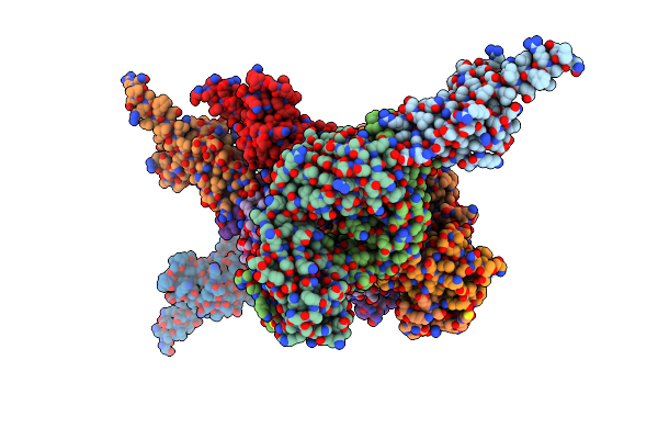 |
Pcrv Fragment With Bound Fab
Organism: Pseudomonas aeruginosa, Homo sapiens
Method: X-RAY DIFFRACTION Resolution:2.78 Å Release Date: 2018-08-01 Classification: IMMUNE SYSTEM |
Organism: Pseudomonas aeruginosa, Homo sapiens
Method: X-RAY DIFFRACTION
Release Date: 2018-08-01
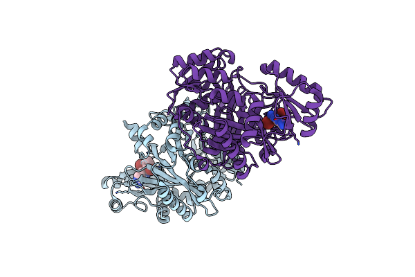 |
Crystal Structure Of Biotin Carboxylase From E.Coli In Complex With Potent Inhibitor 1
Organism: Escherichia coli
Method: X-RAY DIFFRACTION Resolution:2.10 Å Release Date: 2009-01-13 Classification: LIGASE Ligands: LZJ, CL |
Organism: Escherichia coli
Method: X-RAY DIFFRACTION
Release Date: 2009-01-13
Ligands: LZJ, CL
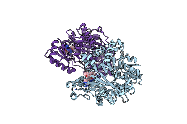 |
Crystal Structure Of Biotin Carboxylase From E.Coli In Complex With Potent Inhibitor 2
Organism: Escherichia coli
Method: X-RAY DIFFRACTION Resolution:2.40 Å Release Date: 2009-01-13 Classification: LIGASE Ligands: LZK |
Organism: Escherichia coli
Method: X-RAY DIFFRACTION
Release Date: 2009-01-13
Ligands: LZK
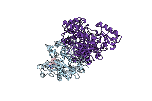 |
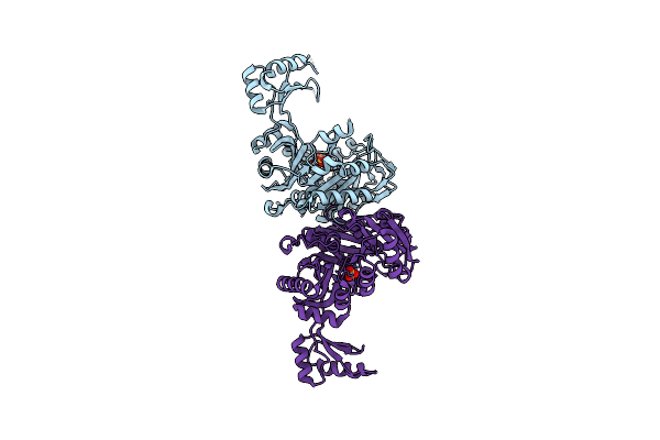
Crystal Structure Of Biotin Carboxylase From E.Coli In Complex With Potent Inhibitor 3
Organism: Escherichia coli
Method: X-RAY DIFFRACTION Resolution:2.31 Å Release Date: 2009-01-13 Classification: LIGASE Ligands: LZL, CL |
Organism: Escherichia coli
Method: X-RAY DIFFRACTION
Release Date: 2009-01-13
Ligands: LZL, CL
 |
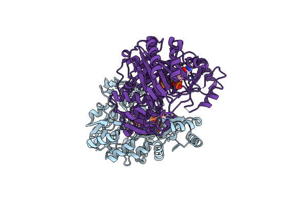
Crystal Structure Of Biotin Carboxylase From Pseudomonas Aeruginosa In Apo Form
Organism: Pseudomonas aeruginosa
Method: X-RAY DIFFRACTION Resolution:2.50 Å Release Date: 2008-09-09 Classification: LIGASE Ligands: SO4 |
Organism: Pseudomonas aeruginosa
Method: X-RAY DIFFRACTION
Release Date: 2008-09-09
Ligands: SO4
 |
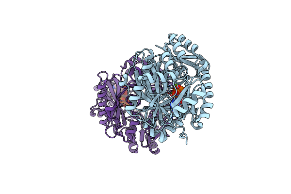
Crystal Structure Of Biotin Carboxylase From E. Coli In Complex With Amppnp And Adp
Organism: Escherichia coli
Method: X-RAY DIFFRACTION Resolution:2.05 Å Release Date: 2008-09-09 Classification: LIGASE Ligands: MG, SO4, ANP, ADP |
Organism: Escherichia coli
Method: X-RAY DIFFRACTION
Release Date: 2008-09-09
Ligands: MG, SO4, ANP, ADP
 |
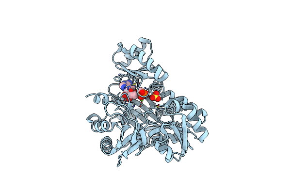
Crystal Structure Of Biotin Carboxylase From S. Aureus Complexed With Amppnp
Organism: Staphylococcus aureus
Method: X-RAY DIFFRACTION Resolution:2.10 Å Release Date: 2008-09-09 Classification: LIGASE Ligands: ANP, MG, CL |
Organism: Staphylococcus aureus
Method: X-RAY DIFFRACTION
Release Date: 2008-09-09
Ligands: ANP, MG, CL
 |
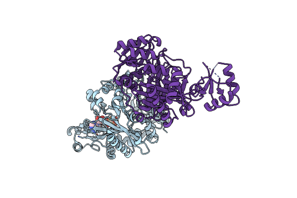
Crystal Structure Of Biotin Carboxylase From Pseudomonas Aeruginosa Complexed With Ampcp
Organism: Pseudomonas aeruginosa
Method: X-RAY DIFFRACTION Resolution:2.41 Å Release Date: 2008-09-09 Classification: LIGASE Ligands: AP2, MG, SO4 |
Organism: Pseudomonas aeruginosa
Method: X-RAY DIFFRACTION
Release Date: 2008-09-09
Ligands: AP2, MG, SO4
 |
Crystal Structure Of Biotin Carboxylase From E. Coli In Complex With Atp Analog, Adpcf2P.
Organism: Escherichia coli
Method: X-RAY DIFFRACTION Resolution:2.60 Å Release Date: 2008-09-09 Classification: LIGASE Ligands: CL, ATF |
Organism: Escherichia coli
Method: X-RAY DIFFRACTION
Release Date: 2008-09-09
Ligands: CL, ATF