

Search Count: 88
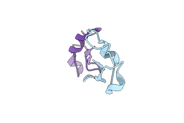 |
High-Resolution Crystal Structure Of Fluoropropylated Cystine Knot, Binding To Alpha-5 Beta-6 Integrin
Organism: Momordica cochinchinensis
Method: X-RAY DIFFRACTION Resolution:1.00 Å Release Date: 2019-08-14 Classification: PROTEIN BINDING |
Organism: Momordica cochinchinensis
Method: X-RAY DIFFRACTION
Release Date: 2019-08-14
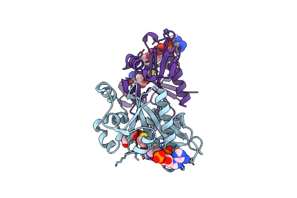 |
The Crystal Structure Of Rv2747 From Mycobacterium Tuberculosis In Complex With Coa And Nlq
Organism: Mycobacterium tuberculosis h37rv
Method: X-RAY DIFFRACTION Resolution:2.30 Å Release Date: 2018-12-26 Classification: TRANSFERASE Ligands: COA, NLQ |
Organism: Mycobacterium tuberculosis h37rv
Method: X-RAY DIFFRACTION
Release Date: 2018-12-26
Ligands: COA, NLQ
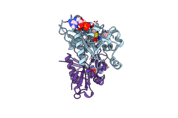 |
The Crystal Structure Of Rv2747 From Mycobacterium Tuberculosis In Complex With Acetyl Coa And L-Arginine
Organism: Mycobacterium tuberculosis h37rv
Method: X-RAY DIFFRACTION Resolution:3.00 Å Release Date: 2018-11-07 Classification: TRANSFERASE Ligands: ARG, ACO |
Organism: Mycobacterium tuberculosis h37rv
Method: X-RAY DIFFRACTION
Release Date: 2018-11-07
Ligands: ARG, ACO
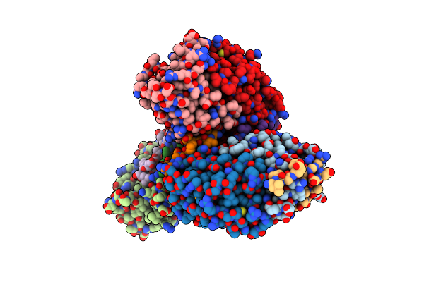 |
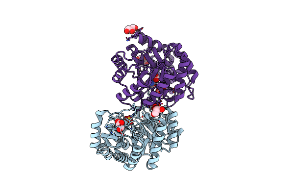
Crystal Structure Of Vapbc15 Complex From Mycobacterium Tuberculosis
Organism: Mycobacterium tuberculosis
Method: X-RAY DIFFRACTION Resolution:2.10 Å Release Date: 2014-11-12 Classification: TOXIN/ANTITOXIN Ligands: MN, MG |
Organism: Mycobacterium tuberculosis
Method: X-RAY DIFFRACTION
Release Date: 2014-11-12
Ligands: MN, MG
 |
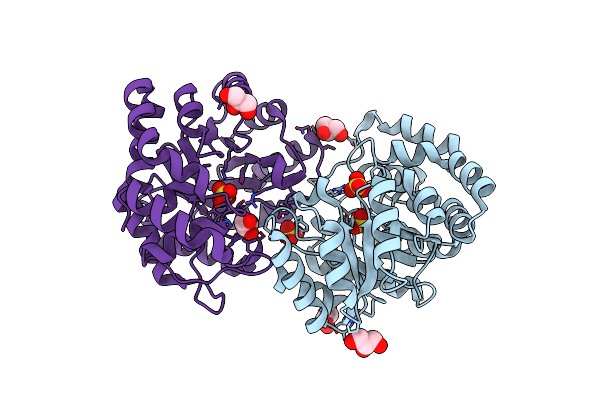
Crystal Structure Of The Complex Of Dhydrodipicolinate Synthase From Acinetobacter Baumannii With Lysine At 2.3A Resolution
Organism: Acinetobacter baumannii
Method: X-RAY DIFFRACTION Resolution:2.30 Å Release Date: 2010-12-29 Classification: LYASE Ligands: GOL, SO4, LYS, ACT |
Organism: Acinetobacter baumannii
Method: X-RAY DIFFRACTION
Release Date: 2010-12-29
Ligands: GOL, SO4, LYS, ACT
 |
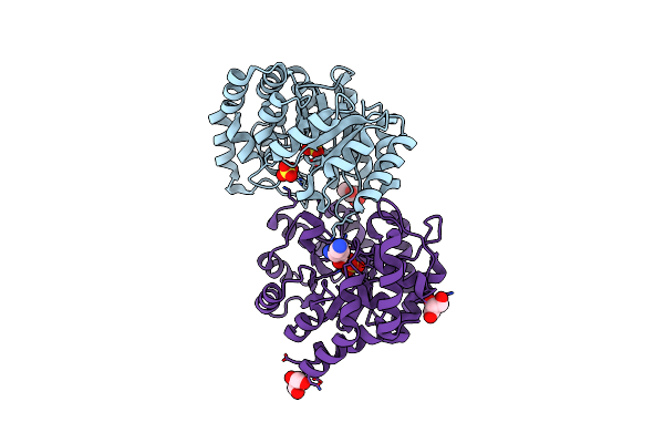
Crystal Structure Of Dhydrodipicolinate Synthase From Acinetobacter Baumannii At 2.8A Resolution
Organism: Acinetobacter baumannii
Method: X-RAY DIFFRACTION Resolution:2.80 Å Release Date: 2010-12-22 Classification: LYASE Ligands: SO4, GOL |
Organism: Acinetobacter baumannii
Method: X-RAY DIFFRACTION
Release Date: 2010-12-22
Ligands: SO4, GOL
 |
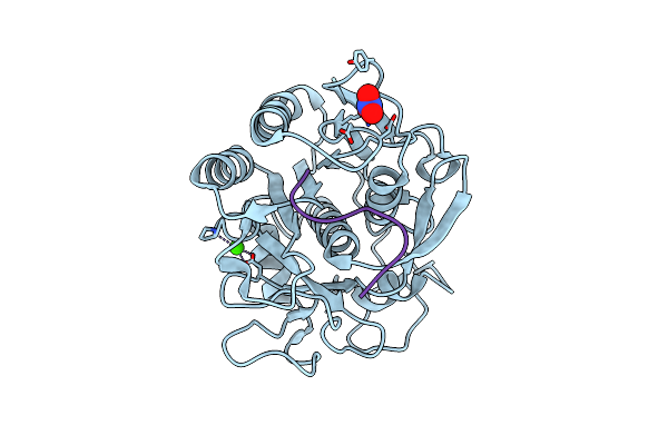
Crystal Structure Of The Complex Of Dhydrodipicolinate Synthase From Acinetobacter Baumannii With Lysine At 2.6A Resolution
Organism: Acinetobacter baumannii
Method: X-RAY DIFFRACTION Resolution:2.60 Å Release Date: 2010-12-22 Classification: LYASE Ligands: SO4, LYS, GOL |
Organism: Acinetobacter baumannii
Method: X-RAY DIFFRACTION
Release Date: 2010-12-22
Ligands: SO4, LYS, GOL
 |
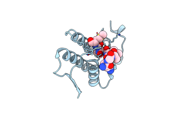
Crystal Structure Of The Complex Of Proteinase K With An Antimicrobial Nonapeptide, At 2.26 A Resolution
Organism: Engyodontium album
Method: X-RAY DIFFRACTION Resolution:2.26 Å Release Date: 2010-10-06 Classification: HYDROLASE Ligands: CA, NO3 |
Organism: Engyodontium album
Method: X-RAY DIFFRACTION
Release Date: 2010-10-06
Ligands: CA, NO3
 |
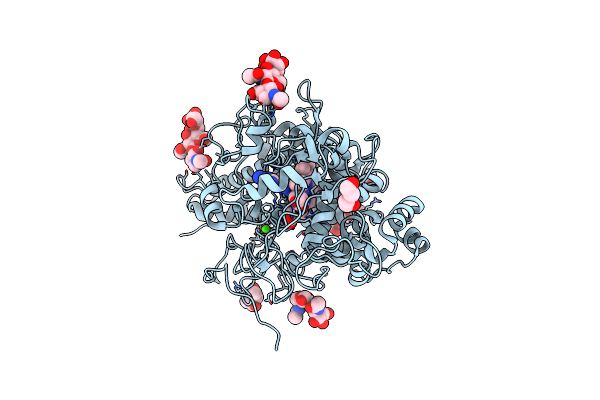
Crystal Structure Of The Complex Formed Between Phospholipase A2 With Beta-Amyloid Fragment, Lys-Gly-Ala-Ile-Ile-Gly-Leu-Met At 1.8 A Resolution
Organism: Homo sapiens, Naja sagittifera
Method: X-RAY DIFFRACTION Resolution:1.80 Å Release Date: 2010-07-21 Classification: HYDROLASE Ligands: CA |
Organism: Homo sapiens, Naja sagittifera
Method: X-RAY DIFFRACTION
Release Date: 2010-07-21
Ligands: CA
 |
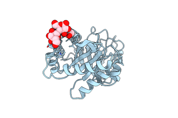
Crystal Structure Of The Complex Of Lactoperoxidase With A Potent Inhibitor Amino-Triazole At 2.2A Resolution
Organism: Bos taurus
Method: X-RAY DIFFRACTION Resolution:2.25 Å Release Date: 2010-05-26 Classification: Oxidoreductase/Oxidoreductase inhibitor Ligands: HEM, CA, IOD, 3TR, MRD, PEG |
Organism: Bos taurus
Method: X-RAY DIFFRACTION
Release Date: 2010-05-26
Ligands: HEM, CA, IOD, 3TR, MRD, PEG
 |
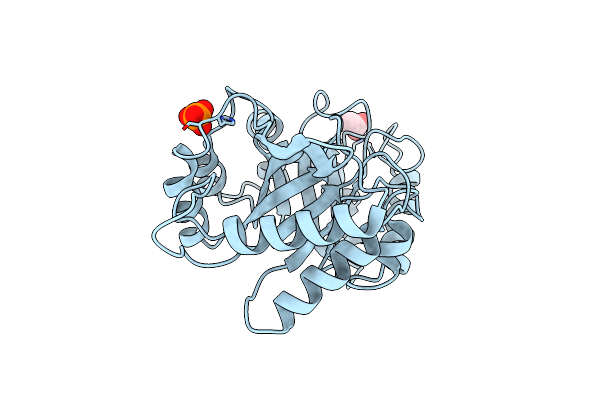
Crystal Structure Of The Complex Of Xylanase Gh-11 And Alpha Amylase Inhibitor Protein With Cellobiose At 2.4 A Resolution
Organism: Scadoxus multiflorus
Method: X-RAY DIFFRACTION Resolution:2.40 Å Release Date: 2010-05-05 Classification: PROTEIN BINDING Ligands: PO4, ACT |
Organism: Scadoxus multiflorus
Method: X-RAY DIFFRACTION
Release Date: 2010-05-05
Ligands: PO4, ACT
 |
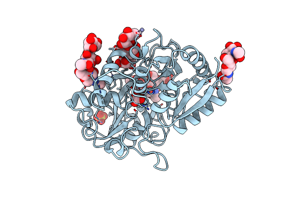
Structural Characterization And Binding Studies Of A Plant Pathogenesis Related Protein Heamanthin From Haemanthus Multiflorus Reveal Its Dual Inhibitory Effects Against Xylanase And Alpha-Amylase
Organism: Scadoxus multiflorus
Method: X-RAY DIFFRACTION Resolution:2.00 Å Release Date: 2010-04-28 Classification: PROTEIN BINDING Ligands: ACT, PO4 |
Organism: Scadoxus multiflorus
Method: X-RAY DIFFRACTION
Release Date: 2010-04-28
Ligands: ACT, PO4
 |
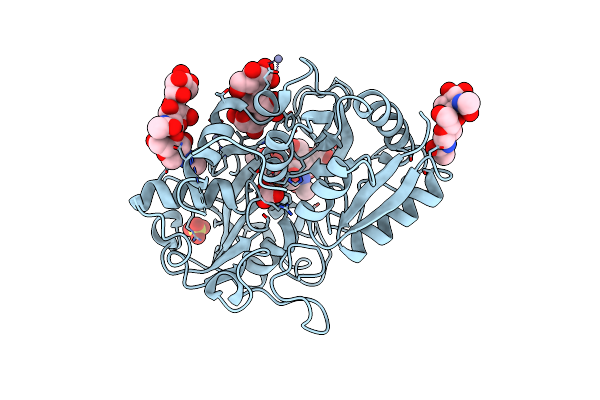
Crystal Structure Of The Complex Of C-Lobe Of Bovine Lactoferrin With Dextrin At 1.9 A Resolution
Organism: Bos taurus
Method: X-RAY DIFFRACTION Resolution:1.91 Å Release Date: 2010-02-16 Classification: HYDROLASE Ligands: SO4, ZN, FE, CO3 |
Organism: Bos taurus
Method: X-RAY DIFFRACTION
Release Date: 2010-02-16
Ligands: SO4, ZN, FE, CO3
 |
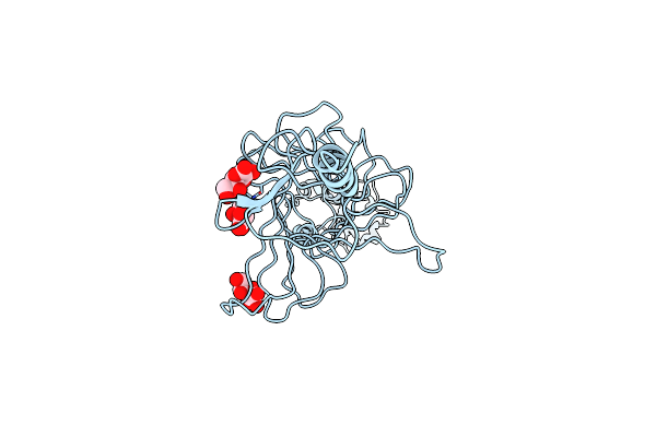
Removal Of Sugars And Sugars-Like Molecules From The Solution By C-Lobe Of Lactoferrin: Crystal Structure Of The Complex Of C-Lobe With Beta-D-Glucopyranosyl-(1->4)-Beta-D-Galactopyranosyl-(1->4)-Alpha-D-Glucopyranose At 1.9 A Resolution
Organism: Bos taurus
Method: X-RAY DIFFRACTION Resolution:1.91 Å Release Date: 2009-10-13 Classification: HYDROLASE Ligands: SO4, ZN, FE, CO3 |
Organism: Bos taurus
Method: X-RAY DIFFRACTION
Release Date: 2009-10-13
Ligands: SO4, ZN, FE, CO3
 |
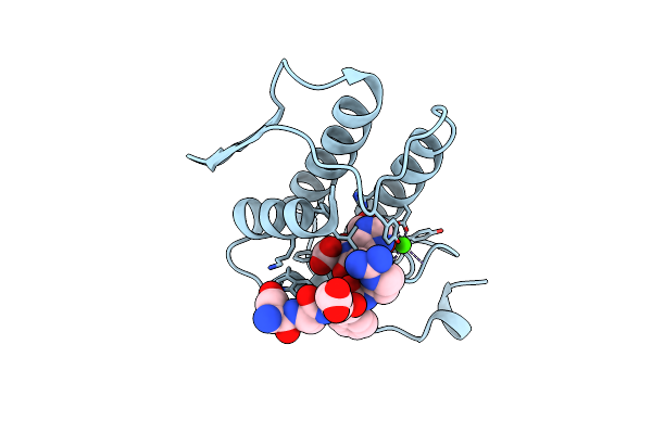
Crystal Structure Of The Complex Of Hyaluranidase Trimer With Ascorbic Acid At 3.1 A Resolution Reveals The Locations Of Three Binding Sites
Organism: Streptococcus pyogenes
Method: X-RAY DIFFRACTION Resolution:3.10 Å Release Date: 2009-09-29 Classification: LYASE Ligands: ASC |
Organism: Streptococcus pyogenes
Method: X-RAY DIFFRACTION
Release Date: 2009-09-29
Ligands: ASC
 |
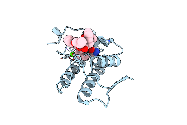
Phospholipase A2 Prevents The Aggregation Of Amyloid Beta Peptides: Crystal Structure Of The Complex Of Phospholipase A2 With Octapeptide Fragment Of Amyloid Beta Peptide, Asp-Ala-Glu-Phe-Arg-His-Asp-Ser At 2 A Resolution
Organism: Naja sagittifera
Method: X-RAY DIFFRACTION Resolution:2.03 Å Release Date: 2009-09-29 Classification: HYDROLASE Ligands: CA |
Organism: Naja sagittifera
Method: X-RAY DIFFRACTION
Release Date: 2009-09-29
Ligands: CA
 |
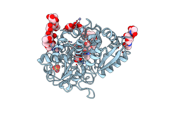
Crystal Structure Of The Complex Formed Between Phospholipase A2 And A Hexapeptide Fragment Of Amyloid Beta Peptide, Lys-Leu-Val-Phe-Phe-Ala At 1.2 A Resolution
Organism: Naja sagittifera
Method: X-RAY DIFFRACTION Resolution:1.20 Å Release Date: 2009-09-29 Classification: HYDROLASE Ligands: CA |
Organism: Naja sagittifera
Method: X-RAY DIFFRACTION
Release Date: 2009-09-29
Ligands: CA
 |
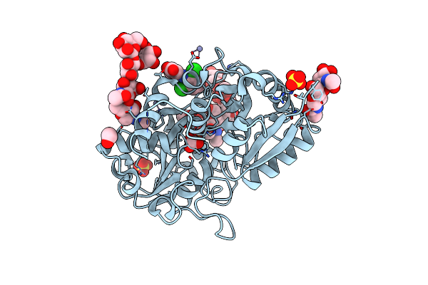
Structural Basis Of The Prevention Of Nsaid-Induced Damage Of The Gastrointestinal Tract By C-Terminal Half (C-Lobe) Of Bovine Colostrum Protein Lactoferrin: Binding And Structural Studies Of The C-Lobe Complex With Aspirin
Organism: Bos taurus
Method: X-RAY DIFFRACTION Resolution:2.00 Å Release Date: 2009-08-11 Classification: METAL BINDING PROTEIN Ligands: AIN, SO4, ZN, FE, CO3, EOH |
Organism: Bos taurus
Method: X-RAY DIFFRACTION
Release Date: 2009-08-11
Ligands: AIN, SO4, ZN, FE, CO3, EOH
 |
Structural Basis Of The Prevention Of Nsaid-Induced Damage Of The Gastrointestinal Tract By C-Terminal Half (C-Lobe) Of Bovine Colostrum Protein Lactoferrin: Binding And Structural Studies Of C-Lobe Complex With Diclofenac
Organism: Bos taurus
Method: X-RAY DIFFRACTION Resolution:1.40 Å Release Date: 2009-08-11 Classification: METAL BINDING PROTEIN Ligands: FE, CO3, DIF, SO4, ZN, EOH |
Organism: Bos taurus
Method: X-RAY DIFFRACTION
Release Date: 2009-08-11
Ligands: FE, CO3, DIF, SO4, ZN, EOH
 |
Structural Basis Of The Prevention Of Nsaid-Induced Damage Of The Gastrointestinal Tract By C-Terminal Half (C-Lobe) Of Bovine Colostrum Protein Lactoferrin: Binding And Structural Studies Of C-Lobe Complex With Indomethacin
Organism: Bos taurus
Method: X-RAY DIFFRACTION Resolution:2.20 Å Release Date: 2009-08-11 Classification: METAL BINDING PROTEIN Ligands: ZN, FE, CO3, SO4, NAG, IMN |
Organism: Bos taurus
Method: X-RAY DIFFRACTION
Release Date: 2009-08-11
Ligands: ZN, FE, CO3, SO4, NAG, IMN