

Search Count: 44
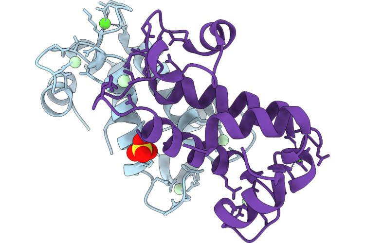 |
X-Ray Crystal Structure Of Ancylobacter Lacus Lanm Bound To Nd(Iii) And Ca(Ii)
Organism: Ancylobacter lacus
Method: X-RAY DIFFRACTION Resolution:0.99 Å Release Date: 2026-08-19 Classification: METAL BINDING PROTEIN Ligands: ND, CA, SO4 |
Organism: Ancylobacter lacus
Method: X-RAY DIFFRACTION
Release Date: 2026-08-19
Ligands: ND, CA, SO4
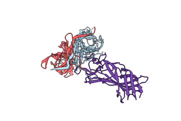 |
Cryo-Em Structure Of Partial Vp4 From Simian Rotavirus Sa11
|
Organism: Rotavirus a
Method: ELECTRON MICROSCOPY
Release Date: 2025-02-12
 |
Cryo-Em Structure Of Simian Rotavirus Sa11 Vp4 In Complex With Nab 7H13
Organism: Mus musculus, Rotavirus a
Method: ELECTRON MICROSCOPY Release Date: 2025-02-12 Classification: VIRUS |
Organism: Mus musculus, Rotavirus a
Method: ELECTRON MICROSCOPY
Release Date: 2025-02-12
 |
Cryo-Em Structure Of Simian Rotavirus Sa11 Vp4 In Complex With Nab 7H13-I54G Mutant (Left Side)
Organism: Mus musculus, Rotavirus a
Method: ELECTRON MICROSCOPY Release Date: 2025-02-12 Classification: VIRUS |
Organism: Mus musculus, Rotavirus a
Method: ELECTRON MICROSCOPY
Release Date: 2025-02-12
 |
Cryo-Em Structure Of Simian Rotavirus Sa11 Vp4 In Complex With Nab 7H13-I54G Mutant (Right Side)
Organism: Mus musculus, Rotavirus a
Method: ELECTRON MICROSCOPY Release Date: 2025-02-12 Classification: VIRUS |
Organism: Mus musculus, Rotavirus a
Method: ELECTRON MICROSCOPY
Release Date: 2025-02-12
 |
Structural Basis For The Linker Histone H5-Nucleosome Binding And Chromatin Compaction
Organism: Synthetic construct, Xenopus laevis, Gallus gallus
Method: ELECTRON MICROSCOPY Release Date: 2024-09-11 Classification: GENE REGULATION |
Organism: Synthetic construct, Xenopus laevis, Gallus gallus
Method: ELECTRON MICROSCOPY
Release Date: 2024-09-11
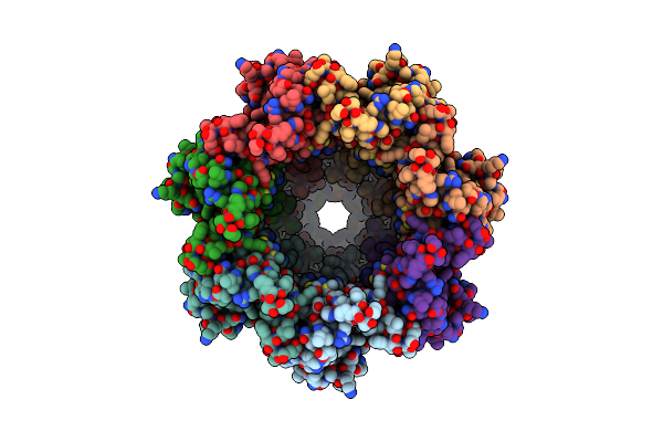 |
The Cryo-Em Structure Of Human Pannexin 1
Organism: Homo sapiens
Method: ELECTRON MICROSCOPY Release Date: 2020-04-15 Classification: TRANSPORT PROTEIN |
Organism: Homo sapiens
Method: ELECTRON MICROSCOPY
Release Date: 2020-04-15
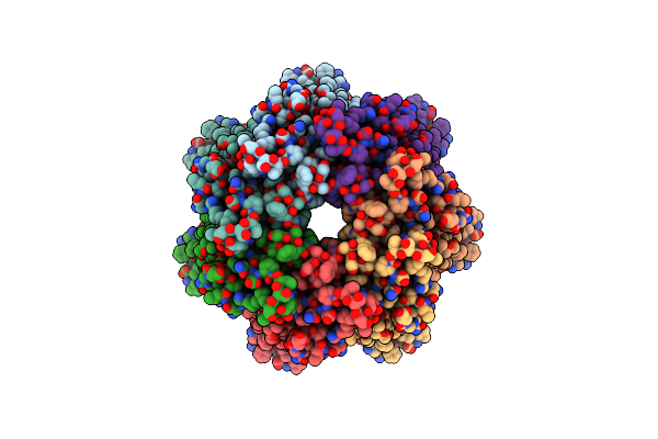 |
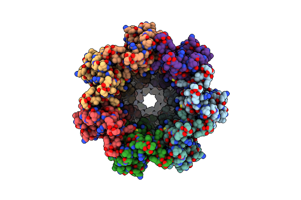
The Cryo-Em Structure Of Human Pannexin 1 With D376E/D379E Mutation
Organism: Homo sapiens
Method: ELECTRON MICROSCOPY Release Date: 2020-04-15 Classification: TRANSPORT PROTEIN |
Organism: Homo sapiens
Method: ELECTRON MICROSCOPY
Release Date: 2020-04-15
 |
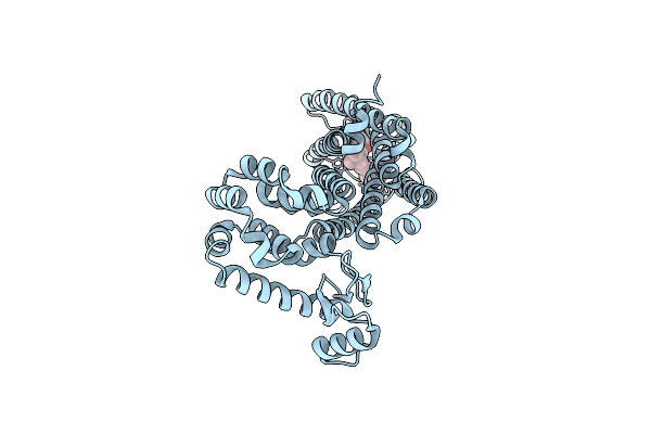
The Cryo-Em Structure Of Human Pannexin 1 In The Presence Of Cbx
Organism: Homo sapiens
Method: ELECTRON MICROSCOPY Release Date: 2020-04-15 Classification: TRANSPORT PROTEIN |
Organism: Homo sapiens
Method: ELECTRON MICROSCOPY
Release Date: 2020-04-15
 |
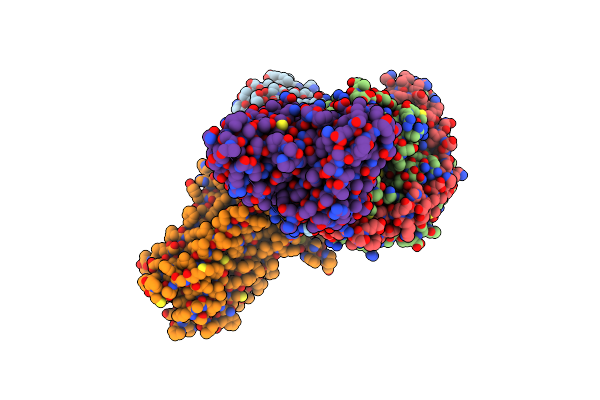
Crystal Structure Of An Agonist Bound Gpcr
Organism: Homo sapiens, Enterobacteria phage rb59
Method: X-RAY DIFFRACTION Resolution:3.20 Å Release Date: 2020-02-12 Classification: MEMBRANE PROTEIN Ligands: E3R |
Organism: Homo sapiens, Enterobacteria phage rb59
Method: X-RAY DIFFRACTION
Release Date: 2020-02-12
Ligands: E3R
 |
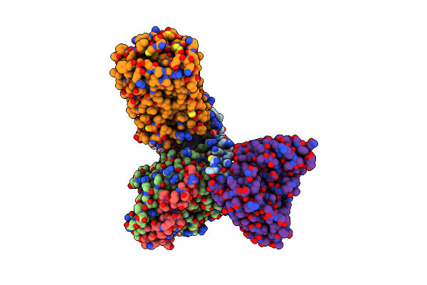
Cryo-Em Structure Of A Class A Gpcr With G Protein Complex
Organism: Homo sapiens
Method: ELECTRON MICROSCOPY Release Date: 2020-02-12 Classification: MEMBRANE PROTEIN Ligands: E3R |
Organism: Homo sapiens
Method: ELECTRON MICROSCOPY
Release Date: 2020-02-12
Ligands: E3R
 |
Cryo-Em Structure Of Cb1-G Protein Complex
Organism: Homo sapiens
Method: ELECTRON MICROSCOPY Release Date: 2020-02-12 Classification: MEMBRANE PROTEIN Ligands: 8D0 |
Organism: Homo sapiens
Method: ELECTRON MICROSCOPY
Release Date: 2020-02-12
Ligands: 8D0
 |
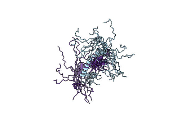
Solution Structure Of Peptide Cq2 From Chenopodium Quinoa
Organism: Chenopodium quinoa
Method: SOLUTION NMR Release Date: 2019-05-15 Classification: PLANT PROTEIN |
Organism: Chenopodium quinoa
Method: SOLUTION NMR
Release Date: 2019-05-15
 |
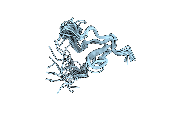
Solution Nmr Structure Of The Colied-Coil Palb2 Homodimer
|
Organism: Mus musculus
Method: SOLUTION NMR
Release Date: 2018-10-17
 |
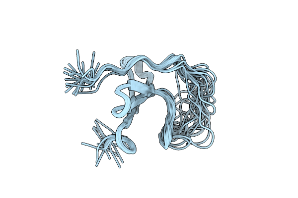
Nmr Structure Of Yap1-2 Ww1 Domain With Lats1 Ppxy Motif Complex
Organism: Homo sapiens
Method: SOLUTION NMR Release Date: 2018-05-02 Classification: SIGNALING PROTEIN |
Organism: Homo sapiens
Method: SOLUTION NMR
Release Date: 2018-05-02
 |
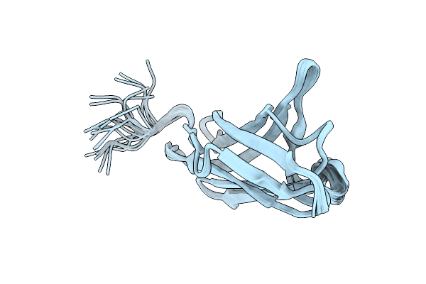
Nmr Structure Of Yap1-2 Ww2 Domain With Lats1 Ppxy Motif Complex
Organism: Homo sapiens
Method: SOLUTION NMR Release Date: 2018-05-02 Classification: SIGNALING PROTEIN |
Organism: Homo sapiens
Method: SOLUTION NMR
Release Date: 2018-05-02
 |
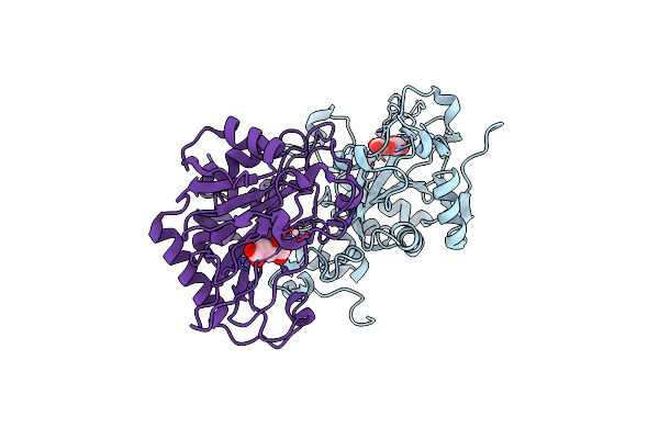
Solution Structure Of The Igi Domain Of Cd147
|
Organism: Homo sapiens
Method: SOLUTION NMR
Release Date: 2018-04-11
 |
Structure Of Ftmox1 With A-Ketoglutarate As Co-Substrate
Organism: Neosartorya fumigata
Method: X-RAY DIFFRACTION Resolution:2.54 Å Release Date: 2015-11-04 Classification: OXIDOREDUCTASE Ligands: AKG, FE2, CO |
Organism: Neosartorya fumigata
Method: X-RAY DIFFRACTION
Release Date: 2015-11-04
Ligands: AKG, FE2, CO
 |
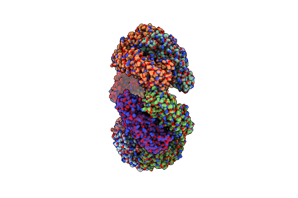
Structure Of Ftmox1 Apo With Metal Iron
Organism: Neosartorya fumigata
Method: X-RAY DIFFRACTION Resolution:1.95 Å Release Date: 2015-11-04 Classification: OXIDOREDUCTASE Ligands: FE2, SO4, CO, MES |
Organism: Neosartorya fumigata
Method: X-RAY DIFFRACTION
Release Date: 2015-11-04
Ligands: FE2, SO4, CO, MES
 |
Crystal Structure Of Dodecameric Iron-Containing Heptosyltransferase Tibc In Complex With Adp-D-Beta-D-Heptose At 3.9 Angstrom Resolution
Organism: Escherichia coli dec13e
Method: X-RAY DIFFRACTION Resolution:3.88 Å Release Date: 2014-11-05 Classification: TRANSFERASE Ligands: FE, AQH, EDO |
Organism: Escherichia coli dec13e
Method: X-RAY DIFFRACTION
Release Date: 2014-11-05
Ligands: FE, AQH, EDO