

Search Count: 21
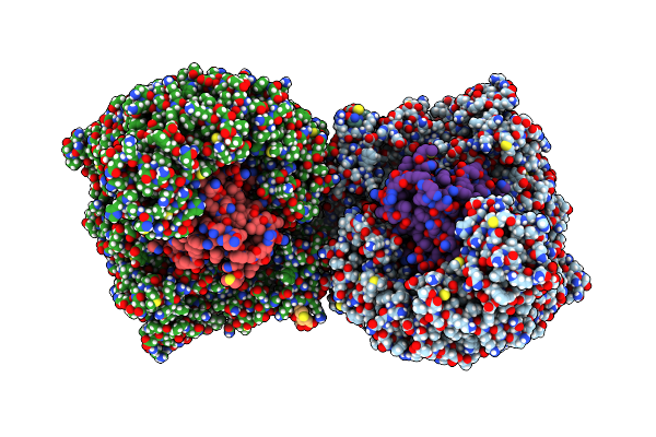 |
Cryo-Em Structure Of The Hiv-1 Nuclear Export Complex
Organism: Homo sapiens, Human immunodeficiency virus 1
Method: ELECTRON MICROSCOPY Release Date: 2025-04-30 Classification: RNA BINDING PROTEIN/RNA |
Organism: Homo sapiens, Human immunodeficiency virus 1
Method: ELECTRON MICROSCOPY
Release Date: 2025-04-30
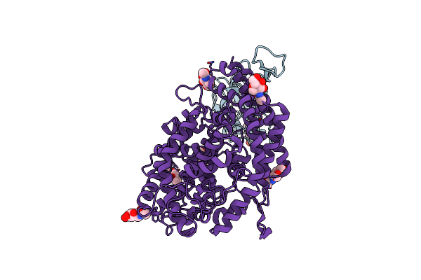 |
Sars-Cov-2 Wuhan-Hu-1-Spike-Rbd Bound To Linker Variant Of Affinity Matured Ace2 Mimetic Cvd432
Organism: Severe acute respiratory syndrome coronavirus 2, Homo sapiens
Method: ELECTRON MICROSCOPY Release Date: 2022-08-31 Classification: VIRAL PROTEIN/ANTIVIRAL PROTEIN Ligands: NAG |
Organism: Severe acute respiratory syndrome coronavirus 2, Homo sapiens
Method: ELECTRON MICROSCOPY
Release Date: 2022-08-31
Ligands: NAG
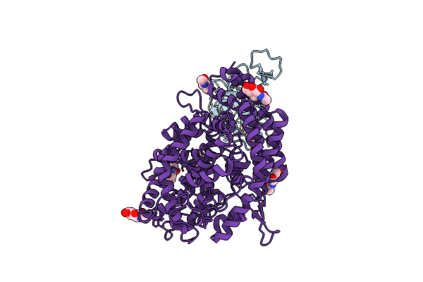 |
Sars-Cov-2 Wuhan-Hu-1-Spike-Rbd Bound To Computationally Engineered Ace2 Mimetic Cvd293
Organism: Severe acute respiratory syndrome coronavirus 2, Homo sapiens
Method: ELECTRON MICROSCOPY Release Date: 2022-08-31 Classification: VIRAL PROTEIN/ANTIVIRAL PROTEIN Ligands: NAG |
Organism: Severe acute respiratory syndrome coronavirus 2, Homo sapiens
Method: ELECTRON MICROSCOPY
Release Date: 2022-08-31
Ligands: NAG
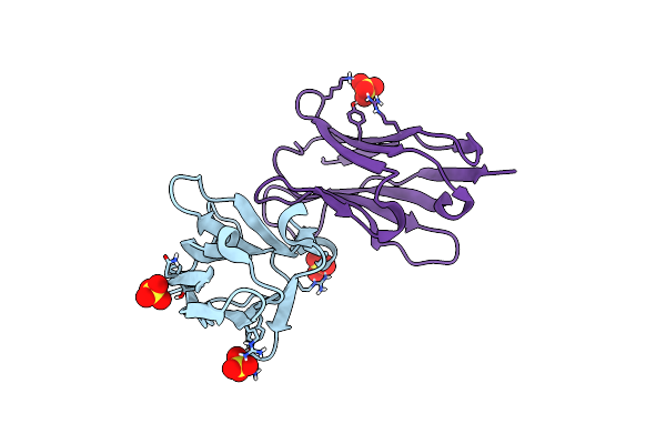 |
Structure Of Anti-Sars-Cov-2 Spike Nanobody Mnb6
Organism: Synthetic construct
Method: X-RAY DIFFRACTION Resolution:2.05 Å Release Date: 2020-11-25 Classification: IMMUNE SYSTEM Ligands: SO4, CL |
Organism: Synthetic construct
Method: X-RAY DIFFRACTION
Release Date: 2020-11-25
Ligands: SO4, CL
 |
Sars-Cov-2 Spike In Complex With Neutralizing Nanobody Nb6
Organism: Severe acute respiratory syndrome coronavirus 2, Synthetic construct
Method: ELECTRON MICROSCOPY Release Date: 2020-11-11 Classification: VIRAL PROTEIN/IMMUNE SYSTEM Ligands: NAG |
Organism: Severe acute respiratory syndrome coronavirus 2, Synthetic construct
Method: ELECTRON MICROSCOPY
Release Date: 2020-11-11
Ligands: NAG
 |
Sars-Cov-2 Spike In Complex With Neutralizing Nanobody Mnb6
Organism: Severe acute respiratory syndrome coronavirus 2, Synthetic construct
Method: ELECTRON MICROSCOPY Release Date: 2020-11-11 Classification: VIRAL PROTEIN/IMMUNE SYSTEM Ligands: NAG |
Organism: Severe acute respiratory syndrome coronavirus 2, Synthetic construct
Method: ELECTRON MICROSCOPY
Release Date: 2020-11-11
Ligands: NAG
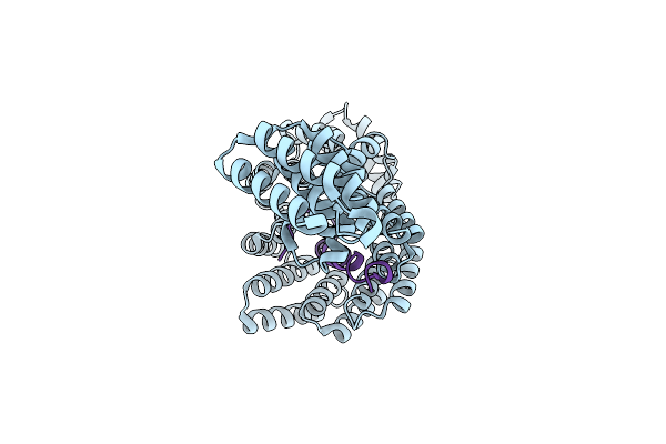 |
Human Tom70 In Complex With Sars Cov2 Orf9B
Organism: Homo sapiens, Severe acute respiratory syndrome coronavirus 2
Method: ELECTRON MICROSCOPY Release Date: 2020-10-21 Classification: VIRAL PROTEIN |
Organism: Homo sapiens, Severe acute respiratory syndrome coronavirus 2
Method: ELECTRON MICROSCOPY
Release Date: 2020-10-21
 |
Plps (Inactive Glutaminase Mutant) Co-Crystallized With Glutamine And R5P.
Organism: Geobacillus kaustophilus
Method: X-RAY DIFFRACTION Resolution:2.70 Å Release Date: 2015-01-14 Classification: TRANSFERASE |
Organism: Geobacillus kaustophilus
Method: X-RAY DIFFRACTION
Release Date: 2015-01-14
 |
Pdxs (G. Stearothermophilus) Co-Crystallized With R5P
Organism: Geobacillus kaustophilus
Method: X-RAY DIFFRACTION Resolution:2.70 Å Release Date: 2015-01-14 Classification: TRANSFERASE |
Organism: Geobacillus kaustophilus
Method: X-RAY DIFFRACTION
Release Date: 2015-01-14
 |
Pdxs (G. Stearothermophilus) Co-Crystallized With R5P In The Presence Of Ammonia.
Organism: Geobacillus kaustophilus (strain hta426)
Method: X-RAY DIFFRACTION Resolution:2.30 Å Release Date: 2015-01-14 Classification: LYASE Ligands: CAC, PO4, R5P, EDO |
Organism: Geobacillus kaustophilus (strain hta426)
Method: X-RAY DIFFRACTION
Release Date: 2015-01-14
Ligands: CAC, PO4, R5P, EDO
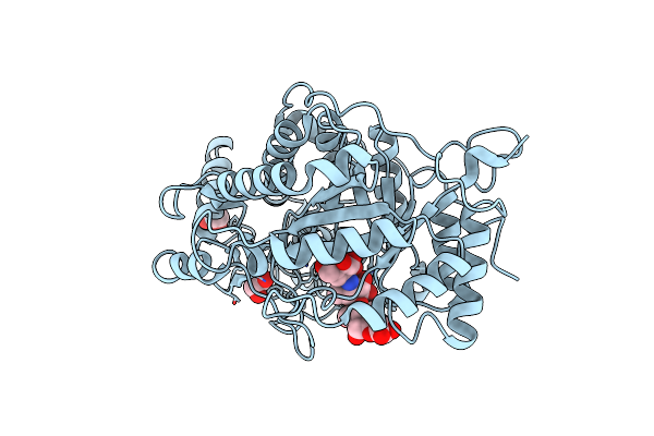 |
Crystal Structure Of Barley Beta-Amylase Complexed With Acarbose
Organism: Hordeum vulgare
Method: X-RAY DIFFRACTION Resolution:1.31 Å Release Date: 2010-12-01 Classification: HYDROLASE Ligands: EDO |
Organism: Hordeum vulgare
Method: X-RAY DIFFRACTION
Release Date: 2010-12-01
Ligands: EDO
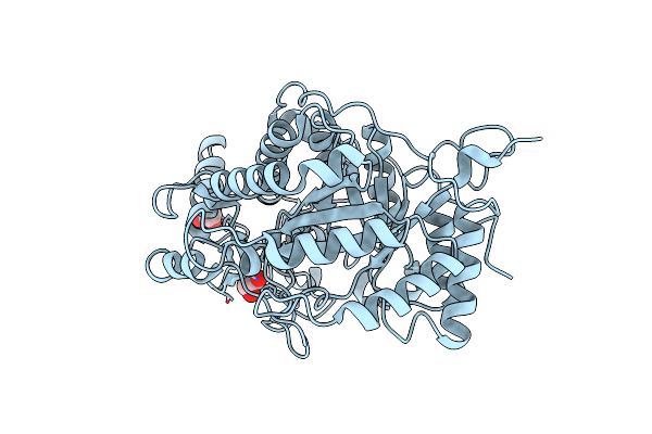 |
Crystal Structure Of Barley Beta-Amylase At Atomic Resolution
Organism: Hordeum vulgare
Method: X-RAY DIFFRACTION Resolution:0.97 Å Release Date: 2010-12-01 Classification: HYDROLASE Ligands: EDO |
Organism: Hordeum vulgare
Method: X-RAY DIFFRACTION
Release Date: 2010-12-01
Ligands: EDO
 |
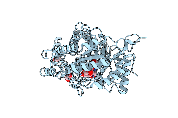
Crystal Structure Of Barley Beta-Amylase Complexed With Alpha- Cyclodextrin
Organism: Hordeum vulgare
Method: X-RAY DIFFRACTION Resolution:1.21 Å Release Date: 2010-12-01 Classification: HYDROLASE Ligands: EDO |
Organism: Hordeum vulgare
Method: X-RAY DIFFRACTION
Release Date: 2010-12-01
Ligands: EDO
 |
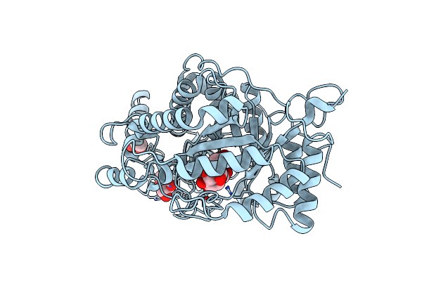
Crystal Structure Of Barley Beta-Amylase Complexed With 4-O-Alpha-D- Glucopyranosylmoranoline
Organism: Hordeum vulgare
Method: X-RAY DIFFRACTION Resolution:1.80 Å Release Date: 2010-12-01 Classification: HYDROLASE Ligands: EDO |
Organism: Hordeum vulgare
Method: X-RAY DIFFRACTION
Release Date: 2010-12-01
Ligands: EDO
 |
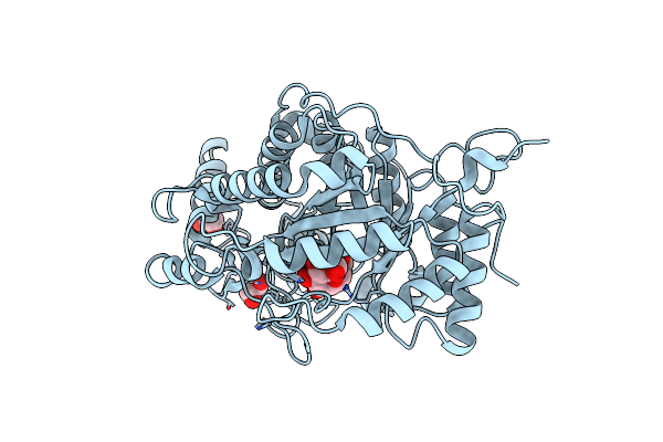
Crystal Structure Of Barley Beta-Amylase Complexed With 2,3- Epoxypropyl-Alpha-D-Glucopyranoside
Organism: Hordeum vulgare
Method: X-RAY DIFFRACTION Resolution:1.20 Å Release Date: 2010-12-01 Classification: HYDROLASE Ligands: EPG, EDO |
Organism: Hordeum vulgare
Method: X-RAY DIFFRACTION
Release Date: 2010-12-01
Ligands: EPG, EDO
 |
Crystal Structure Of Barley Beta-Amylase Complexed With 3,4- Epoxybutyl Alpha-D-Glucopyranoside
Organism: Hordeum vulgare
Method: X-RAY DIFFRACTION Resolution:1.30 Å Release Date: 2010-12-01 Classification: HYDROLASE Ligands: EDO, J5B, EBQ |
Organism: Hordeum vulgare
Method: X-RAY DIFFRACTION
Release Date: 2010-12-01
Ligands: EDO, J5B, EBQ
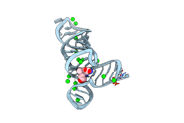 |
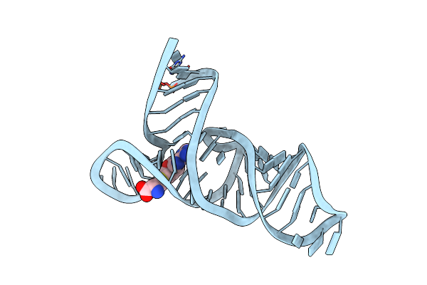
Crystal Structure Of The Smk Box (Sam-Iii) Riboswitch With Sam
Method: X-RAY DIFFRACTION
Resolution:2.25 Å Release Date: 2008-10-07 Classification: RNA Ligands: SR, SAM |
Method: X-RAY DIFFRACTION
Release Date: 2008-10-07
Ligands: SR, SAM
 |
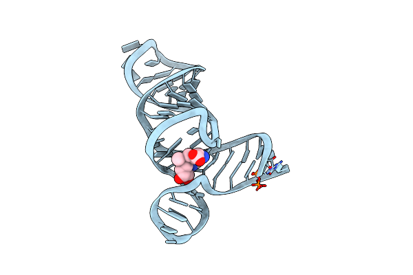
Crystal Structures Of The Smk Box (Sam-Iii) Riboswitch With Sah
Method: X-RAY DIFFRACTION
Resolution:2.90 Å Release Date: 2008-10-07 Classification: RNA Ligands: SR, SAH |
Method: X-RAY DIFFRACTION
Release Date: 2008-10-07
Ligands: SR, SAH
 |
Crystal Structures Of The Smk Box (Sam-Iii) Riboswitch With Se-Sam
Method: X-RAY DIFFRACTION
Resolution:2.70 Å Release Date: 2008-10-07 Classification: RNA Ligands: SR, EEM |
Method: X-RAY DIFFRACTION
Release Date: 2008-10-07
Ligands: SR, EEM
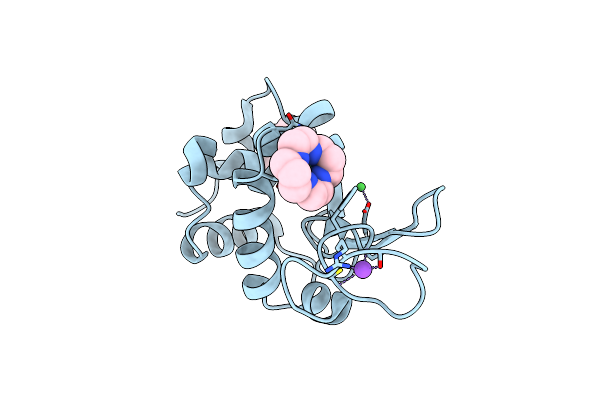 |
Structure Of Hen Egg White Lysozyme Soaked With Ni2-Xylylbicyclam
Organism: Gallus gallus
Method: X-RAY DIFFRACTION Resolution:1.75 Å Release Date: 2007-04-10 Classification: HYDROLASE Ligands: CL, NA, NI, MM5 |
Organism: Gallus gallus
Method: X-RAY DIFFRACTION
Release Date: 2007-04-10
Ligands: CL, NA, NI, MM5