

Search Count: 36
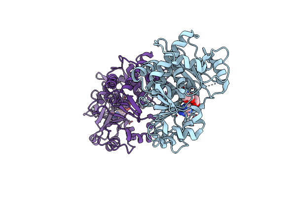 |
Crystal Structure Of The L. Plantarum D-Alanine Ligase Dlta
Organism: Lactiplantibacillus plantarum subsp. plantarum nc8
Method: X-RAY DIFFRACTION Resolution:2.01 Å Release Date: 2022-08-03 Classification: CYTOSOLIC PROTEIN Ligands: DAL, AMP |
Organism: Lactiplantibacillus plantarum subsp. plantarum nc8
Method: X-RAY DIFFRACTION
Release Date: 2022-08-03
Ligands: DAL, AMP
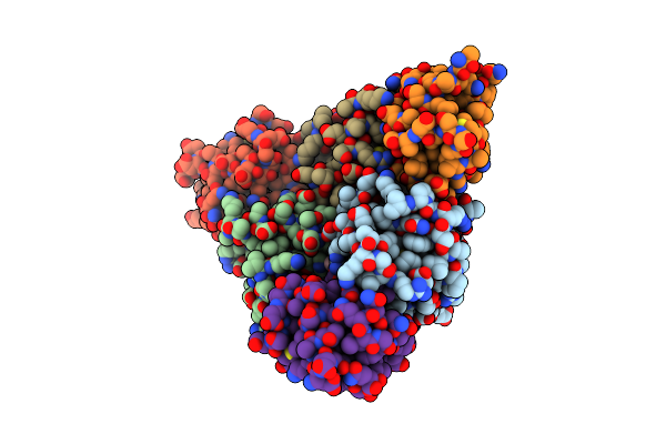 |
Crystal Structure Of The L. Plantarum Acyl Carrier Protein Synthase (Acps)In Complex With D-Alanyl Carrier Protein (Dltc1)
Organism: Lactiplantibacillus plantarum subsp. plantarum nc8
Method: X-RAY DIFFRACTION Resolution:1.88 Å Release Date: 2022-08-03 Classification: CYTOSOLIC PROTEIN Ligands: PNS, TRS |
Organism: Lactiplantibacillus plantarum subsp. plantarum nc8
Method: X-RAY DIFFRACTION
Release Date: 2022-08-03
Ligands: PNS, TRS
 |
Hyperlytic Variant Of Tae1, Type Vi Secretion Amidase Effector 1, From Pseudomonas Aeruginosa (Cys110Ser)
Organism: Pseudomonas aeruginosa (strain atcc 15692 / dsm 22644 / cip 104116 / jcm 14847 / lmg 12228 / 1c / prs 101 / pao1)
Method: X-RAY DIFFRACTION Resolution:1.71 Å Release Date: 2022-07-06 Classification: HYDROLASE |
 |
Crystal Structure Of The Helical Cell Shape Determining Protein Pgp2 From Campylobacter Jejuni
Organism: Campylobacter jejuni subsp. jejuni
Method: X-RAY DIFFRACTION Resolution:1.50 Å Release Date: 2021-03-17 Classification: HYDROLASE |
Organism: Campylobacter jejuni subsp. jejuni
Method: X-RAY DIFFRACTION
Release Date: 2021-03-17
 |
Crystal Structure Of The Helical Cell Shape Determining Protein Pgp2 (K307A Mutant) From Campylobacter Jejuni
Organism: Campylobacter jejuni subsp. jejuni
Method: X-RAY DIFFRACTION Resolution:1.85 Å Release Date: 2021-03-17 Classification: HYDROLASE |
Organism: Campylobacter jejuni subsp. jejuni
Method: X-RAY DIFFRACTION
Release Date: 2021-03-17
 |
Spor Protein Dedd
|
Organism: Escherichia coli
Method: SOLUTION NMR
Release Date: 2020-11-11
 |
Structure Of The Globular C-Terminal Domain Of P. Aeruginosa Lpop
Organism: Pseudomonas aeruginosa
Method: X-RAY DIFFRACTION Resolution:2.20 Å Release Date: 2020-05-06 Classification: PROTEIN BINDING Ligands: SO4, PGE |
Organism: Pseudomonas aeruginosa
Method: X-RAY DIFFRACTION
Release Date: 2020-05-06
Ligands: SO4, PGE
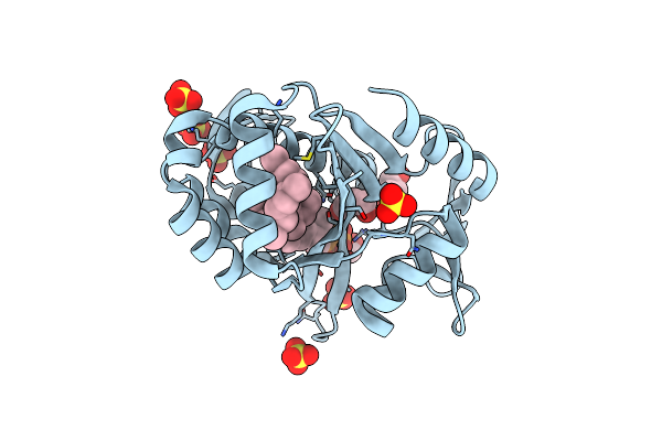 |
Crystal Structure Of S. Aureus Lcpa In Complex With Octaprenyl-Pyrophosphate-Glcnac
Organism: Staphylococcus aureus (strain n315)
Method: X-RAY DIFFRACTION Resolution:1.90 Å Release Date: 2020-01-29 Classification: TRANSFERASE Ligands: Q5S, SO4, GOL |
Organism: Staphylococcus aureus (strain n315)
Method: X-RAY DIFFRACTION
Release Date: 2020-01-29
Ligands: Q5S, SO4, GOL
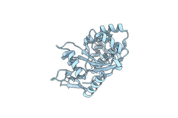 |
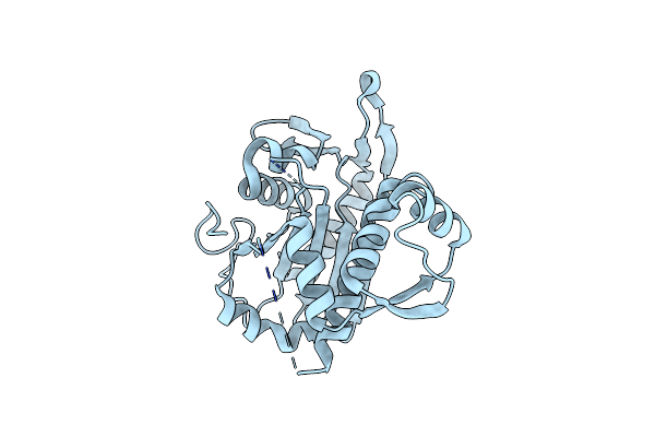
Crystal Structure Of B. Subtilis Tagv
Organism: Bacillus subtilis (strain 168)
Method: X-RAY DIFFRACTION Resolution:1.60 Å Release Date: 2020-01-29 Classification: TRANSFERASE |
Organism: Bacillus subtilis (strain 168)
Method: X-RAY DIFFRACTION
Release Date: 2020-01-29
 |
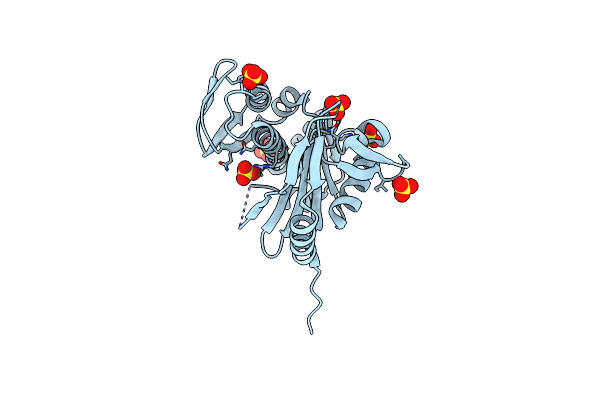
Crystal Structure Of B. Subtilis Tagt
Organism: Bacillus subtilis (strain 168)
Method: X-RAY DIFFRACTION Resolution:2.80 Å Release Date: 2020-01-29 Classification: TRANSFERASE |
Organism: Bacillus subtilis (strain 168)
Method: X-RAY DIFFRACTION
Release Date: 2020-01-29
 |
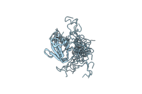
Crystal Structure Of B. Subtilis Tagu
Organism: Bacillus subtilis (strain 168)
Method: X-RAY DIFFRACTION Resolution:2.20 Å Release Date: 2020-01-29 Classification: TRANSFERASE Ligands: SO4, GOL |
Organism: Bacillus subtilis (strain 168)
Method: X-RAY DIFFRACTION
Release Date: 2020-01-29
Ligands: SO4, GOL
 |
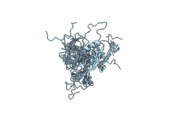
Nmr Structure Of Ub2H, Regulatory Domain Of Pbp1B From E. Coli
Organism: Escherichia coli k-12
Method: SOLUTION NMR Release Date: 2019-02-20 Classification: TRANSFERASE |
Organism: Escherichia coli k-12
Method: SOLUTION NMR
Release Date: 2019-02-20
 |
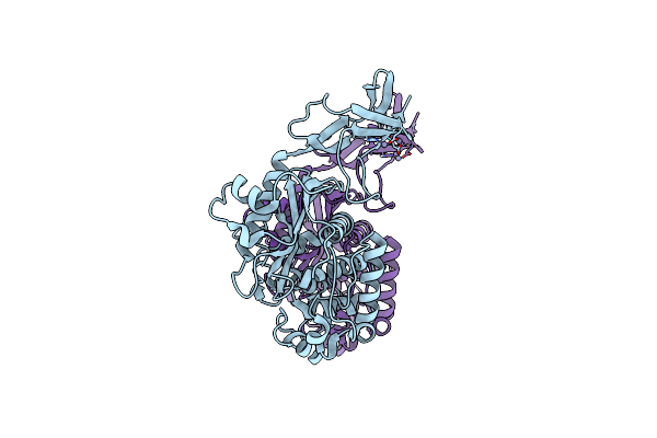
Structure Of The Ub2H Domain Of E.Coli Pbp1B In Complex With Lpob
Organism: Escherichia coli (strain k12)
Method: SOLUTION NMR Release Date: 2019-02-20 Classification: TRANSFERASE |
Organism: Escherichia coli (strain k12)
Method: SOLUTION NMR
Release Date: 2019-02-20
 |
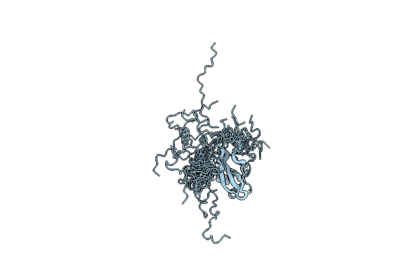
Crystal Structure Of S. Aureus Penicillin Binding Protein 4 (Pbp4) Mutant (S75C)
Organism: Staphylococcus aureus (strain col)
Method: X-RAY DIFFRACTION Resolution:1.86 Å Release Date: 2018-12-19 Classification: HYDROLASE Ligands: ZN |
Organism: Staphylococcus aureus (strain col)
Method: X-RAY DIFFRACTION
Release Date: 2018-12-19
Ligands: ZN
 |
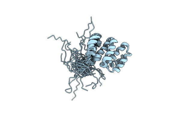
Nmr Solution Structure Of Murine Cxcl12 Gamma Isoform
|
Organism: Mus musculus
Method: SOLUTION NMR
Release Date: 2018-10-10
 |
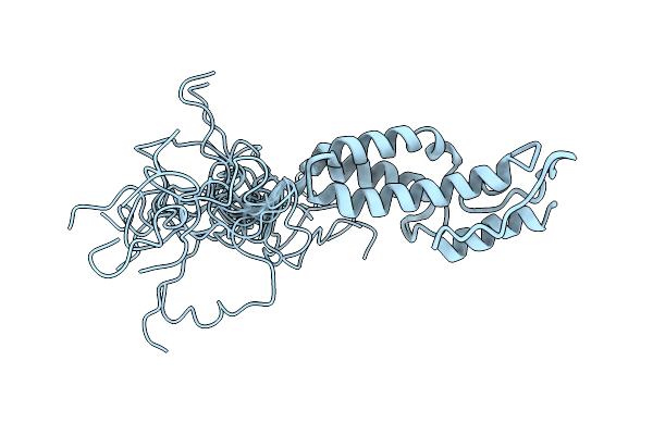
Solution Structure Of The Tpr Domain Of The Cell Division Coordinator, Cpob
Organism: Escherichia coli (strain k12)
Method: SOLUTION NMR Release Date: 2018-08-08 Classification: CELL CYCLE |
Organism: Escherichia coli (strain k12)
Method: SOLUTION NMR
Release Date: 2018-08-08
 |
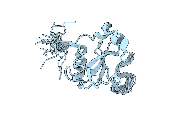
Solution Structure Of Mapz Extracellular Domain First Subdomain
Organism: Streptococcus pneumoniae r6
Method: SOLUTION NMR Release Date: 2016-06-29 Classification: CELL CYCLE |
Organism: Streptococcus pneumoniae r6
Method: SOLUTION NMR
Release Date: 2016-06-29
 |
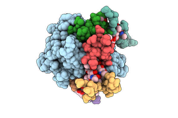
Solution Structure Of Mapz Extracellular Domain Second Subdomain
Organism: Streptococcus pneumoniae r6
Method: SOLUTION NMR Release Date: 2016-06-29 Classification: CELL CYCLE |
Organism: Streptococcus pneumoniae r6
Method: SOLUTION NMR
Release Date: 2016-06-29
 |
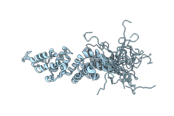
Haddock Model Of Bacillus Subtilis L,D-Transpeptidase In Complex With A Peptidoglycan Hexamuropeptide
Organism: Bacillus subtilis
Method: SOLID-STATE NMR Release Date: 2015-01-14 Classification: TRANSFERASE/STRUCTURAL PROTEIN |
Organism: Bacillus subtilis
Method: SOLID-STATE NMR
Release Date: 2015-01-14
 |
E. Coli Lpoa N-Terminal Domain
Organism: Escherichia coli
Method: SOLUTION NMR Release Date: 2014-06-25 Classification: PROTEIN BINDING |
Organism: Escherichia coli
Method: SOLUTION NMR
Release Date: 2014-06-25