

Search Count: 13
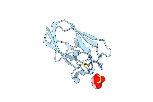 |
The 1.5 Angstrom Crystal Structure Of Oxidized (Cuii) Poplar Plastocyanin B At Ph 4.0
Organism: Populus nigra
Method: X-RAY DIFFRACTION Resolution:1.50 Å Release Date: 2013-02-13 Classification: ELECTRON TRANSPORT Ligands: CU, SO4, GOL |
Organism: Populus nigra
Method: X-RAY DIFFRACTION
Release Date: 2013-02-13
Ligands: CU, SO4, GOL
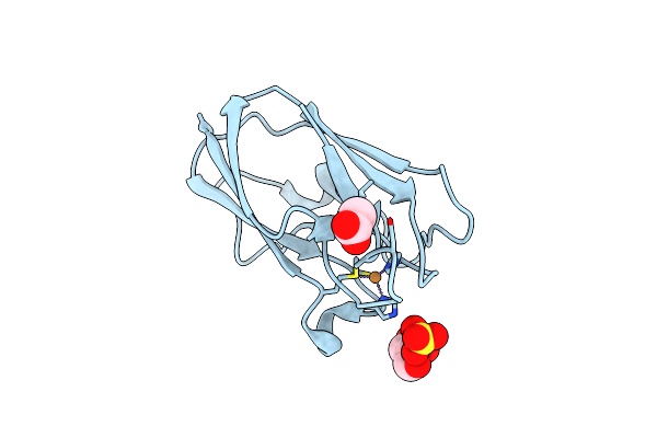 |
The 1.35 Angstrom Crystal Structure Of Reduced (Cui) Poplar Plastocyanin B At Ph 4.0
Organism: Populus nigra
Method: X-RAY DIFFRACTION Resolution:1.35 Å Release Date: 2013-02-13 Classification: ELECTRON TRANSPORT Ligands: CU1, SO4, GOL, ACT |
Organism: Populus nigra
Method: X-RAY DIFFRACTION
Release Date: 2013-02-13
Ligands: CU1, SO4, GOL, ACT
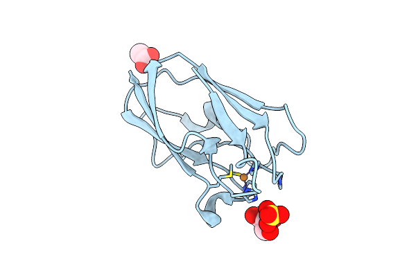 |
The 1.8 Angstrom Crystal Structure Of Oxidized (Cuii) Poplar Plastocyanin B At Ph 6.0
Organism: Populus nigra
Method: X-RAY DIFFRACTION Resolution:1.80 Å Release Date: 2013-02-13 Classification: ELECTRON TRANSPORT Ligands: CU, SO4, GOL, ACT |
Organism: Populus nigra
Method: X-RAY DIFFRACTION
Release Date: 2013-02-13
Ligands: CU, SO4, GOL, ACT
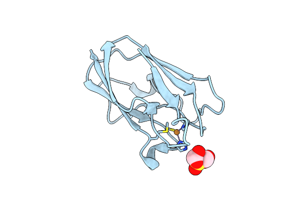 |
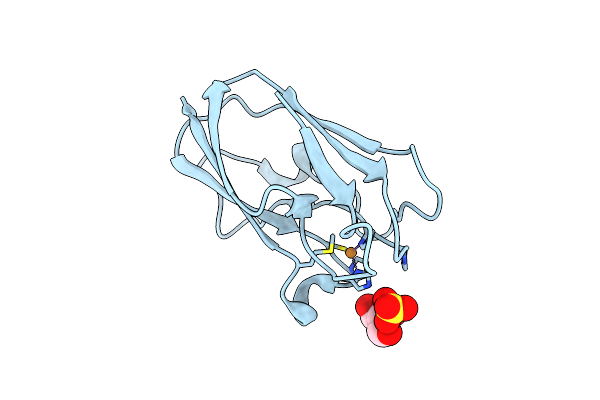
The 1.54 Angstrom Crystal Structure Of Reduced (Cui) Poplar Plastocyanin B At Ph 6.0
Organism: Populus nigra
Method: X-RAY DIFFRACTION Resolution:1.54 Å Release Date: 2013-02-13 Classification: ELECTRON TRANSPORT Ligands: CU1, SO4, GOL |
Organism: Populus nigra
Method: X-RAY DIFFRACTION
Release Date: 2013-02-13
Ligands: CU1, SO4, GOL
 |
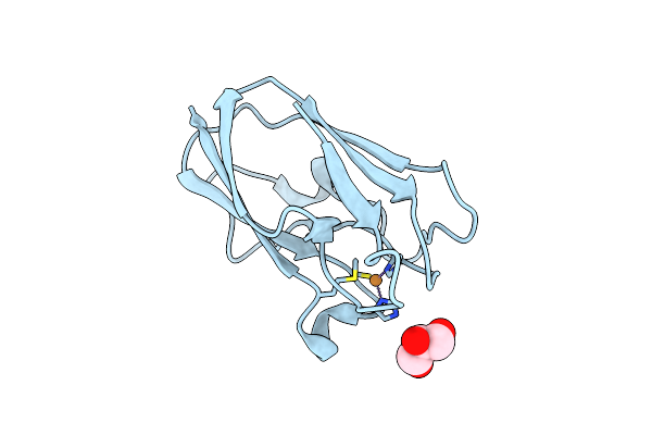
The 1.88 Angstrom Crystal Structure Of Oxidized (Cuii) Poplar Plastocyanin B At Ph 8.0
Organism: Populus nigra
Method: X-RAY DIFFRACTION Resolution:1.88 Å Release Date: 2013-02-13 Classification: ELECTRON TRANSPORT Ligands: CU, SO4, GOL |
Organism: Populus nigra
Method: X-RAY DIFFRACTION
Release Date: 2013-02-13
Ligands: CU, SO4, GOL
 |
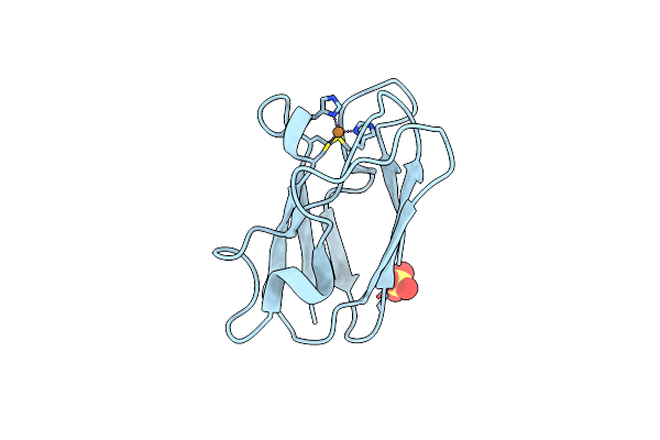
The 1.67 Angstrom Crystal Structure Of Reduced (Cui) Poplar Plastocyanin B At Ph 8.0
Organism: Populus nigra
Method: X-RAY DIFFRACTION Resolution:1.67 Å Release Date: 2013-02-13 Classification: ELECTRON TRANSPORT Ligands: CU1, GOL |
Organism: Populus nigra
Method: X-RAY DIFFRACTION
Release Date: 2013-02-13
Ligands: CU1, GOL
 |
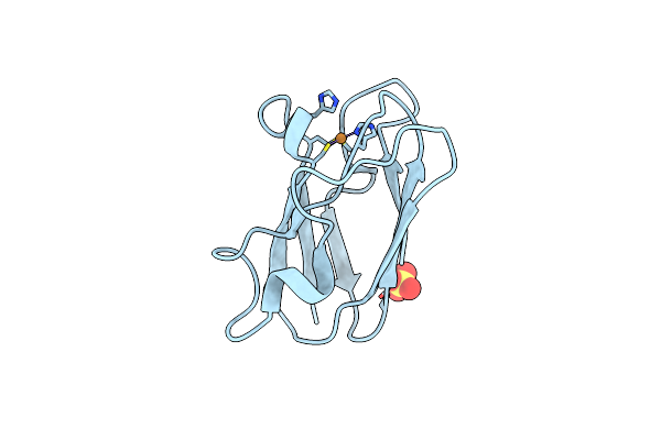
The 1.08 Angstrom Crystal Structure Of Oxidized (Cuii) Poplar Plastocyanin A At Ph 4.0
Organism: Populus nigra
Method: X-RAY DIFFRACTION Resolution:1.08 Å Release Date: 2013-02-13 Classification: ELECTRON TRANSPORT Ligands: CU, SO4 |
Organism: Populus nigra
Method: X-RAY DIFFRACTION
Release Date: 2013-02-13
Ligands: CU, SO4
 |
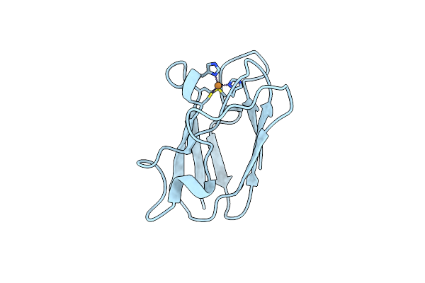
The 1.07 Angstrom Crystal Structure Of Reduced (Cui) Poplar Plastocyanin A At Ph 4.0
Organism: Populus nigra
Method: X-RAY DIFFRACTION Resolution:1.07 Å Release Date: 2013-02-13 Classification: ELECTRON TRANSPORT Ligands: CU1, SO4 |
Organism: Populus nigra
Method: X-RAY DIFFRACTION
Release Date: 2013-02-13
Ligands: CU1, SO4
 |
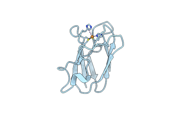
The 1.00 Angstrom Crystal Structure Of Oxidized (Cuii) Poplar Plastocyanin A At Ph 6.0
Organism: Populus nigra
Method: X-RAY DIFFRACTION Resolution:1.00 Å Release Date: 2013-02-13 Classification: ELECTRON TRANSPORT Ligands: CU |
Organism: Populus nigra
Method: X-RAY DIFFRACTION
Release Date: 2013-02-13
Ligands: CU
 |
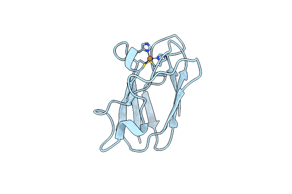
The 1.05 Angstrom Crystal Structure Of Reduced (Cui) Poplar Plastocyanin A At Ph 6.0
Organism: Populus nigra
Method: X-RAY DIFFRACTION Resolution:1.05 Å Release Date: 2013-02-13 Classification: ELECTRON TRANSPORT Ligands: CU1 |
Organism: Populus nigra
Method: X-RAY DIFFRACTION
Release Date: 2013-02-13
Ligands: CU1
 |
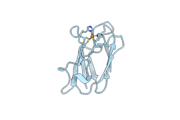
The 1.00 Angstrom Crystal Structure Of Oxidized (Cuii) Poplar Plastocyanin A At Ph 8.0
Organism: Populus nigra
Method: X-RAY DIFFRACTION Resolution:1.00 Å Release Date: 2013-02-13 Classification: ELECTRON TRANSPORT Ligands: CU |
Organism: Populus nigra
Method: X-RAY DIFFRACTION
Release Date: 2013-02-13
Ligands: CU
 |
The 1.06 Angstrom Crystal Structure Of Reduced (Cui) Poplar Plastocyanin A At Ph 8.0
Organism: Populus nigra
Method: X-RAY DIFFRACTION Resolution:1.06 Å Release Date: 2013-02-13 Classification: ELECTRON TRANSPORT Ligands: CU1 |
Organism: Populus nigra
Method: X-RAY DIFFRACTION
Release Date: 2013-02-13
Ligands: CU1
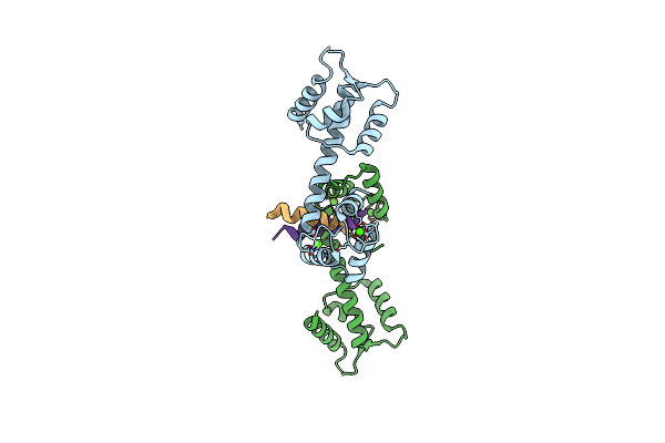 |
Centrin-Xpc Peptide
Organism: Homo sapiens
Method: X-RAY DIFFRACTION Resolution:1.80 Å Release Date: 2007-10-09 Classification: CELL CYCLE Ligands: CA |
Organism: Homo sapiens
Method: X-RAY DIFFRACTION
Release Date: 2007-10-09
Ligands: CA