

Search Count: 25
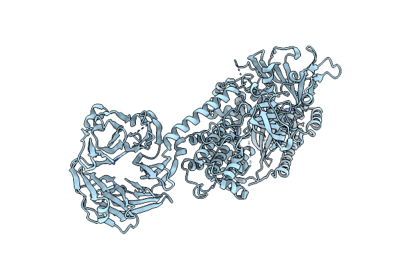 |
Cryo-Em Structure Of The C-Terminal Half Of The Parkinson'S Disease-Linked Protein Leucine Rich Repeat Kinase 2 (Lrrk2)
Organism: Homo sapiens
Method: ELECTRON MICROSCOPY Release Date: 2020-08-26 Classification: SIGNALING PROTEIN Ligands: GDP, MG |
Organism: Homo sapiens
Method: ELECTRON MICROSCOPY
Release Date: 2020-08-26
Ligands: GDP, MG
 |
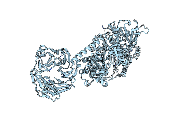
Cryo-Em Structure Of The C-Terminal Half Of The Parkinson'S Disease-Linked Protein Leucine Rich Repeat Kinase 2 (Lrrk2)
Organism: Homo sapiens
Method: ELECTRON MICROSCOPY Release Date: 2020-08-26 Classification: SIGNALING PROTEIN Ligands: GDP, MG |
Organism: Homo sapiens
Method: ELECTRON MICROSCOPY
Release Date: 2020-08-26
Ligands: GDP, MG
 |
Cryo-Em Structure Of The C-Terminal Half Of The Parkinson'S Disease-Linked Protein Leucine Rich Repeat Kinase 2 (Lrrk2)
Organism: Homo sapiens
Method: ELECTRON MICROSCOPY Release Date: 2020-08-26 Classification: SIGNALING PROTEIN |
Organism: Homo sapiens
Method: ELECTRON MICROSCOPY
Release Date: 2020-08-26
 |
Cryo-Em Structure Of The C-Terminal Half Of The Parkinson'S Disease-Linked Protein Leucine Rich Repeat Kinase 2 (Lrrk2)
Organism: Homo sapiens
Method: ELECTRON MICROSCOPY Release Date: 2020-08-26 Classification: SIGNALING PROTEIN |
Organism: Homo sapiens
Method: ELECTRON MICROSCOPY
Release Date: 2020-08-26
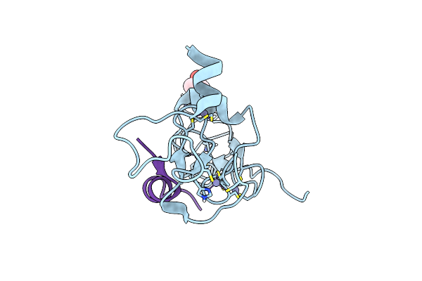 |
Structure Of Human Dpf3 Double-Phd Domain Bound To Histone H3 Tail Peptide With Acetylated K14
Organism: Homo sapiens
Method: X-RAY DIFFRACTION Resolution:1.20 Å Release Date: 2017-08-16 Classification: PEPTIDE BINDING PROTEIN Ligands: ZN, MPD |
Organism: Homo sapiens
Method: X-RAY DIFFRACTION
Release Date: 2017-08-16
Ligands: ZN, MPD
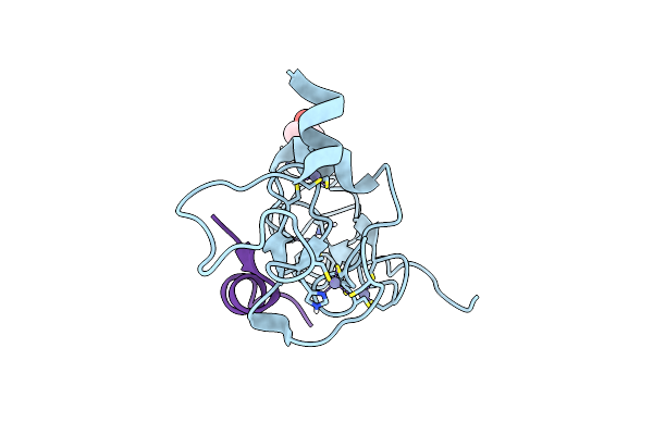 |
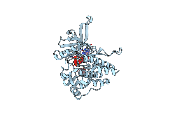
Structure Of Human Dpf3 Double-Phd Domain Bound To Histone H3 Tail Peptide With Monomethylated K4 And Acetylated K14
Organism: Homo sapiens
Method: X-RAY DIFFRACTION Resolution:1.19 Å Release Date: 2017-08-16 Classification: PEPTIDE BINDING PROTEIN Ligands: ZN, MPD |
Organism: Homo sapiens
Method: X-RAY DIFFRACTION
Release Date: 2017-08-16
Ligands: ZN, MPD
 |
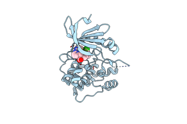
Crystal Structure Of The E596A V617F Mutant Jak2 Pseudokinase Domain Bound To Mg-Atp
Organism: Homo sapiens
Method: X-RAY DIFFRACTION Resolution:1.54 Å Release Date: 2016-04-13 Classification: TRANSFERASE Ligands: ATP, MG, GOL |
Organism: Homo sapiens
Method: X-RAY DIFFRACTION
Release Date: 2016-04-13
Ligands: ATP, MG, GOL
 |
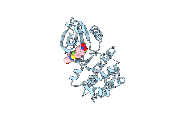
Crystal Structure Of Aurora A Kinase Domain Bound To Mk-5108
Organism: Homo sapiens
Method: X-RAY DIFFRACTION Resolution:2.18 Å Release Date: 2016-01-20 Classification: TRANSFERASE/TRANSFERASE INHIBITOR Ligands: 5VC |
Organism: Homo sapiens
Method: X-RAY DIFFRACTION
Release Date: 2016-01-20
Ligands: 5VC
 |
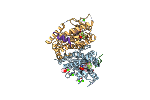
Crystal Structure Of Plk4 Kinase Domain Bound To Centrinone
Organism: Homo sapiens
Method: X-RAY DIFFRACTION Resolution:2.65 Å Release Date: 2015-06-17 Classification: Transferase/Transferase Inhibitor Ligands: 4J7 |
Organism: Homo sapiens
Method: X-RAY DIFFRACTION
Release Date: 2015-06-17
Ligands: 4J7
 |
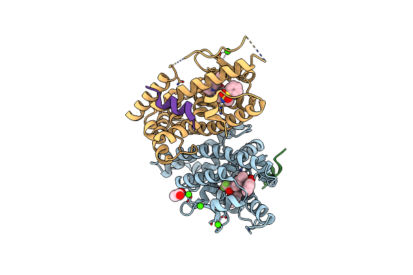
Crystal Structure Of Lxr Ligand Binding Domain In Complex With Full Agonist 1
Organism: Homo sapiens
Method: X-RAY DIFFRACTION Resolution:2.45 Å Release Date: 2012-03-21 Classification: TRANSCRIPTION/Peptide/Agonist Ligands: 0KS, ACT, CA |
Organism: Homo sapiens
Method: X-RAY DIFFRACTION
Release Date: 2012-03-21
Ligands: 0KS, ACT, CA
 |
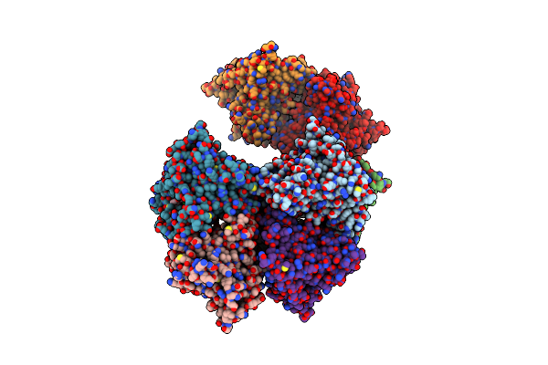
Crystal Structure Of Lxr Ligand Binding Domain In Complex With Partial Agonist 5
Organism: Homo sapiens
Method: X-RAY DIFFRACTION Resolution:2.75 Å Release Date: 2012-03-21 Classification: TRANSCRIPTION/Peptide/Agonist Ligands: 0KT, ACT, CA |
Organism: Homo sapiens
Method: X-RAY DIFFRACTION
Release Date: 2012-03-21
Ligands: 0KT, ACT, CA
 |
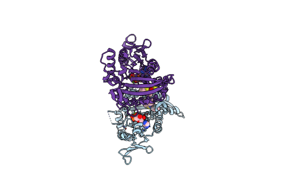
Catalytic Domain Of Human Phosphodiesterase 4B In Complex With A Coumarin-Based Inhibitor
Organism: Homo sapiens
Method: X-RAY DIFFRACTION Resolution:2.60 Å Release Date: 2010-04-28 Classification: HYDROLASE/HYDROLASE INHIBITOR Ligands: Z72, ZN, MG, SO4 |
Organism: Homo sapiens
Method: X-RAY DIFFRACTION
Release Date: 2010-04-28
Ligands: Z72, ZN, MG, SO4
 |
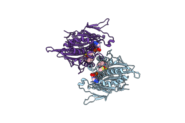
Crystal Structure Of Ksp In Complex With Adp And Thiophene Containing Inhibitor 15
Organism: Homo sapiens
Method: X-RAY DIFFRACTION Resolution:1.85 Å Release Date: 2007-06-05 Classification: MOTOR PROTEIN,CELL CYCLE Ligands: MG, ADP, K01 |
Organism: Homo sapiens
Method: X-RAY DIFFRACTION
Release Date: 2007-06-05
Ligands: MG, ADP, K01
 |
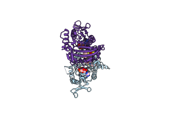
Crystal Structure Of Ksp In Complex With Adp And Thiophene Containing Inhibitor 33
Organism: Homo sapiens
Method: X-RAY DIFFRACTION Resolution:2.10 Å Release Date: 2007-05-22 Classification: CELL CYCLE Ligands: ADP, MG, K02 |
Organism: Homo sapiens
Method: X-RAY DIFFRACTION
Release Date: 2007-05-22
Ligands: ADP, MG, K02
 |
Crystal Structure Of Ksp In Complex With Adp And Thiophene Containing Inhibitor 37
Organism: Homo sapiens
Method: X-RAY DIFFRACTION Resolution:2.11 Å Release Date: 2007-05-22 Classification: CELL CYCLE Ligands: ADP, MG, K03 |
Organism: Homo sapiens
Method: X-RAY DIFFRACTION
Release Date: 2007-05-22
Ligands: ADP, MG, K03
 |
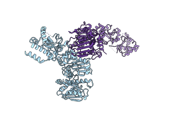
Crystal Structure Of Full-Length Htpg, The Escherichia Coli Hsp90, Bound To Adp
Organism: Escherichia coli
Method: X-RAY DIFFRACTION Resolution:3.55 Å Release Date: 2006-11-21 Classification: CHAPERONE Ligands: ADP |
Organism: Escherichia coli
Method: X-RAY DIFFRACTION
Release Date: 2006-11-21
Ligands: ADP
 |
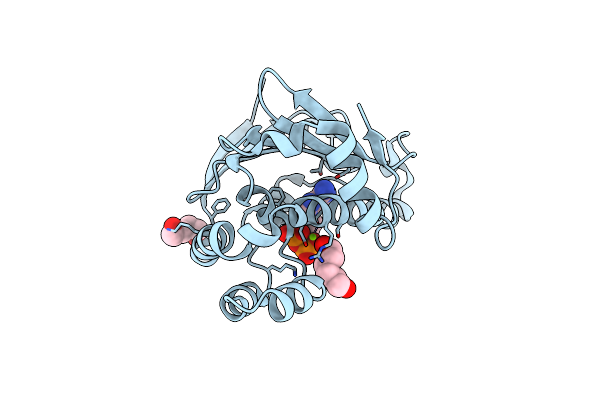
Crystal Structure Of Full-Length Htpg, The Escherichia Coli Hsp90
Organism: Escherichia coli
Method: X-RAY DIFFRACTION Resolution:3.50 Å Release Date: 2006-11-21 Classification: CHAPERONE |
Organism: Escherichia coli
Method: X-RAY DIFFRACTION
Release Date: 2006-11-21
 |
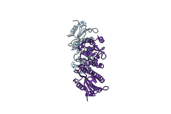
Crystal Structure Of The N-Terminal Domain Of Htpg, The Escherichia Coli Hsp90, Bound To Adp
Organism: Escherichia coli
Method: X-RAY DIFFRACTION Resolution:1.65 Å Release Date: 2006-11-21 Classification: CHAPERONE Ligands: MG, ADP, HEZ |
Organism: Escherichia coli
Method: X-RAY DIFFRACTION
Release Date: 2006-11-21
Ligands: MG, ADP, HEZ
 |
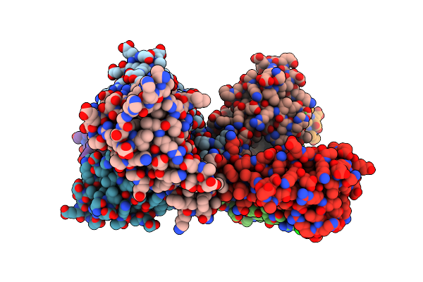
Crystal Structure Of The Middle Domain Of Htpg, The E. Coli Hsp90
Organism: Escherichia coli
Method: X-RAY DIFFRACTION Resolution:1.90 Å Release Date: 2006-10-31 Classification: Chaperone, hydrolase |
Organism: Escherichia coli
Method: X-RAY DIFFRACTION
Release Date: 2006-10-31
 |
Crystal Structure Of The Carboxy-Terminal Domain Of Htpg, The E. Coli Hsp90
Organism: Escherichia coli
Method: X-RAY DIFFRACTION Resolution:2.60 Å Release Date: 2004-06-15 Classification: CHAPERONE Ligands: NI, CL |
Organism: Escherichia coli
Method: X-RAY DIFFRACTION
Release Date: 2004-06-15
Ligands: NI, CL