

Search Count: 30
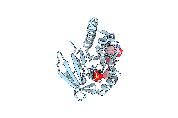 |
Humrada1 In Complex With 3-Amino-2-Naphthoic Acid
Organism: Pyrococcus furiosus
Method: X-RAY DIFFRACTION Resolution:1.50 Å Release Date: 2021-01-27 Classification: DNA BINDING PROTEIN Ligands: NYE, PO4, GOL |
Organism: Pyrococcus furiosus
Method: X-RAY DIFFRACTION
Release Date: 2021-01-27
Ligands: NYE, PO4, GOL
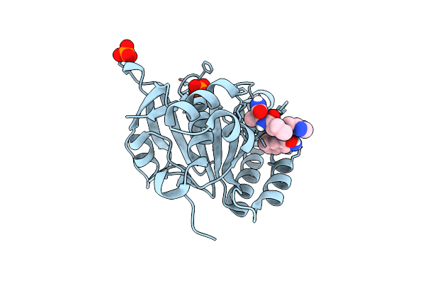 |
Humrada2 In Complex With Naphthyl-Hpa Fragment-Peptide Chimera
Organism: Pyrococcus furiosus (strain atcc 43587 / dsm 3638 / jcm 8422 / vc1)
Method: X-RAY DIFFRACTION Resolution:1.35 Å Release Date: 2021-01-27 Classification: DNA BINDING PROTEIN Ligands: PO4, O0E |
Organism: Pyrococcus furiosus (strain atcc 43587 / dsm 3638 / jcm 8422 / vc1)
Method: X-RAY DIFFRACTION
Release Date: 2021-01-27
Ligands: PO4, O0E
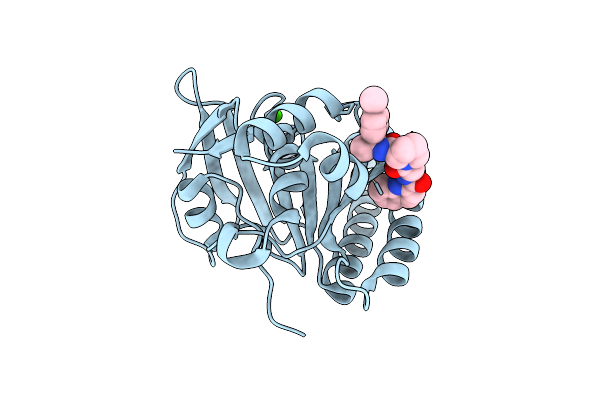 |
Humrada22F In Complex With Compound 6
Organism: Pyrococcus furiosus (strain atcc 43587 / dsm 3638 / jcm 8422 / vc1)
Method: X-RAY DIFFRACTION Resolution:1.73 Å Release Date: 2021-01-27 Classification: DNA BINDING PROTEIN Ligands: NZW, CA |
Organism: Pyrococcus furiosus (strain atcc 43587 / dsm 3638 / jcm 8422 / vc1)
Method: X-RAY DIFFRACTION
Release Date: 2021-01-27
Ligands: NZW, CA
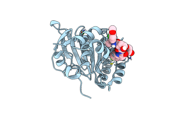 |
Humrada22F In Complex With Cam833
Organism: Pyrococcus furiosus (strain atcc 43587 / dsm 3638 / jcm 8422 / vc1)
Method: X-RAY DIFFRACTION Resolution:1.52 Å Release Date: 2021-01-27 Classification: DNA BINDING PROTEIN Ligands: O08, CA, GOL |
Organism: Pyrococcus furiosus (strain atcc 43587 / dsm 3638 / jcm 8422 / vc1)
Method: X-RAY DIFFRACTION
Release Date: 2021-01-27
Ligands: O08, CA, GOL
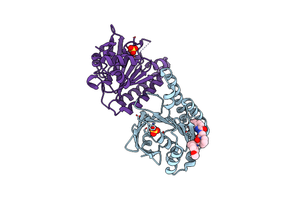 |
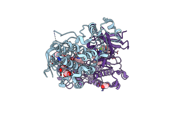
Humrada33F In Complex With Peptidic Inhibitor 6
Organism: Pyrococcus furiosus (strain atcc 43587 / dsm 3638 / jcm 8422 / vc1)
Method: X-RAY DIFFRACTION Resolution:2.31 Å Release Date: 2021-01-27 Classification: DNA BINDING PROTEIN Ligands: NZW, SO4 |
Organism: Pyrococcus furiosus (strain atcc 43587 / dsm 3638 / jcm 8422 / vc1)
Method: X-RAY DIFFRACTION
Release Date: 2021-01-27
Ligands: NZW, SO4
 |
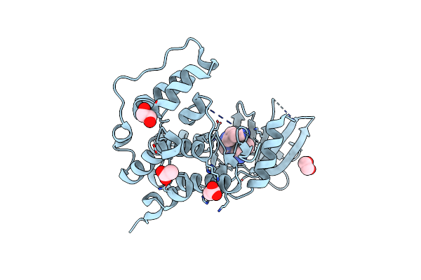
Crystal Structure Of The Pctaire1 Kinase In Complex With Inhibitor
Organism: Homo sapiens
Method: X-RAY DIFFRACTION Resolution:2.20 Å Release Date: 2016-11-23 Classification: TRANSFERASE Ligands: 919, EDO |
Organism: Homo sapiens
Method: X-RAY DIFFRACTION
Release Date: 2016-11-23
Ligands: 919, EDO
 |
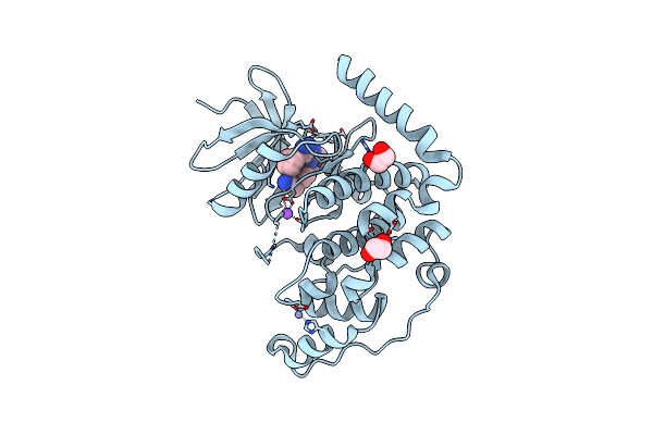
Crystal Structure Of The Human Cdkl5 Kinase Domain
Organism: Homo sapiens
Method: X-RAY DIFFRACTION Resolution:2.00 Å Release Date: 2013-05-08 Classification: TRANSFERASE |
Organism: Homo sapiens
Method: X-RAY DIFFRACTION
Release Date: 2013-05-08
 |
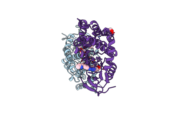
Crystal Structure Of The Human Cdkl3 Kinase Domain
Organism: Homo sapiens
Method: X-RAY DIFFRACTION Resolution:2.20 Å Release Date: 2013-03-20 Classification: TRANSFERASE Ligands: 38R, ZN, NA, EDO |
Organism: Homo sapiens
Method: X-RAY DIFFRACTION
Release Date: 2013-03-20
Ligands: 38R, ZN, NA, EDO
 |
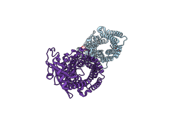
Crystal Structure Of The Human Cdkl2 Kinase Domain With Bound Tcs 2312
Organism: Homo sapiens
Method: X-RAY DIFFRACTION Resolution:2.00 Å Release Date: 2012-10-17 Classification: TRANSFERASE Ligands: TC0, CL, EDO |
Organism: Homo sapiens
Method: X-RAY DIFFRACTION
Release Date: 2012-10-17
Ligands: TC0, CL, EDO
 |
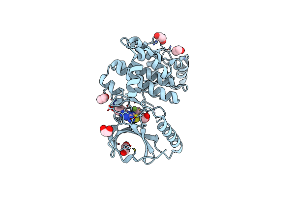
Crystal Structure Of The Human Cdkl1 Kinase Domain
Organism: Homo sapiens
Method: X-RAY DIFFRACTION Resolution:2.40 Å Release Date: 2012-02-08 Classification: TRANSFERASE Ligands: D15 |
Organism: Homo sapiens
Method: X-RAY DIFFRACTION
Release Date: 2012-02-08
Ligands: D15
 |
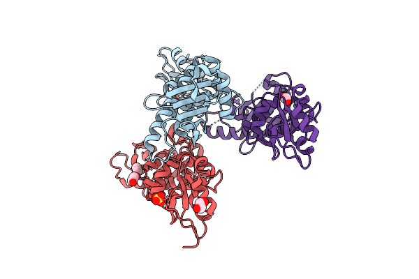
Crystal Structure Of Full Length Mouse Strap (Ttc5)
Organism: Mus musculus
Method: X-RAY DIFFRACTION Resolution:2.05 Å Release Date: 2012-01-25 Classification: GENE REGULATION Ligands: EDO |
Organism: Mus musculus
Method: X-RAY DIFFRACTION
Release Date: 2012-01-25
Ligands: EDO
 |
Crystal Structure Of The Human Cdkl2 Kinase Domain
Organism: Homo sapiens
Method: X-RAY DIFFRACTION Resolution:1.53 Å Release Date: 2011-12-21 Classification: TRANSFERASE Ligands: DKI, EDO |
Organism: Homo sapiens
Method: X-RAY DIFFRACTION
Release Date: 2011-12-21
Ligands: DKI, EDO
 |
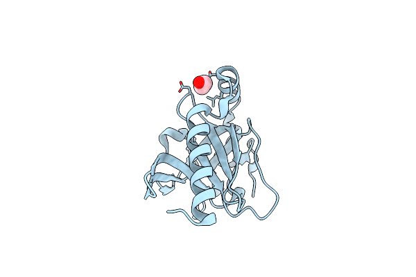
Crystal Structure Of The Middle Domain Of Human Hsp90-Beta Refined At 2.3 A Resolution
Organism: Homo sapiens
Method: X-RAY DIFFRACTION Resolution:2.28 Å Release Date: 2010-12-15 Classification: CHAPERONE Ligands: SO4, GOL, EDO |
Organism: Homo sapiens
Method: X-RAY DIFFRACTION
Release Date: 2010-12-15
Ligands: SO4, GOL, EDO
 |
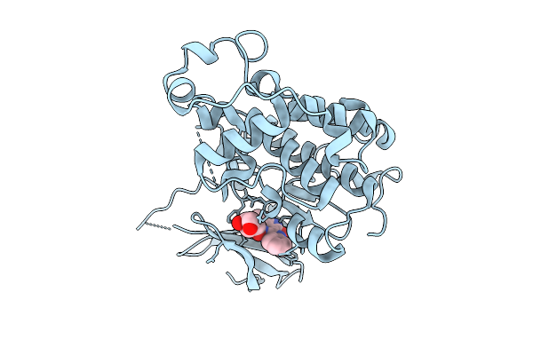
Crystal Structure Of Human Ttc5 (Strap) C-Terminal Ob Domain
Organism: Homo sapiens
Method: X-RAY DIFFRACTION Resolution:1.80 Å Release Date: 2010-11-17 Classification: ANTITUMOR PROTEIN Ligands: IOD, EDO, CL |
Organism: Homo sapiens
Method: X-RAY DIFFRACTION
Release Date: 2010-11-17
Ligands: IOD, EDO, CL
 |
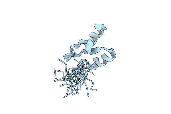
Crystal Structure Of The Pctaire1 Kinase In Complex With Indirubin E804
Organism: Homo sapiens
Method: X-RAY DIFFRACTION Resolution:2.40 Å Release Date: 2010-06-09 Classification: TRANSFERASE Ligands: FEF |
Organism: Homo sapiens
Method: X-RAY DIFFRACTION
Release Date: 2010-06-09
Ligands: FEF
 |
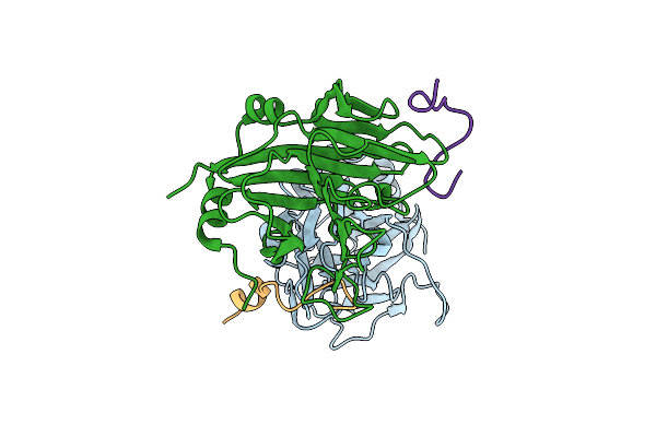
The Folding Mechanism Of Bbl: Plasticity Of Transition-State Structure Observed Within An Ultrafast Folding Protein Family.
Organism: Escherichia coli
Method: SOLUTION NMR Release Date: 2009-11-17 Classification: TRANSFERASE |
Organism: Escherichia coli
Method: SOLUTION NMR
Release Date: 2009-11-17
 |
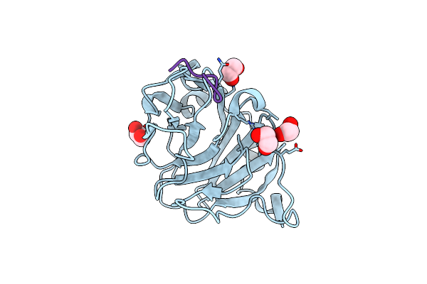
Crystal Structure Of Human Spla/Ryanodine Receptor Domain And Socs Box Containing 1 (Spsb1) In Complex With A 20-Residue Vasa Peptide
Organism: Homo sapiens
Method: X-RAY DIFFRACTION Resolution:2.05 Å Release Date: 2008-12-09 Classification: APOPTOSIS/HYDROLASE |
Organism: Homo sapiens
Method: X-RAY DIFFRACTION
Release Date: 2008-12-09
 |
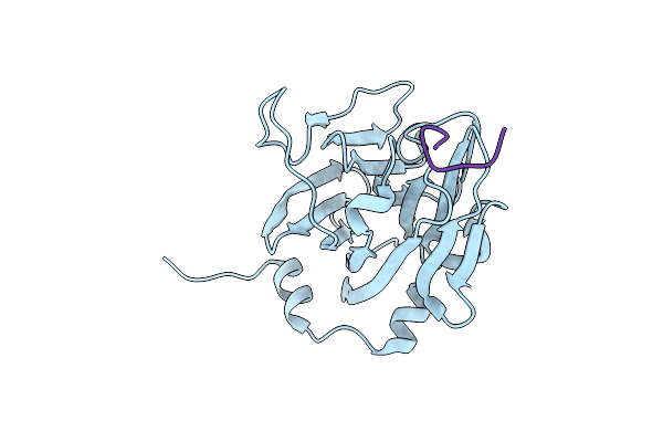
Crystal Structure Of Human Spla/Ryanodine Receptor Domain And Socs Box Containing 2 (Spsb2) In Complex With A 20-Residue Vasa Peptide
Organism: Homo sapiens
Method: X-RAY DIFFRACTION Resolution:1.80 Å Release Date: 2008-10-28 Classification: APOPTOSIS Ligands: EDO |
Organism: Homo sapiens
Method: X-RAY DIFFRACTION
Release Date: 2008-10-28
Ligands: EDO
 |
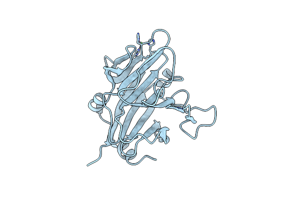
The Structure Of Spla-Ryanodine Receptor Domain And Socs Box Containing 1 In Complex With A Par-4 Peptide
Organism: Homo sapiens
Method: X-RAY DIFFRACTION Resolution:1.79 Å Release Date: 2008-09-16 Classification: APOPTOSIS |
Organism: Homo sapiens
Method: X-RAY DIFFRACTION
Release Date: 2008-09-16
 |
Structure Of The Human Spry Domain-Containing Socs Box Protein Ssb-4
Organism: Homo sapiens
Method: X-RAY DIFFRACTION Resolution:2.20 Å Release Date: 2007-07-17 Classification: PROTEIN BINDING Ligands: NI |
Organism: Homo sapiens
Method: X-RAY DIFFRACTION
Release Date: 2007-07-17
Ligands: NI