

Search Count: 16
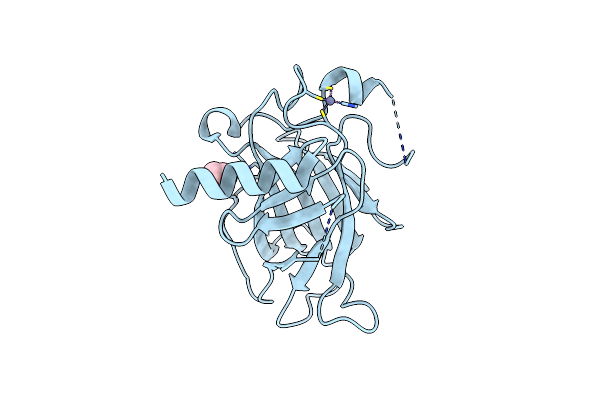 |
Structure Of P53 Cancer Mutant Y126C
Organism: Homo sapiens
Method: X-RAY DIFFRACTION Resolution:1.69 Å Release Date: 2024-06-19 Classification: DNA BINDING PROTEIN Ligands: ZN, EDO |
Organism: Homo sapiens
Method: X-RAY DIFFRACTION
Release Date: 2024-06-19
Ligands: ZN, EDO
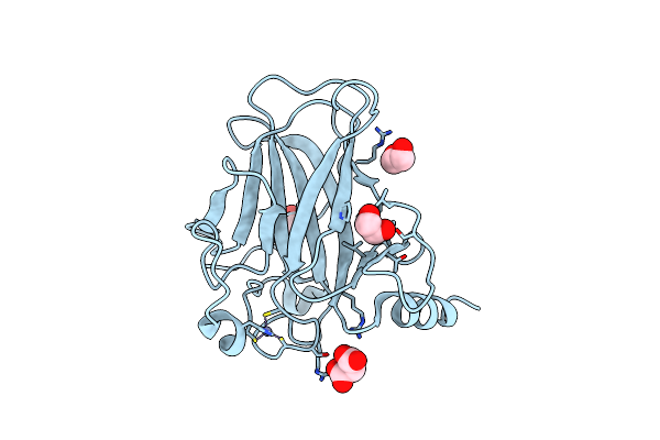 |
Structure Of P53 Cancer Mutant Y163C
Organism: Homo sapiens
Method: X-RAY DIFFRACTION Resolution:1.65 Å Release Date: 2024-06-19 Classification: DNA BINDING PROTEIN Ligands: ZN, EDO, MLI |
Organism: Homo sapiens
Method: X-RAY DIFFRACTION
Release Date: 2024-06-19
Ligands: ZN, EDO, MLI
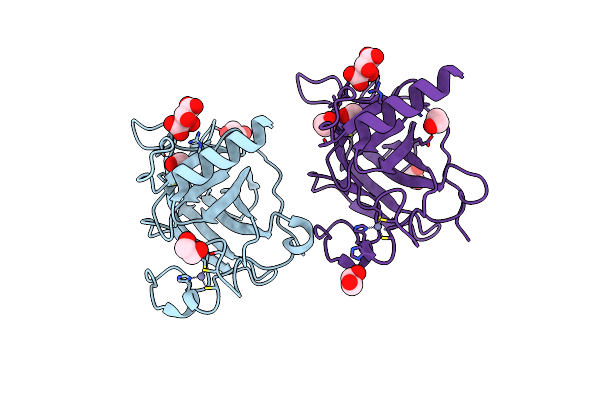 |
Structure Of P53 Cancer Mutant Y205C
Organism: Homo sapiens
Method: X-RAY DIFFRACTION Resolution:1.54 Å Release Date: 2024-06-19 Classification: DNA BINDING PROTEIN Ligands: ZN, TLA, EDO |
Organism: Homo sapiens
Method: X-RAY DIFFRACTION
Release Date: 2024-06-19
Ligands: ZN, TLA, EDO
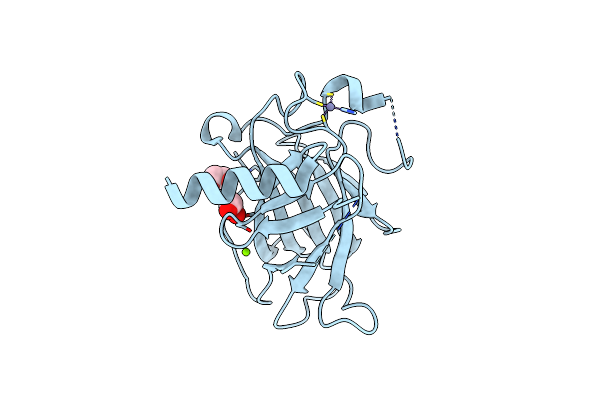 |
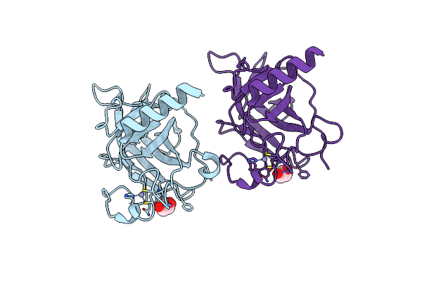
Structure Of P53 Cancer Mutant Y220C (Hexagonal Crystal Form)
Organism: Homo sapiens
Method: X-RAY DIFFRACTION Resolution:1.44 Å Release Date: 2024-06-19 Classification: DNA BINDING PROTEIN Ligands: ZN, EDO, MG, CL |
Organism: Homo sapiens
Method: X-RAY DIFFRACTION
Release Date: 2024-06-19
Ligands: ZN, EDO, MG, CL
 |
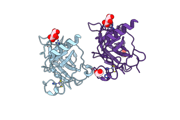
Structure Of P53 Cancer Mutant Y234C
Organism: Homo sapiens
Method: X-RAY DIFFRACTION Resolution:1.38 Å Release Date: 2024-06-19 Classification: DNA BINDING PROTEIN Ligands: ZN, GOL |
Organism: Homo sapiens
Method: X-RAY DIFFRACTION
Release Date: 2024-06-19
Ligands: ZN, GOL
 |
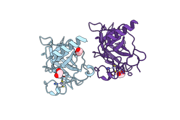
Structure Of P53 Cancer Mutant Y236C
Organism: Homo sapiens
Method: X-RAY DIFFRACTION Resolution:2.10 Å Release Date: 2024-06-19 Classification: DNA BINDING PROTEIN Ligands: ZN, TLA, EDO |
Organism: Homo sapiens
Method: X-RAY DIFFRACTION
Release Date: 2024-06-19
Ligands: ZN, TLA, EDO
 |
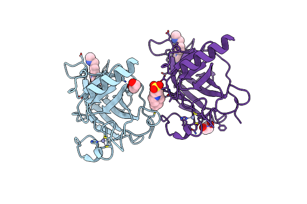
P53 Cancer Mutant Y220C
Organism: Homo sapiens
Method: X-RAY DIFFRACTION Resolution:1.24 Å Release Date: 2020-02-19 Classification: DNA BINDING PROTEIN Ligands: EDO, ZN, GOL |
Organism: Homo sapiens
Method: X-RAY DIFFRACTION
Release Date: 2020-02-19
Ligands: EDO, ZN, GOL
 |
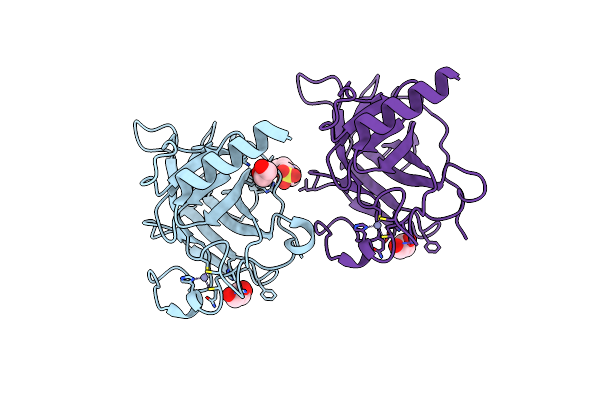
P53 Cancer Mutant Y220C In Complex With Small-Molecule Stabilizer Pk9323
Organism: Homo sapiens
Method: X-RAY DIFFRACTION Resolution:1.53 Å Release Date: 2020-02-19 Classification: DNA BINDING PROTEIN Ligands: ZN, LEK, EDO, GOL, EPE |
Organism: Homo sapiens
Method: X-RAY DIFFRACTION
Release Date: 2020-02-19
Ligands: ZN, LEK, EDO, GOL, EPE
 |
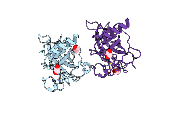
P53 Cancer Mutant Y220H
Organism: Homo sapiens
Method: X-RAY DIFFRACTION Resolution:1.44 Å Release Date: 2020-02-19 Classification: DNA BINDING PROTEIN Ligands: ZN, EDO, GOL, EPE |
Organism: Homo sapiens
Method: X-RAY DIFFRACTION
Release Date: 2020-02-19
Ligands: ZN, EDO, GOL, EPE
 |
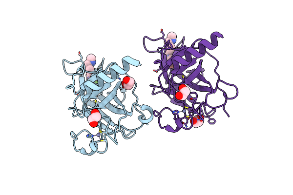
P53 Cancer Mutant Y220S
Organism: Homo sapiens
Method: X-RAY DIFFRACTION Resolution:1.50 Å Release Date: 2020-02-19 Classification: DNA BINDING PROTEIN Ligands: ZN, EDO, GOL |
Organism: Homo sapiens
Method: X-RAY DIFFRACTION
Release Date: 2020-02-19
Ligands: ZN, EDO, GOL
 |
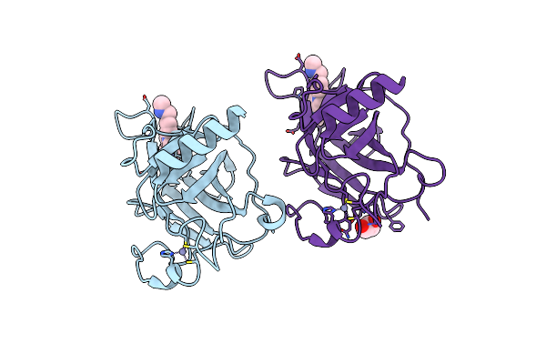
P53 Cancer Mutant Y220S In Complex With Small-Molecule Stabilizer Pk9301
Organism: Homo sapiens
Method: X-RAY DIFFRACTION Resolution:1.40 Å Release Date: 2020-02-19 Classification: DNA BINDING PROTEIN Ligands: ZN, LEB, EDO, GOL |
Organism: Homo sapiens
Method: X-RAY DIFFRACTION
Release Date: 2020-02-19
Ligands: ZN, LEB, EDO, GOL
 |
P53 Cancer Mutant Y220S In Complex With Small-Molecule Stabilizer Pk9323
Organism: Homo sapiens
Method: X-RAY DIFFRACTION Resolution:1.80 Å Release Date: 2020-02-19 Classification: DNA BINDING PROTEIN Ligands: ZN, LEK, GOL |
Organism: Homo sapiens
Method: X-RAY DIFFRACTION
Release Date: 2020-02-19
Ligands: ZN, LEK, GOL
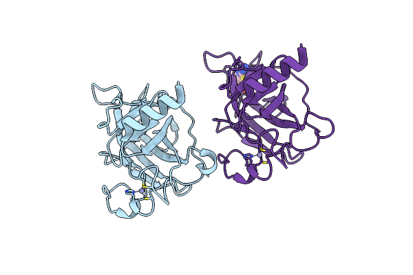 |
Structure Of The P53 Core Domain Mutant Y220C Bound To A 2-Amino Substituted Benzothiazole Scaffold
Organism: Homo sapiens
Method: X-RAY DIFFRACTION Resolution:1.60 Å Release Date: 2010-01-26 Classification: CELL CYCLE Ligands: ZN, X0U |
Organism: Homo sapiens
Method: X-RAY DIFFRACTION
Release Date: 2010-01-26
Ligands: ZN, X0U
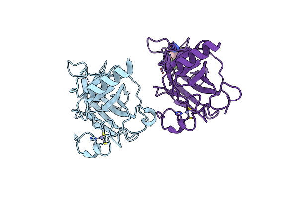 |
Structure Of The P53 Core Domain Mutant Y220C Bound To 4-(Trifluoromethyl)Benzene-1,2-Diamine
Organism: Homo sapiens
Method: X-RAY DIFFRACTION Resolution:1.80 Å Release Date: 2010-01-26 Classification: CELL CYCLE Ligands: ZN, X0V |
Organism: Homo sapiens
Method: X-RAY DIFFRACTION
Release Date: 2010-01-26
Ligands: ZN, X0V
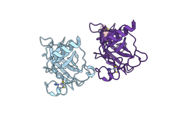 |
Structure Of The P53 Core Domain Mutant Y220C Bound To 5,6-Dimethoxy- 2-Methylbenzothiazole
Organism: Homo sapiens
Method: X-RAY DIFFRACTION Resolution:2.10 Å Release Date: 2010-01-26 Classification: CELL CYCLE Ligands: ZN, X0W |
Organism: Homo sapiens
Method: X-RAY DIFFRACTION
Release Date: 2010-01-26
Ligands: ZN, X0W
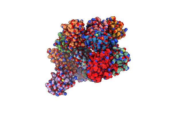 |
Structure Of Human Mdm4 N-Terminal Domain Bound To A Single Domain Antibody
Organism: Homo sapiens
Method: X-RAY DIFFRACTION Resolution:2.00 Å Release Date: 2008-11-25 Classification: IMMUNE SYSTEM Ligands: SO4 |
Organism: Homo sapiens
Method: X-RAY DIFFRACTION
Release Date: 2008-11-25
Ligands: SO4