

Search Count: 16
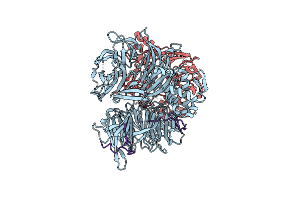 |
The Ternary Complex Of Ddb1, Dda1, Det1
Organism: Homo sapiens
Method: ELECTRON MICROSCOPY Release Date: 2025-02-05 Classification: PROTEIN BINDING |
Organism: Homo sapiens
Method: ELECTRON MICROSCOPY
Release Date: 2025-02-05
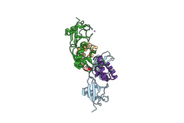 |
The N4Bp1 Cue-Like Domain In Complex With Linear Di-Ubiquitin
Organism: Homo sapiens
Method: X-RAY DIFFRACTION Resolution:2.00 Å Release Date: 2024-03-13 Classification: PROTEIN BINDING Ligands: GOL, LI |
Organism: Homo sapiens
Method: X-RAY DIFFRACTION
Release Date: 2024-03-13
Ligands: GOL, LI
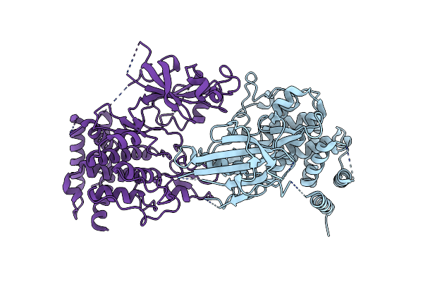 |
Structure Of Dimeric Phosphorylated Pediculus Humanus (Ph) Pink1 With Kinked Alpha-C Helix In Chain B
Organism: Pediculus humanus corporis
Method: ELECTRON MICROSCOPY Release Date: 2022-01-12 Classification: TRANSFERASE |
Organism: Pediculus humanus corporis
Method: ELECTRON MICROSCOPY
Release Date: 2022-01-12
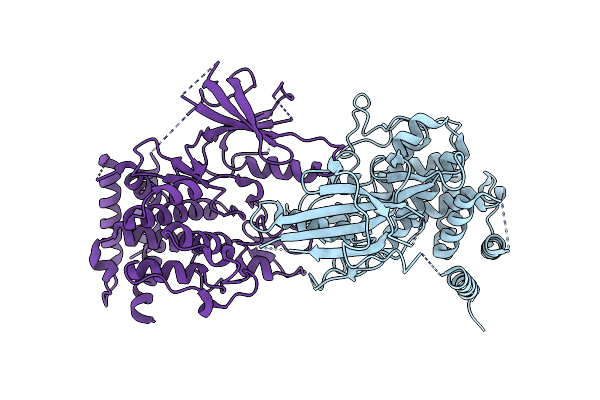 |
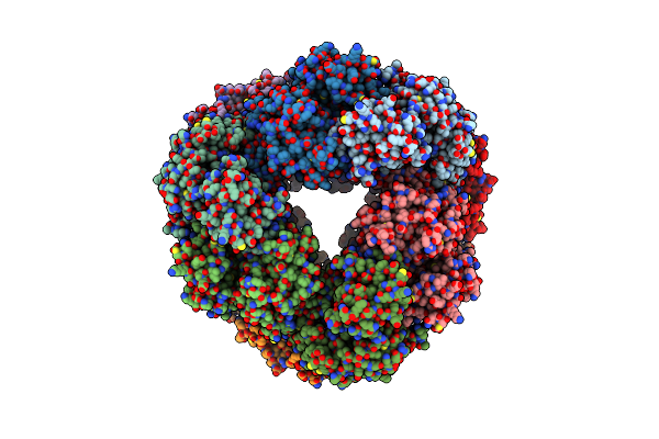
Structure Of Dimeric Phosphorylated Pediculus Humanus (Ph) Pink1 With Extended Alpha-C Helix In Chain B
Organism: Pediculus humanus corporis
Method: ELECTRON MICROSCOPY Release Date: 2022-01-12 Classification: TRANSFERASE |
Organism: Pediculus humanus corporis
Method: ELECTRON MICROSCOPY
Release Date: 2022-01-12
 |
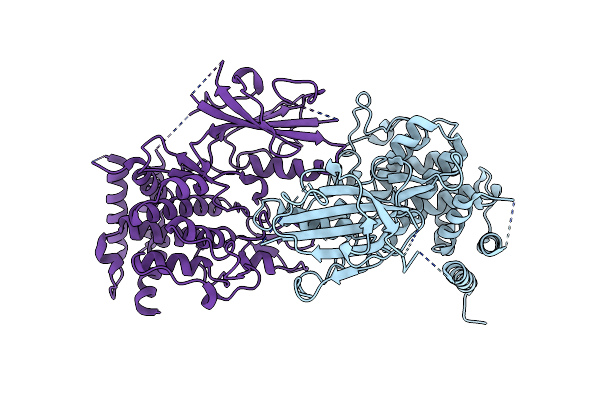
Structure Of Dodecameric Unphosphorylated Pediculus Humanus (Ph) Pink1 D357A Mutant
Organism: Pediculus humanus corporis
Method: ELECTRON MICROSCOPY Release Date: 2022-01-12 Classification: TRANSFERASE |
Organism: Pediculus humanus corporis
Method: ELECTRON MICROSCOPY
Release Date: 2022-01-12
 |
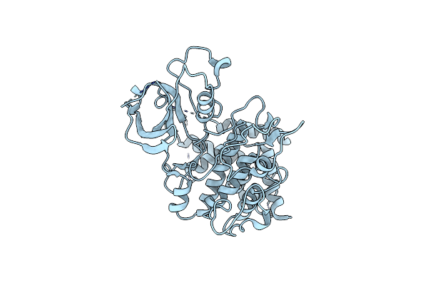
Structure Of Dimeric Unphosphorylated Pediculus Humanus (Ph) Pink1 D357A Mutant
Organism: Pediculus humanus corporis
Method: ELECTRON MICROSCOPY Release Date: 2022-01-12 Classification: TRANSFERASE |
Organism: Pediculus humanus corporis
Method: ELECTRON MICROSCOPY
Release Date: 2022-01-12
 |
Structure Of Unphosphorylated Pediculus Humanus (Ph) Pink1 D334A Mutant
Organism: Pediculus humanus corporis
Method: X-RAY DIFFRACTION Resolution:3.53 Å Release Date: 2021-12-22 Classification: TRANSFERASE |
Organism: Pediculus humanus corporis
Method: X-RAY DIFFRACTION
Release Date: 2021-12-22
 |
Crystal Structure Of An Otu Deubiquitinase From Wolbachia Pipientis Wmel
Organism: Wolbachia pipientis wmel
Method: X-RAY DIFFRACTION Resolution:1.47 Å Release Date: 2020-07-01 Classification: HYDROLASE Ligands: ACT |
Organism: Wolbachia pipientis wmel
Method: X-RAY DIFFRACTION
Release Date: 2020-07-01
Ligands: ACT
 |
Crystal Structure Of An Otu Deubiquitinase From Wolbachia Pipientis Wmel Bound To Ubiquitin
Organism: Homo sapiens, Wolbachia pipientis wmel
Method: X-RAY DIFFRACTION Resolution:1.82 Å Release Date: 2020-07-01 Classification: HYDROLASE Ligands: FLC |
Organism: Homo sapiens, Wolbachia pipientis wmel
Method: X-RAY DIFFRACTION
Release Date: 2020-07-01
Ligands: FLC
 |
Crystal Structure Of An Otu Deubiquitinase From Escherichia Albertii Bound To Ubiquitin
Organism: Escherichia albertii (strain tw07627), Homo sapiens
Method: X-RAY DIFFRACTION Resolution:2.10 Å Release Date: 2020-07-01 Classification: HYDROLASE Ligands: FMT |
Organism: Escherichia albertii (strain tw07627), Homo sapiens
Method: X-RAY DIFFRACTION
Release Date: 2020-07-01
Ligands: FMT
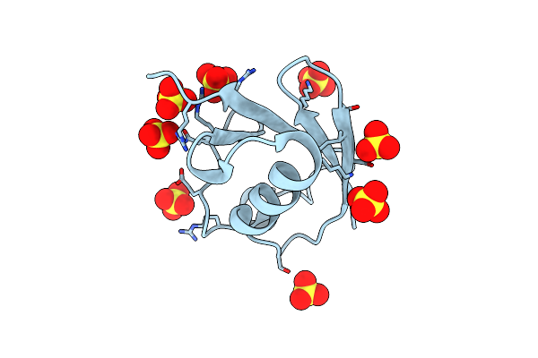 |
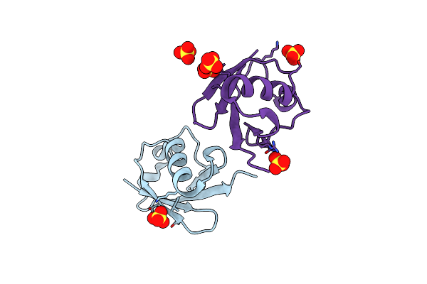
C-Terminally Retracted Ubiquitin T66V/L67N Mutant
Organism: Homo sapiens
Method: X-RAY DIFFRACTION Resolution:1.60 Å Release Date: 2017-11-22 Classification: SIGNALING PROTEIN Ligands: SO4 |
Organism: Homo sapiens
Method: X-RAY DIFFRACTION
Release Date: 2017-11-22
Ligands: SO4
 |
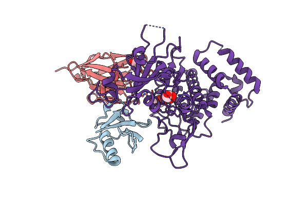
C-Terminally Retracted Ubiquitin L67S Mutant
Organism: Homo sapiens
Method: X-RAY DIFFRACTION Resolution:1.63 Å Release Date: 2017-11-22 Classification: SIGNALING PROTEIN Ligands: SO4 |
Organism: Homo sapiens
Method: X-RAY DIFFRACTION
Release Date: 2017-11-22
Ligands: SO4
 |
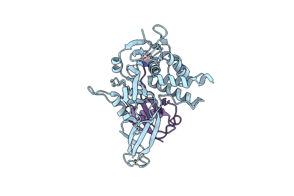
Structure Of Pink1 Bound To Ubiquitin
Organism: Homo sapiens, Lama glama, Pediculus humanus corporis
Method: X-RAY DIFFRACTION Resolution:3.10 Å Release Date: 2017-11-08 Classification: TRANSFERASE Ligands: GOL |
Organism: Homo sapiens, Lama glama, Pediculus humanus corporis
Method: X-RAY DIFFRACTION
Release Date: 2017-11-08
Ligands: GOL
 |
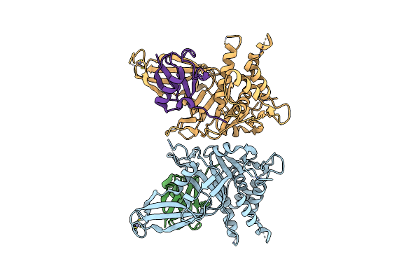
Crystal Structure Of Usp30 In Covalent Complex With Ubiquitin Propargylamide (High Resolution)
Organism: Homo sapiens
Method: X-RAY DIFFRACTION Resolution:2.34 Å Release Date: 2017-09-20 Classification: HYDROLASE Ligands: ZN, AYE |
Organism: Homo sapiens
Method: X-RAY DIFFRACTION
Release Date: 2017-09-20
Ligands: ZN, AYE
 |
Crystal Structure Of Usp30 In Covalent Complex With Ubiquitin Propargylamide (Low Resolution)
Organism: Homo sapiens
Method: X-RAY DIFFRACTION Resolution:3.60 Å Release Date: 2017-09-20 Classification: HYDROLASE Ligands: ZN |
Organism: Homo sapiens
Method: X-RAY DIFFRACTION
Release Date: 2017-09-20
Ligands: ZN
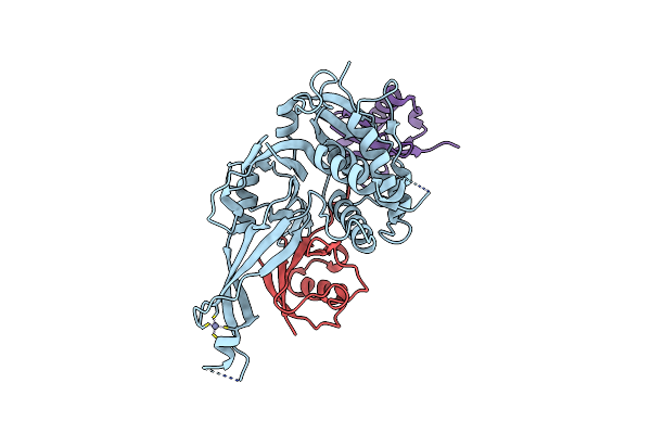 |
Crystal Structure Of Usp30 (C77A) In Complex With Lys6-Linked Diubiquitin
Organism: Homo sapiens
Method: X-RAY DIFFRACTION Resolution:2.80 Å Release Date: 2017-09-20 Classification: HYDROLASE Ligands: ZN |
Organism: Homo sapiens
Method: X-RAY DIFFRACTION
Release Date: 2017-09-20
Ligands: ZN