

Search Count: 55
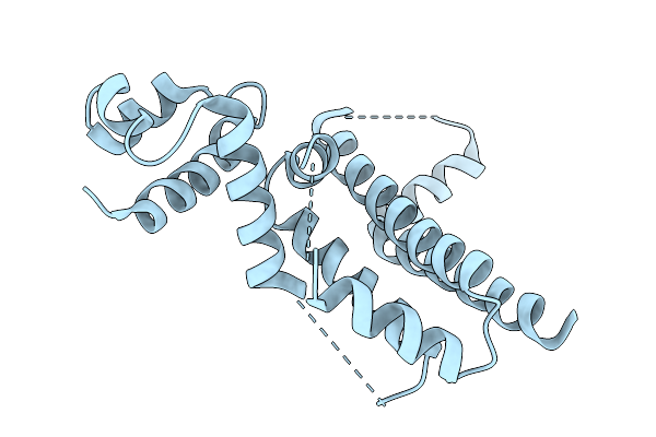 |
Crystal Structure Of The Engineered Sulfonylurea Repressor Esr L7-D01, Apo Form
Organism: Escherichia coli
Method: X-RAY DIFFRACTION Resolution:3.10 Å Release Date: 2025-10-08 Classification: DNA BINDING PROTEIN |
Organism: Escherichia coli
Method: X-RAY DIFFRACTION
Release Date: 2025-10-08
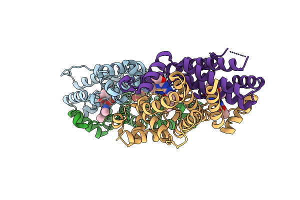 |
Crystal Structure Of The Engineered Sulfonylurea Repressor Esr L11-C06, Bound To Ethametsulfuron-Methyl
Organism: Escherichia coli
Method: X-RAY DIFFRACTION Resolution:2.80 Å Release Date: 2025-10-08 Classification: DNA BINDING PROTEIN Ligands: RXF |
Organism: Escherichia coli
Method: X-RAY DIFFRACTION
Release Date: 2025-10-08
Ligands: RXF
 |
Crystal Structure Of The Engineered Sulfonylurea Repressor Csr L4.2-20, Apo Form
Organism: Escherichia coli
Method: X-RAY DIFFRACTION Resolution:2.40 Å Release Date: 2025-10-08 Classification: DNA BINDING PROTEIN |
Organism: Escherichia coli
Method: X-RAY DIFFRACTION
Release Date: 2025-10-08
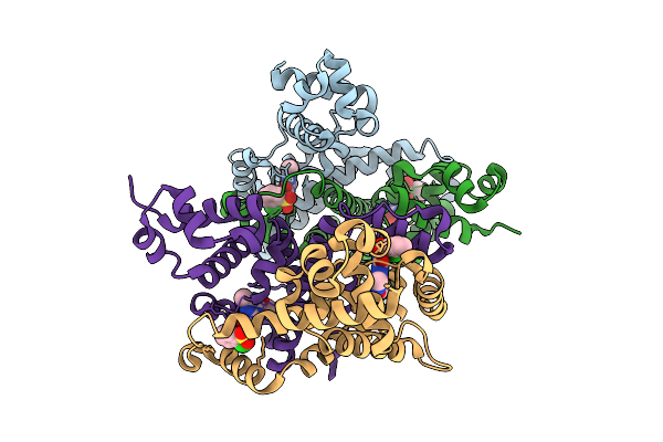 |
Crystal Structure Of The Engineered Sulfonylurea Repressor Csr L4.2-20, Bound To Chlorsulfuron
Organism: Escherichia coli
Method: X-RAY DIFFRACTION Resolution:2.00 Å Release Date: 2025-10-08 Classification: DNA BINDING PROTEIN Ligands: 1CS |
Organism: Escherichia coli
Method: X-RAY DIFFRACTION
Release Date: 2025-10-08
Ligands: 1CS
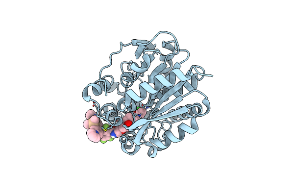 |
Crystal Structure Of Halotag7 Bound To Jf669-Halotag Ligand
Organism: Rhodococcus sp. (in: high g+c gram-positive bacteria)
Method: X-RAY DIFFRACTION Resolution:1.90 Å Release Date: 2024-06-05 Classification: HYDROLASE Ligands: XSR, CL |
Organism: Rhodococcus sp. (in: high g+c gram-positive bacteria)
Method: X-RAY DIFFRACTION
Release Date: 2024-06-05
Ligands: XSR, CL
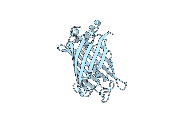 |
Genetically Encoded Ph Sensor Lime At Ph10
Organism: Aequorea victoria
Method: X-RAY DIFFRACTION Resolution:2.00 Å Release Date: 2023-08-16 Classification: FLUORESCENT PROTEIN |
Organism: Aequorea victoria
Method: X-RAY DIFFRACTION
Release Date: 2023-08-16
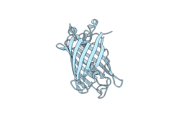 |
Genetically Encoded Ph Sensor Lime At Ph6
Organism: Aequorea victoria
Method: X-RAY DIFFRACTION Resolution:2.85 Å Release Date: 2023-08-16 Classification: FLUORESCENT PROTEIN |
Organism: Aequorea victoria
Method: X-RAY DIFFRACTION
Release Date: 2023-08-16
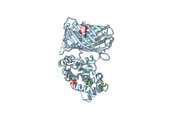 |
Calcium-Saturated Jgcamp8.410.80
Organism: Synthetic construct
Method: X-RAY DIFFRACTION Resolution:2.00 Å Release Date: 2022-11-16 Classification: FLUORESCENT PROTEIN Ligands: CA, GOL, TLA |
Organism: Synthetic construct
Method: X-RAY DIFFRACTION
Release Date: 2022-11-16
Ligands: CA, GOL, TLA
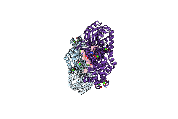 |
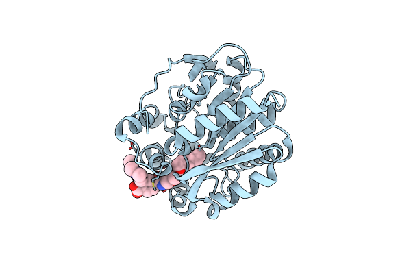
Crystal Structure Of A Halotag-Based Calcium Indicator, Halocamp V2, Bound To Jf635
Organism: Rhodococcus sp.
Method: X-RAY DIFFRACTION Resolution:2.00 Å Release Date: 2020-09-30 Classification: HYDROLASE Ligands: PUJ, CL, CA |
Organism: Rhodococcus sp.
Method: X-RAY DIFFRACTION
Release Date: 2020-09-30
Ligands: PUJ, CL, CA
 |
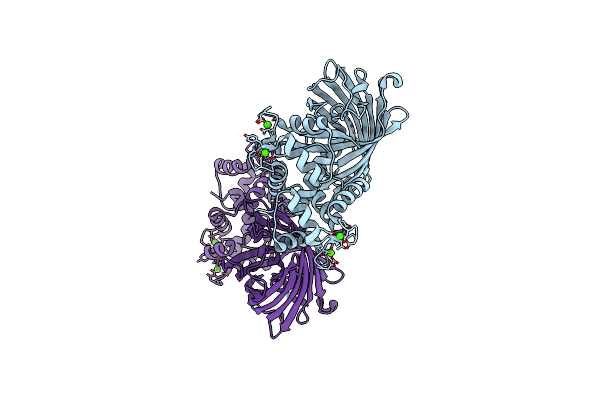
Crystal Structure Of Halotag Bound To Tetramethylrhodamine-Halotag Ligand
Organism: Rhodococcus sp.
Method: X-RAY DIFFRACTION Resolution:1.80 Å Release Date: 2020-09-30 Classification: HYDROLASE Ligands: PVY, CL |
Organism: Rhodococcus sp.
Method: X-RAY DIFFRACTION
Release Date: 2020-09-30
Ligands: PVY, CL
 |
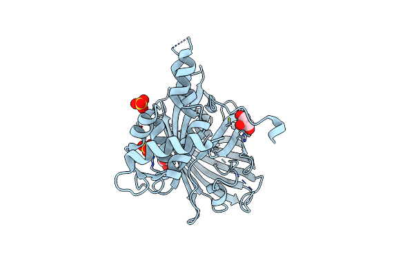
Crystal Structure Of The Genetically Encoded Calcium Indicator K-Geco
Organism: Entacmaea quadricolor
Method: X-RAY DIFFRACTION Resolution:2.36 Å Release Date: 2018-02-07 Classification: FLUORESCENT PROTEIN Ligands: CA |
Organism: Entacmaea quadricolor
Method: X-RAY DIFFRACTION
Release Date: 2018-02-07
Ligands: CA
 |
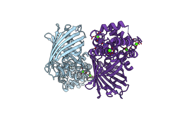
Calcium-Free Campari V0.2
Organism: Rattus norvegicus, Lobophyllia hemprichii, Gallus gallus
Method: X-RAY DIFFRACTION Resolution:2.03 Å Release Date: 2015-02-18 Classification: Calcium Binding, Fluorescent, Transferase Ligands: SO4, GOL |
Organism: Rattus norvegicus, Lobophyllia hemprichii, Gallus gallus
Method: X-RAY DIFFRACTION
Release Date: 2015-02-18
Ligands: SO4, GOL
 |
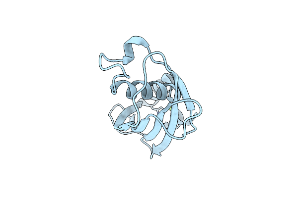
Crystal Structure Of The Genetically Encoded Calcium Indicator Rgeco1
Organism: Gallus gallus, Discosoma sp., Rattus norvegicus
Method: X-RAY DIFFRACTION Resolution:2.20 Å Release Date: 2013-07-24 Classification: FLUORESCENT PROTEIN Ligands: CA |
Organism: Gallus gallus, Discosoma sp., Rattus norvegicus
Method: X-RAY DIFFRACTION
Release Date: 2013-07-24
Ligands: CA
 |
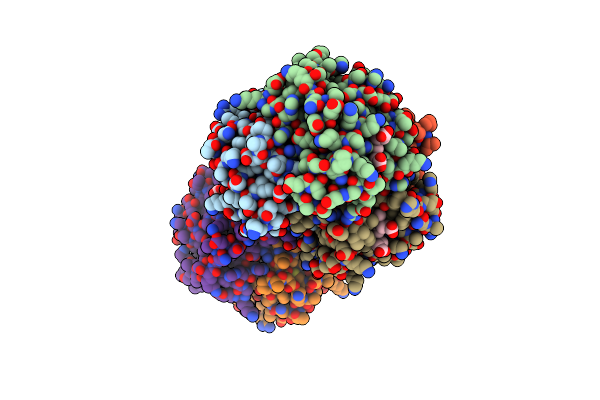
Crystal Structure Of Orf6 Protein From Photobacterium Profundum, Mg2+-Bound Form
Organism: Photobacterium profundum
Method: X-RAY DIFFRACTION Resolution:1.40 Å Release Date: 2013-02-27 Classification: HYDROLASE Ligands: MG |
Organism: Photobacterium profundum
Method: X-RAY DIFFRACTION
Release Date: 2013-02-27
Ligands: MG
 |
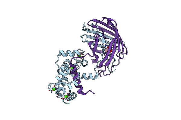
Human Zeta-2 Beta-2-S Hemoglobin
Organism: Homo sapiens
Method: X-RAY DIFFRACTION Resolution:1.95 Å Release Date: 2013-02-06 Classification: OXYGEN TRANSPORT Ligands: HEM, CMO |
Organism: Homo sapiens
Method: X-RAY DIFFRACTION
Release Date: 2013-02-06
Ligands: HEM, CMO
 |
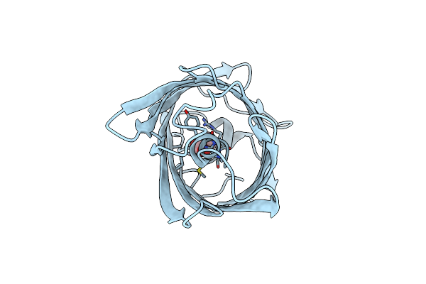
Crystal Structure Of The Genetically Encoded Calcium Indicator Rcamp
Organism: Entacmaea quadricolor
Method: X-RAY DIFFRACTION Resolution:2.10 Å Release Date: 2012-10-03 Classification: FLUORESCENT PROTEIN Ligands: ACT, CA |
Organism: Entacmaea quadricolor
Method: X-RAY DIFFRACTION
Release Date: 2012-10-03
Ligands: ACT, CA
 |
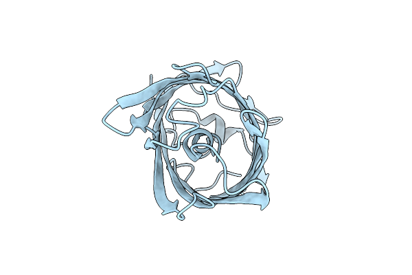
Crystal Structure Of The Engineered Fluorescent Protein Mruby, Crystal Form 1, Ph 4.5
Organism: Entacmaea quadricolor
Method: X-RAY DIFFRACTION Resolution:1.25 Å Release Date: 2012-10-03 Classification: FLUORESCENT PROTEIN Ligands: ACT |
Organism: Entacmaea quadricolor
Method: X-RAY DIFFRACTION
Release Date: 2012-10-03
Ligands: ACT
 |
Crystal Structure Of The Engineered Fluorescent Protein Mruby, Crystal Form 1, Ph 8.5
Organism: Entacmaea quadricolor
Method: X-RAY DIFFRACTION Resolution:1.65 Å Release Date: 2012-10-03 Classification: FLUORESCENT PROTEIN |
Organism: Entacmaea quadricolor
Method: X-RAY DIFFRACTION
Release Date: 2012-10-03
 |
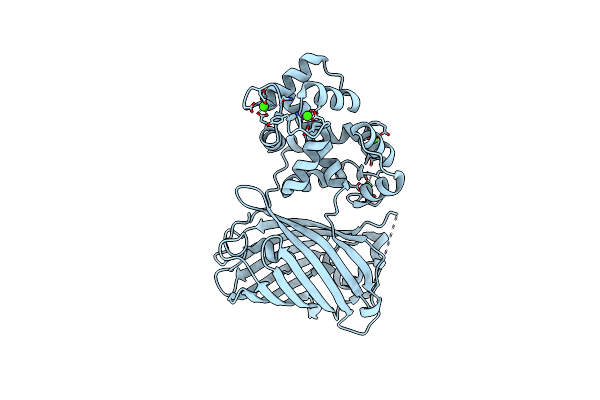
Crystal Structure Of The Engineered Fluorescent Protein Mruby, Crystal Form 2
Organism: Entacmaea quadricolor
Method: X-RAY DIFFRACTION Resolution:1.60 Å Release Date: 2012-10-03 Classification: FLUORESCENT PROTEIN Ligands: SO4 |
Organism: Entacmaea quadricolor
Method: X-RAY DIFFRACTION
Release Date: 2012-10-03
Ligands: SO4
 |
Crystal Structure Of Gcamp2-T116V,D381Y
Organism: Gallus gallus, Aequorea victoria, Rattus norvegicus
Method: X-RAY DIFFRACTION Resolution:2.00 Å Release Date: 2012-06-20 Classification: FLUORESCENT PROTEIN Ligands: CA |
Organism: Gallus gallus, Aequorea victoria, Rattus norvegicus
Method: X-RAY DIFFRACTION
Release Date: 2012-06-20
Ligands: CA