

Search Count: 164
 |
Prescottella Amidase Wt
Organism: Prescottella equi
Method: X-RAY DIFFRACTION Resolution:1.25 Å Release Date: 2026-09-16 Classification: HYDROLASE Ligands: DMS, MG, EDO |
Organism: Prescottella equi
Method: X-RAY DIFFRACTION
Release Date: 2026-09-16
Ligands: DMS, MG, EDO
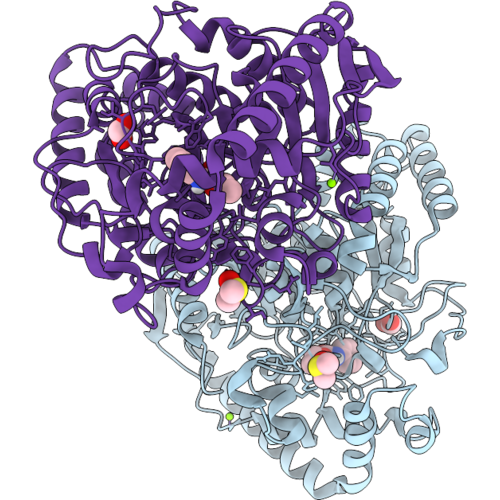 |
Prescottella Amidase S181A Mutant, Ethyl N-Phenylcarbamate Soak.
Organism: Prescottella equi
Method: X-RAY DIFFRACTION Resolution:1.30 Å Release Date: 2026-09-16 Classification: HYDROLASE Ligands: DMS, FMT, A1J75, MG |
Organism: Prescottella equi
Method: X-RAY DIFFRACTION
Release Date: 2026-09-16
Ligands: DMS, FMT, A1J75, MG
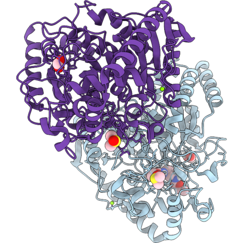 |
Prescottella Amidase, S181A Mutant, Diethyl Toluene-2,4-Dicarbamate Soak
Organism: Prescottella equi
Method: X-RAY DIFFRACTION Resolution:1.20 Å Release Date: 2026-09-16 Classification: HYDROLASE Ligands: FMT, A1J91, MG, DMS |
Organism: Prescottella equi
Method: X-RAY DIFFRACTION
Release Date: 2026-09-16
Ligands: FMT, A1J91, MG, DMS
 |
Cryo-Em Structure Of The Flotillin-Associated Rhodopsin Psfar In Detergent Micelle
Organism: Candidatus pseudothioglobus
Method: ELECTRON MICROSCOPY Resolution:2.56 Å Release Date: 2025-07-23 Classification: MEMBRANE PROTEIN Ligands: LFA |
Organism: Candidatus pseudothioglobus
Method: ELECTRON MICROSCOPY
Release Date: 2025-07-23
Ligands: LFA
 |
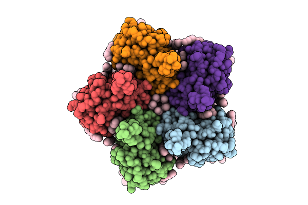
Cryo-Em Structure Of The Light-Driven Proton Pump Pspr In Detergent Micelle
Organism: Candidatus pseudothioglobus sp.
Method: ELECTRON MICROSCOPY Resolution:2.48 Å Release Date: 2025-07-23 Classification: MEMBRANE PROTEIN Ligands: LFA, RET, LMT |
Organism: Candidatus pseudothioglobus sp.
Method: ELECTRON MICROSCOPY
Release Date: 2025-07-23
Ligands: LFA, RET, LMT
 |
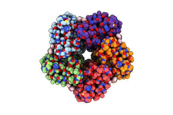
Cryo-Em Structure Of The Double Mutant H84V/E120G Of The Flotillin-Associated Rhodopsin Psfar In Detergent Micelle
Organism: Candidatus pseudothioglobus sp.
Method: ELECTRON MICROSCOPY Resolution:2.81 Å Release Date: 2025-07-23 Classification: MEMBRANE PROTEIN Ligands: LFA, RET |
Organism: Candidatus pseudothioglobus sp.
Method: ELECTRON MICROSCOPY
Release Date: 2025-07-23
Ligands: LFA, RET
 |
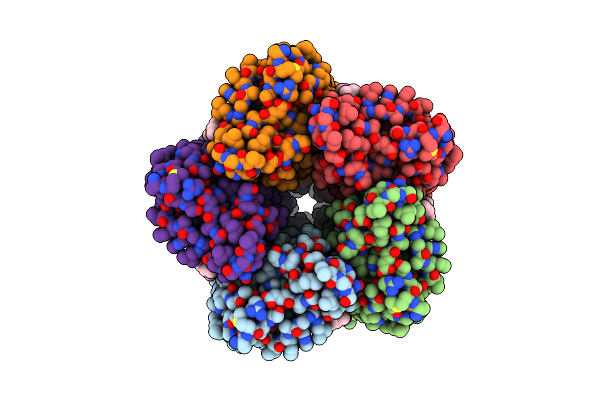
Cryo-Em Structure Of The Microbial Rhodopsin Cryor1 At Ph 4.3 In Detergent
Organism: Cryobacterium levicorallinum
Method: ELECTRON MICROSCOPY Resolution:2.94 Å Release Date: 2025-05-14 Classification: MEMBRANE PROTEIN Ligands: LFA, RET |
Organism: Cryobacterium levicorallinum
Method: ELECTRON MICROSCOPY
Release Date: 2025-05-14
Ligands: LFA, RET
 |
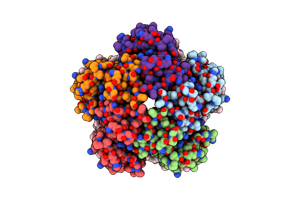
Cryo-Em Structure Of The Microbial Rhodopsin Cryor1 At Ph 8.0 In Nanodisc
Organism: Cryobacterium levicorallinum
Method: ELECTRON MICROSCOPY Resolution:2.43 Å Release Date: 2025-05-14 Classification: MEMBRANE PROTEIN Ligands: LFA, RET |
Organism: Cryobacterium levicorallinum
Method: ELECTRON MICROSCOPY
Release Date: 2025-05-14
Ligands: LFA, RET
 |
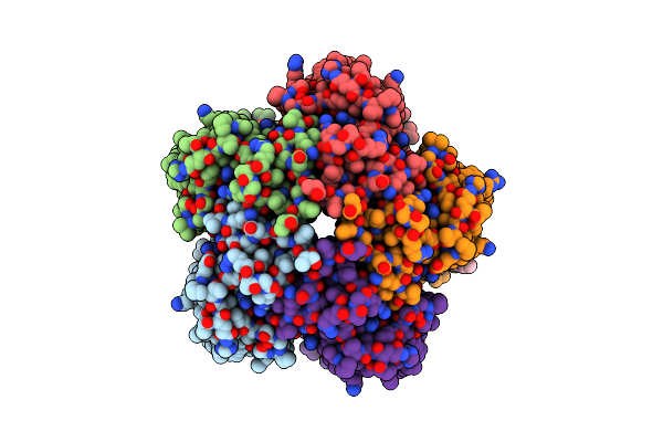
Cryo-Em Structure Of The Microbial Rhodopsin Cryor1 At Ph 8.0 In Detergent
Organism: Cryobacterium levicorallinum
Method: ELECTRON MICROSCOPY Resolution:2.87 Å Release Date: 2025-05-14 Classification: MEMBRANE PROTEIN Ligands: LMT, LFA, RET |
Organism: Cryobacterium levicorallinum
Method: ELECTRON MICROSCOPY
Release Date: 2025-05-14
Ligands: LMT, LFA, RET
 |
Cryo-Em Structure Of The Microbial Rhodopsin Cryor1 At Ph 10.5 In Detergent In The Ground State
Organism: Cryobacterium levicorallinum
Method: ELECTRON MICROSCOPY Resolution:2.70 Å Release Date: 2025-05-14 Classification: MEMBRANE PROTEIN Ligands: LMT, RET, LFA |
Organism: Cryobacterium levicorallinum
Method: ELECTRON MICROSCOPY
Release Date: 2025-05-14
Ligands: LMT, RET, LFA
 |
Cryo-Em Structure Of The Microbial Rhodopsin Cryor1 At Ph 10.5 In Detergent In The M State
Organism: Cryobacterium levicorallinum
Method: ELECTRON MICROSCOPY Resolution:2.30 Å Release Date: 2025-05-14 Classification: MEMBRANE PROTEIN Ligands: LMT, RET |
Organism: Cryobacterium levicorallinum
Method: ELECTRON MICROSCOPY
Release Date: 2025-05-14
Ligands: LMT, RET
 |
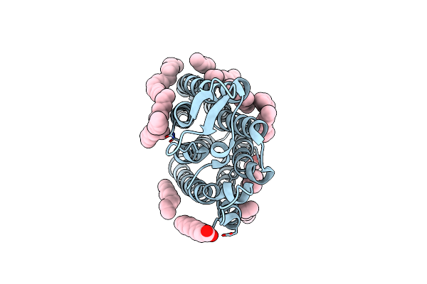
Cryo-Em Structure Of The Microbial Rhodopsin Cryor2 At Ph 8.0 In Detergent
Organism: Subtercola endophyticus
Method: ELECTRON MICROSCOPY Resolution:2.44 Å Release Date: 2025-05-14 Classification: MEMBRANE PROTEIN Ligands: LFA, RET |
Organism: Subtercola endophyticus
Method: ELECTRON MICROSCOPY
Release Date: 2025-05-14
Ligands: LFA, RET
 |
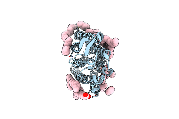
Crystal Structure Of The Light-Driven Sodium Pump Ernar In The Monomeric Form At Ph 4.6
Organism: Erythrobacter
Method: X-RAY DIFFRACTION Resolution:1.70 Å Release Date: 2024-04-24 Classification: MEMBRANE PROTEIN Ligands: LFA, OLA |
Organism: Erythrobacter
Method: X-RAY DIFFRACTION
Release Date: 2024-04-24
Ligands: LFA, OLA
 |
Crystal Structure Of The Light-Driven Sodium Pump Ernar In The Monomeric Form At Ph 8.8
Organism: Erythrobacter
Method: X-RAY DIFFRACTION Resolution:1.71 Å Release Date: 2024-04-24 Classification: MEMBRANE PROTEIN Ligands: LFA, OLA |
Organism: Erythrobacter
Method: X-RAY DIFFRACTION
Release Date: 2024-04-24
Ligands: LFA, OLA
 |
Cryo-Em Structure Of The Light-Driven Sodium Pump Ernar In The Pentameric Form At Ph 8.0
Organism: Erythrobacter
Method: ELECTRON MICROSCOPY Resolution:2.63 Å Release Date: 2024-04-24 Classification: MEMBRANE PROTEIN Ligands: LFA, LMT |
Organism: Erythrobacter
Method: ELECTRON MICROSCOPY
Release Date: 2024-04-24
Ligands: LFA, LMT
 |
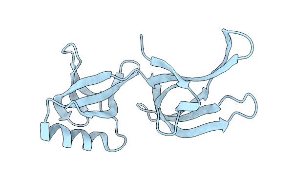
Cryo-Em Structure Of The Light-Driven Sodium Pump Ernar In The Pentameric Form At Ph 4.3
Organism: Erythrobacter
Method: ELECTRON MICROSCOPY Resolution:2.50 Å Release Date: 2024-04-24 Classification: MEMBRANE PROTEIN Ligands: LMT, LFA |
Organism: Erythrobacter
Method: ELECTRON MICROSCOPY
Release Date: 2024-04-24
Ligands: LMT, LFA
 |
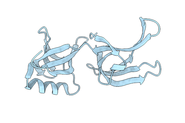
Structure Of Hex-1 From N. Crassa Crystallized In Cellulo (Cytosol), Diffracted At 100K And Resolved Using Crystfel
Organism: Neurospora crassa
Method: X-RAY DIFFRACTION Resolution:1.83 Å Release Date: 2024-02-21 Classification: STRUCTURAL PROTEIN |
Organism: Neurospora crassa
Method: X-RAY DIFFRACTION
Release Date: 2024-02-21
 |
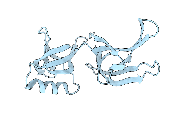
Structure Of Hex-1 From N. Crassa Crystallized In Cellulo, Diffracted At 100K And Resolved Using Crystfel
Organism: Neurospora crassa
Method: X-RAY DIFFRACTION Resolution:1.56 Å Release Date: 2024-02-21 Classification: STRUCTURAL PROTEIN |
Organism: Neurospora crassa
Method: X-RAY DIFFRACTION
Release Date: 2024-02-21
 |
Structure Of Hex-1 (Cyto V2) From N. Crassa Grown In Living Insect Cells, Diffracted At 100K And Resolved Using Crystfel
Organism: Neurospora crassa
Method: X-RAY DIFFRACTION Resolution:1.85 Å Release Date: 2024-02-21 Classification: STRUCTURAL PROTEIN |
Organism: Neurospora crassa
Method: X-RAY DIFFRACTION
Release Date: 2024-02-21
 |
Structure Of Hex-1 From N. Crassa Crystallized In Cellulo, Diffracted At 100K And Resolved Using Xds
Organism: Neurospora crassa
Method: X-RAY DIFFRACTION Resolution:1.85 Å Release Date: 2024-02-21 Classification: STRUCTURAL PROTEIN |
Organism: Neurospora crassa
Method: X-RAY DIFFRACTION
Release Date: 2024-02-21