

Search Count: 17
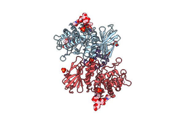 |
Human Alpha-Galactosidase A In Complex With Saposin B
Organism: Homo sapiens
Method: X-RAY DIFFRACTION Resolution:3.53 Å Release Date: 2024-04-17 Classification: HYDROLASE Ligands: SO4, NAG |
Organism: Homo sapiens
Method: X-RAY DIFFRACTION
Release Date: 2024-04-17
Ligands: SO4, NAG
 |
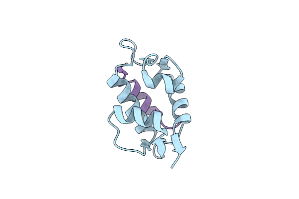
Human Saposin B In The Presence Of Globotriaosylceramide-Nbd
Organism: Homo sapiens
Method: X-RAY DIFFRACTION Resolution:2.68 Å Release Date: 2024-03-13 Classification: LIPID BINDING PROTEIN Ligands: MLI |
Organism: Homo sapiens
Method: X-RAY DIFFRACTION
Release Date: 2024-03-13
Ligands: MLI
 |
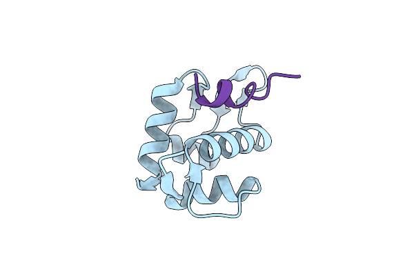
Stapled Peptide Alrn-6924 Bound To Mdmx
Organism: Danio rerio
Method: X-RAY DIFFRACTION Resolution:1.75 Å Release Date: 2023-07-26 Classification: APOPTOSIS |
Organism: Danio rerio
Method: X-RAY DIFFRACTION
Release Date: 2023-07-26
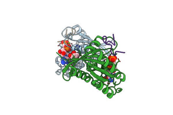 |
Kras G12D Mutant In Complex With Gmppcp And Cyclic Peptide Mp-9903
Organism: Homo sapiens, Synthetic construct
Method: X-RAY DIFFRACTION Resolution:1.32 Å Release Date: 2021-09-22 Classification: HYDROLASE Ligands: GCP, MG, GOL |
Organism: Homo sapiens, Synthetic construct
Method: X-RAY DIFFRACTION
Release Date: 2021-09-22
Ligands: GCP, MG, GOL
 |
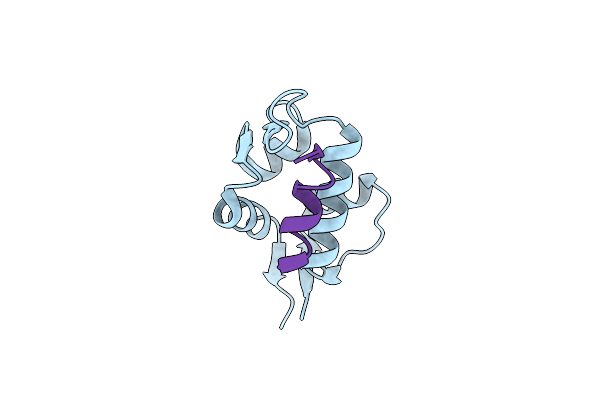
Macrocyclization Of An All-D Linear Peptide Improves Target Affinity And Imparts Cellular Activity: A Novel Stapled Alpha-Helical Peptide Modality
Organism: Homo sapiens, Phage 13
Method: X-RAY DIFFRACTION Resolution:1.79 Å Release Date: 2019-10-16 Classification: LIGASE |
Organism: Homo sapiens, Phage 13
Method: X-RAY DIFFRACTION
Release Date: 2019-10-16
 |
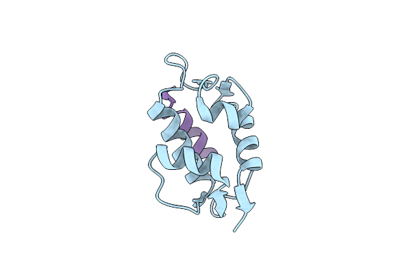
Mdm2 In Complex With A D Amino Acid Containing Stapled Peptide
Organism: Homo sapiens, Enterobacteria phage m13
Method: X-RAY DIFFRACTION Resolution:2.00 Å Release Date: 2019-07-24 Classification: LIGASE |
Organism: Homo sapiens, Enterobacteria phage m13
Method: X-RAY DIFFRACTION
Release Date: 2019-07-24
 |
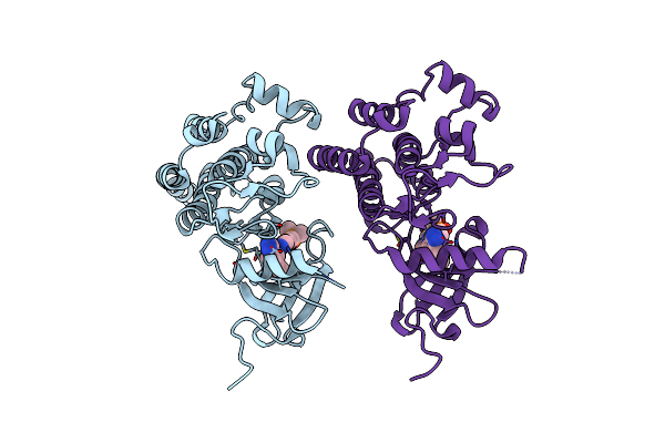
The 1.7A Crystal Structure Of Mdmx With A Stapled Peptide, Atsp-7041
Organism: Danio rerio, Synthetic construct
Method: X-RAY DIFFRACTION Resolution:1.70 Å Release Date: 2013-11-20 Classification: CELL CYCLE/CELL CYCLE INHIBITOR |
Organism: Danio rerio, Synthetic construct
Method: X-RAY DIFFRACTION
Release Date: 2013-11-20
 |
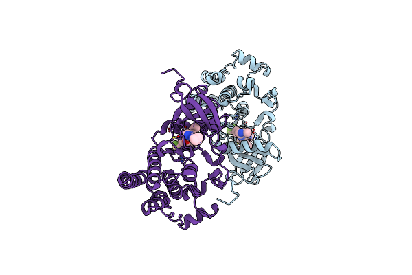
Structural Analysis Of Dfg-In And Dfg-Out Dual Src-Abl Inhibitors Sharing A Common Vinyl Purine Template
Organism: Mus musculus
Method: X-RAY DIFFRACTION Resolution:1.90 Å Release Date: 2009-12-22 Classification: TRANSFERASE Ligands: B90 |
Organism: Mus musculus
Method: X-RAY DIFFRACTION
Release Date: 2009-12-22
Ligands: B90
 |
Structural Analysis Of Dfg-In And Dfg-Out Dual Src-Abl Inhibitors Sharing A Common Vinyl Purine Template
Organism: Mus musculus
Method: X-RAY DIFFRACTION Resolution:1.22 Å Release Date: 2009-12-22 Classification: TRANSFERASE Ligands: B91 |
Organism: Mus musculus
Method: X-RAY DIFFRACTION
Release Date: 2009-12-22
Ligands: B91
 |
Ap24534, A Pan-Bcr-Abl Inhibitor For Chronic Myeloid Leukemia, Potently Inhibits The T315I Mutant And Overcomes Mutation-Based Resistance
Organism: Mus musculus
Method: X-RAY DIFFRACTION Resolution:1.90 Å Release Date: 2009-11-03 Classification: TRANSFERASE Ligands: 0LI |
Organism: Mus musculus
Method: X-RAY DIFFRACTION
Release Date: 2009-11-03
Ligands: 0LI
 |
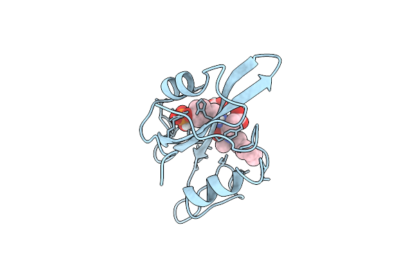
Crystal Structure Of Lck Sh2 Complexed With Nonpeptide Phosphotyrosine Mimetic
Organism: Homo sapiens
Method: X-RAY DIFFRACTION Resolution:2.20 Å Release Date: 2002-05-01 Classification: TRANSFERASE/SIGNALING PROTEIN Ligands: CC0 |
Organism: Homo sapiens
Method: X-RAY DIFFRACTION
Release Date: 2002-05-01
Ligands: CC0
 |
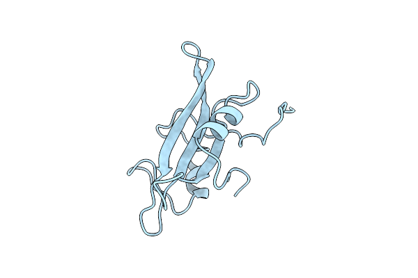
Cocrystal Structure Of Urea-Substituted Phosphopeptide Complex
Organism: Rous sarcoma virus
Method: X-RAY DIFFRACTION Resolution:2.00 Å Release Date: 1998-02-25 Classification: TYROSINE-PROTEIN KINASE Ligands: UR2 |
Organism: Rous sarcoma virus
Method: X-RAY DIFFRACTION
Release Date: 1998-02-25
Ligands: UR2
 |
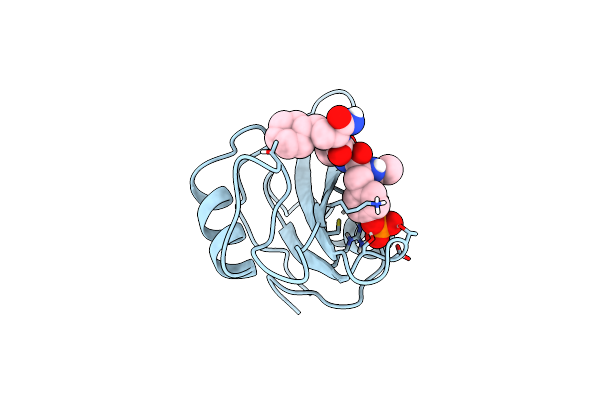
Self-Associated Apo Src Sh2 Domain
Organism: Rous sarcoma virus
Method: X-RAY DIFFRACTION Resolution:2.10 Å Release Date: 1997-07-23 Classification: V-SRC SH2 DOMAIN |
Organism: Rous sarcoma virus
Method: X-RAY DIFFRACTION
Release Date: 1997-07-23
 |
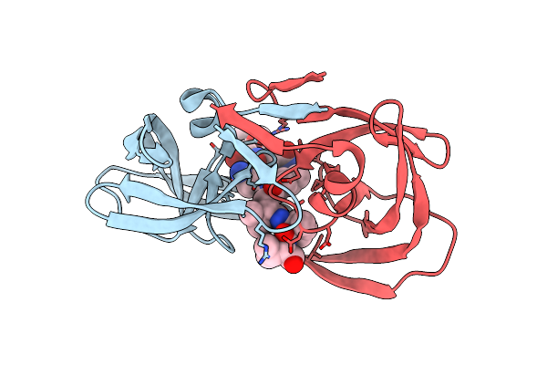
Cocrystal Structure Of D-Amino Acid Substituted Phosphopeptide Complex
Organism: Rous sarcoma virus
Method: X-RAY DIFFRACTION Resolution:2.00 Å Release Date: 1997-07-07 Classification: TRANSFORMING PROTEIN Ligands: 1C5 |
Organism: Rous sarcoma virus
Method: X-RAY DIFFRACTION
Release Date: 1997-07-07
Ligands: 1C5
 |
Structure At 2.5-Angstroms Resolution Of Chemically Synthesized Human Immunodeficiency Virus Type 1 Protease Complexed With A Hydroxyethylene*-Based Inhibitor
Organism: Human immunodeficiency virus 1
Method: X-RAY DIFFRACTION Resolution:2.50 Å Release Date: 1993-10-31 Classification: HYDROLASE/HYDROLASE INHIBITOR |
Organism: Human immunodeficiency virus 1
Method: X-RAY DIFFRACTION
Release Date: 1993-10-31
 |
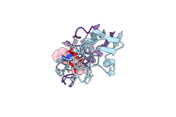
The Crystallographic Structure Of The Protease From Human Immunodeficiency Virus Type 2 With Two Synthetic Peptidic Transition State Analog Inhibitors
Organism: Human immunodeficiency virus 2
Method: X-RAY DIFFRACTION Resolution:2.50 Å Release Date: 1993-07-15 Classification: HYDROLASE/HYDROLASE INHIBITOR Ligands: 1ZK |
Organism: Human immunodeficiency virus 2
Method: X-RAY DIFFRACTION
Release Date: 1993-07-15
Ligands: 1ZK
 |
The Crystallographic Structure Of The Protease From Human Immunodeficiency Virus Type 2 With Two Synthetic Peptidic Transition State Analog Inhibitors
Organism: Human immunodeficiency virus 2
Method: X-RAY DIFFRACTION Resolution:2.60 Å Release Date: 1993-07-15 Classification: HYDROLASE/HYDROLASE INHIBITOR Ligands: 0PX |
Organism: Human immunodeficiency virus 2
Method: X-RAY DIFFRACTION
Release Date: 1993-07-15
Ligands: 0PX