

Search Count: 15
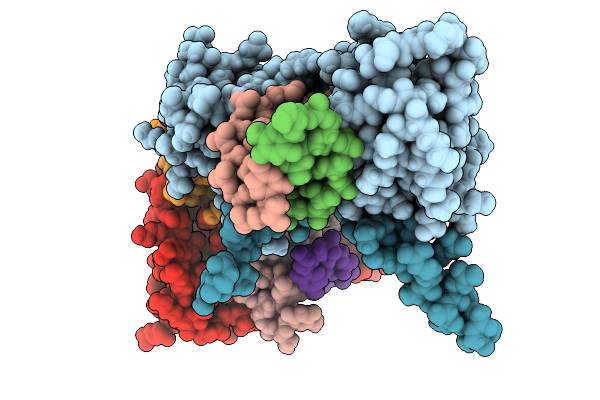 |
Structure Of Pou2F3 Pou Domains Bound To Coactivator Oca-T2 And Dna (2.8 Angstrom Resolution)
Organism: Homo sapiens
Method: X-RAY DIFFRACTION Resolution:2.79 Å Release Date: 2025-10-01 Classification: TRANSCRIPTION |
Organism: Homo sapiens
Method: X-RAY DIFFRACTION
Release Date: 2025-10-01
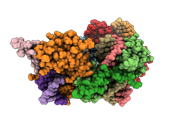 |
Structure Of Pou2F3 Pou Domains Bound To Coactivator Oca-T2 And Dna (2.1 Angstrom Resolution)
Organism: Homo sapiens
Method: X-RAY DIFFRACTION Resolution:2.10 Å Release Date: 2025-10-01 Classification: TRANSCRIPTION |
Organism: Homo sapiens
Method: X-RAY DIFFRACTION
Release Date: 2025-10-01
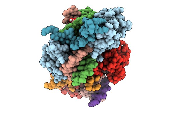 |
Structure Of Pou2F3 Pou Domains Bound To Coactivator Oca-T1 And Dna
Organism: Homo sapiens
Method: X-RAY DIFFRACTION Resolution:1.70 Å Release Date: 2025-10-01 Classification: TRANSCRIPTION Ligands: BO3 |
Organism: Homo sapiens
Method: X-RAY DIFFRACTION
Release Date: 2025-10-01
Ligands: BO3
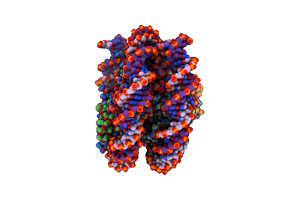 |
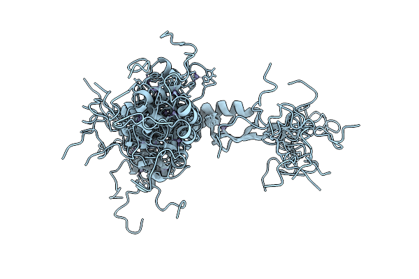
Arabidopsis Ddm1 Bound To Nucleosome (H2A.W, H2B, H3.3, H4, With 147 Bp Dna)
Organism: Arabidopsis thaliana, Xenopus laevis, Synthetic construct
Method: ELECTRON MICROSCOPY Release Date: 2023-08-09 Classification: GENE REGULATION |
Organism: Arabidopsis thaliana, Xenopus laevis, Synthetic construct
Method: ELECTRON MICROSCOPY
Release Date: 2023-08-09
 |
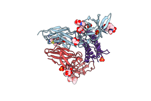
Nmr Solution Structure Of Asterix/Gtsf1 From Mouse (Chhc Zinc Finger Domains)
Organism: Mus musculus
Method: SOLUTION NMR Release Date: 2021-03-03 Classification: RNA BINDING PROTEIN Ligands: ZN |
Organism: Mus musculus
Method: SOLUTION NMR
Release Date: 2021-03-03
Ligands: ZN
 |
Crystal Structure Of A Interleukin-2 Variant In Complex With Interleukin-2 Receptor
Organism: Homo sapiens
Method: X-RAY DIFFRACTION Resolution:2.30 Å Release Date: 2017-05-03 Classification: IMMUNE SYSTEM Ligands: MAN, SO4, CYS, NAG |
Organism: Homo sapiens
Method: X-RAY DIFFRACTION
Release Date: 2017-05-03
Ligands: MAN, SO4, CYS, NAG
 |
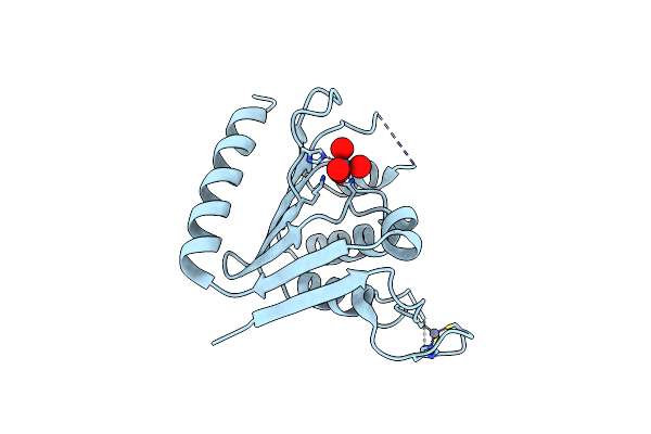
Crystal Structure Of Zucchini From Mouse (Mzuc / Pld6 / Mitopld)
Organism: Mus musculus
Method: X-RAY DIFFRACTION Resolution:1.75 Å Release Date: 2012-10-10 Classification: HYDROLASE Ligands: ZN |
Organism: Mus musculus
Method: X-RAY DIFFRACTION
Release Date: 2012-10-10
Ligands: ZN
 |
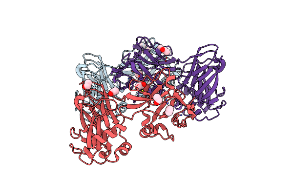
Crystal Structure Of Zucchini From Mouse (Mzuc / Pld6 / Mitopld) Bound To Tungstate
Organism: Mus musculus
Method: X-RAY DIFFRACTION Resolution:2.10 Å Release Date: 2012-10-10 Classification: HYDROLASE Ligands: ZN, WO4 |
Organism: Mus musculus
Method: X-RAY DIFFRACTION
Release Date: 2012-10-10
Ligands: ZN, WO4
 |
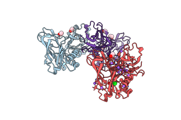
Crystal Structure Of Zu5A-Zu5B Domains Of Human Erythrocyte Ankyrin
Organism: Homo sapiens
Method: X-RAY DIFFRACTION Resolution:2.00 Å Release Date: 2012-02-22 Classification: PROTEIN BINDING Ligands: EOH |
Organism: Homo sapiens
Method: X-RAY DIFFRACTION
Release Date: 2012-02-22
Ligands: EOH
 |
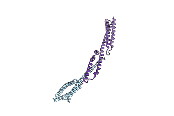
Crystal Structure Of Selenomethionine Zu5A-Zu5B Protein Domains Of Human Erythrocyte Ankyrin
Organism: Homo sapiens
Method: X-RAY DIFFRACTION Resolution:2.21 Å Release Date: 2012-02-22 Classification: PROTEIN BINDING |
Organism: Homo sapiens
Method: X-RAY DIFFRACTION
Release Date: 2012-02-22
 |
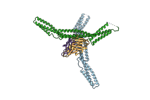
Crystal Structure Of The Erythrocyte Spectrin Tetramerization Domain Complex
Organism: Homo sapiens
Method: X-RAY DIFFRACTION Resolution:2.80 Å Release Date: 2010-03-09 Classification: STRUCTURAL PROTEIN |
Organism: Homo sapiens
Method: X-RAY DIFFRACTION
Release Date: 2010-03-09
 |
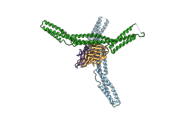
Crystal Structure Of The Ankyrin Binding Domain Of Human Erythroid Beta Spectrin (Repeats 13-15) In Complex With The Spectrin Binding Domain Of Human Erythroid Ankyrin (Zu5-Ank)
Organism: Homo sapiens
Method: X-RAY DIFFRACTION Resolution:2.75 Å Release Date: 2010-02-02 Classification: STRUCTURAL PROTEIN |
Organism: Homo sapiens
Method: X-RAY DIFFRACTION
Release Date: 2010-02-02
 |
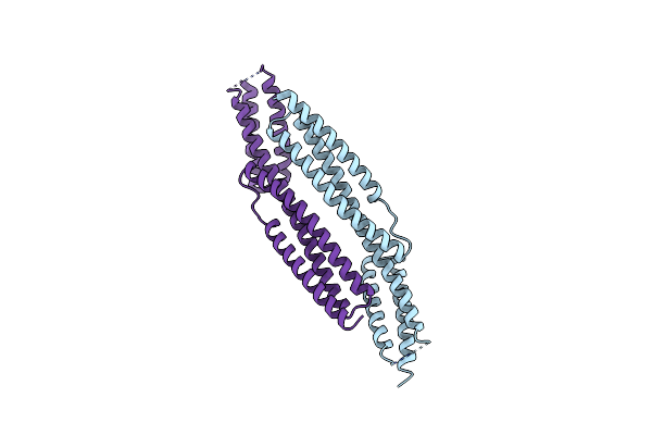
Crystal Structure Of The Ankyrin Binding Domain Of Human Erythroid Beta Spectrin (Repeats 13-15) In Complex With The Spectrin Binding Domain Of Human Erythroid Ankyrin (Zu5-Ank), Emts Derivative
Organism: Homo sapiens
Method: X-RAY DIFFRACTION Resolution:2.75 Å Release Date: 2010-02-02 Classification: STRUCTURAL PROTEIN Ligands: HG |
Organism: Homo sapiens
Method: X-RAY DIFFRACTION
Release Date: 2010-02-02
Ligands: HG
 |
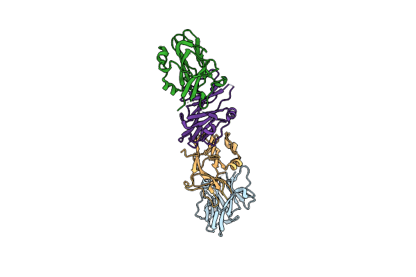
Crystal Structure Of Human Erythroid Beta Spectrin Repeats 14 And 15 (Ankyrin Binding Domain)
Organism: Homo sapiens
Method: X-RAY DIFFRACTION Resolution:2.90 Å Release Date: 2009-02-24 Classification: STRUCTURAL PROTEIN |
Organism: Homo sapiens
Method: X-RAY DIFFRACTION
Release Date: 2009-02-24
 |
Crystal Structure Of Zu5-Ank, The Spectrin Binding Region Of Human Erythroid Ankyrin
Organism: Homo sapiens
Method: X-RAY DIFFRACTION Resolution:2.00 Å Release Date: 2009-02-24 Classification: STRUCTURAL PROTEIN Ligands: BR |
Organism: Homo sapiens
Method: X-RAY DIFFRACTION
Release Date: 2009-02-24
Ligands: BR