

Search Count: 33
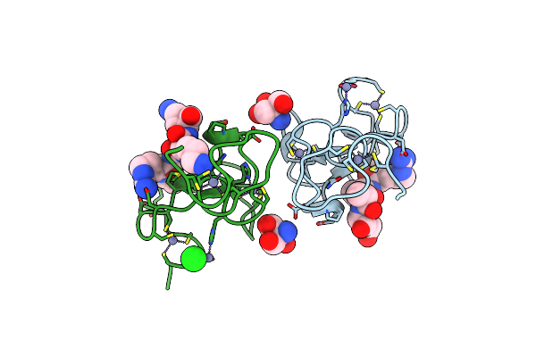 |
Phd Finger Of Human Uhrf1 In Complex With Unmodified Histone H3 N- Terminal Tail
Organism: Homo sapiens
Method: X-RAY DIFFRACTION Resolution:1.95 Å Release Date: 2011-12-07 Classification: LIGASE/PEPTIDE Ligands: ZN, TRS, CL |
Organism: Homo sapiens
Method: X-RAY DIFFRACTION
Release Date: 2011-12-07
Ligands: ZN, TRS, CL
 |
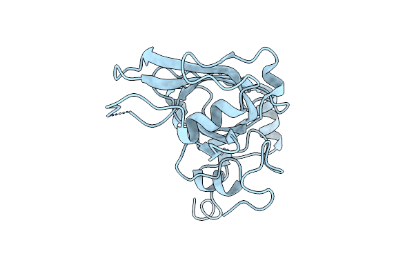
Phd Finger Of Human Uhrf1
Organism: Homo sapiens
Method: X-RAY DIFFRACTION Resolution:1.45 Å Release Date: 2011-11-30 Classification: LIGASE Ligands: ZN |
Organism: Homo sapiens
Method: X-RAY DIFFRACTION
Release Date: 2011-11-30
Ligands: ZN
 |
Crystal Structure Of The Sra Domain Of The Human Uhrf1 Protein
Organism: Homo sapiens
Method: X-RAY DIFFRACTION Resolution:1.90 Å Release Date: 2008-04-22 Classification: LIGASE |
Organism: Homo sapiens
Method: X-RAY DIFFRACTION
Release Date: 2008-04-22
 |
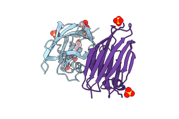
X-Ray Crystal Structure Of Wild-Type Xerocomus Chrysenteron Lectin Xcl
Organism: Xerocomus chrysenteron
Method: X-RAY DIFFRACTION Resolution:2.00 Å Release Date: 2005-08-30 Classification: SUGAR BINDING PROTEIN |
Organism: Xerocomus chrysenteron
Method: X-RAY DIFFRACTION
Release Date: 2005-08-30
 |
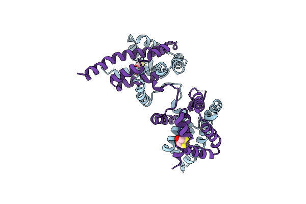
X-Ray Crystal Structure Of Xerocomus Chrysenteron Lectin Xcl At 1.4 Angstroms Resolution, Mutated At Q46M, V54M, L58M
Organism: Xerocomus chrysenteron
Method: X-RAY DIFFRACTION Resolution:1.40 Å Release Date: 2004-12-14 Classification: SUGAR BINDING PROTEIN Ligands: SO4, EDO |
Organism: Xerocomus chrysenteron
Method: X-RAY DIFFRACTION
Release Date: 2004-12-14
Ligands: SO4, EDO
 |
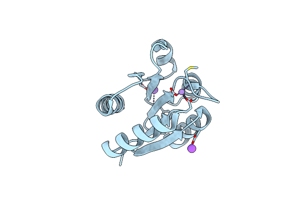
Crystal Structure Of The Dimeric Tord Chaperone From Shewanella Massilia
Organism: Shewanella massilia
Method: X-RAY DIFFRACTION Resolution:2.40 Å Release Date: 2003-05-13 Classification: CHAPERONE Ligands: DTT |
Organism: Shewanella massilia
Method: X-RAY DIFFRACTION
Release Date: 2003-05-13
Ligands: DTT
 |
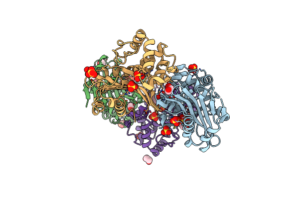
Crystal Structure Of The Phop Receiver Domain From Bacillus Subtilis
Organism: Bacillus subtilis
Method: X-RAY DIFFRACTION Resolution:1.60 Å Release Date: 2002-10-16 Classification: TRANSCRIPTION Ligands: MN, NA |
Organism: Bacillus subtilis
Method: X-RAY DIFFRACTION
Release Date: 2002-10-16
Ligands: MN, NA
 |
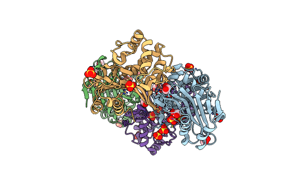
Oxa-10 Class D Beta-Lactamase Partially Acylated With Reacted 6Beta-(1-Hydroxy-1-Methylethyl) Penicillanic Acid
Organism: Pseudomonas aeruginosa
Method: X-RAY DIFFRACTION Resolution:1.70 Å Release Date: 2001-11-07 Classification: HYDROLASE Ligands: SO4, HOQ, EDO |
Organism: Pseudomonas aeruginosa
Method: X-RAY DIFFRACTION
Release Date: 2001-11-07
Ligands: SO4, HOQ, EDO
 |
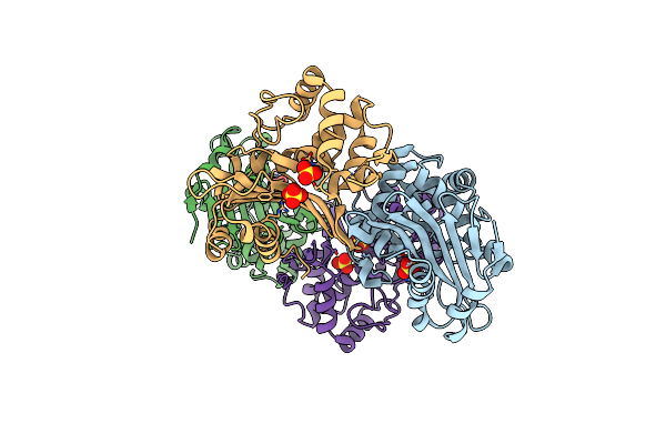
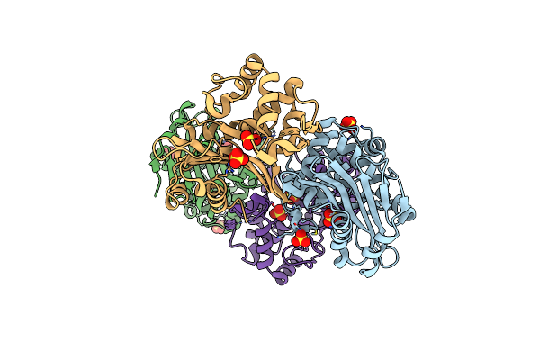
Oxa 10 Class D Beta-Lactamase At Ph 7.5
Organism: Pseudomonas aeruginosa
Method: X-RAY DIFFRACTION Resolution:1.39 Å Release Date: 2001-11-07 Classification: HYDROLASE Ligands: SO4, EDO |
Organism: Pseudomonas aeruginosa
Method: X-RAY DIFFRACTION
Release Date: 2001-11-07
Ligands: SO4, EDO
 |
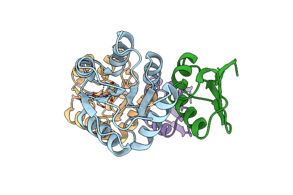
Oxa 10 Class D Beta-Lactamase At Ph 6.5
Organism: Pseudomonas aeruginosa
Method: X-RAY DIFFRACTION Resolution:1.70 Å Release Date: 2001-11-07 Classification: HYDROLASE Ligands: SO4 |
Organism: Pseudomonas aeruginosa
Method: X-RAY DIFFRACTION
Release Date: 2001-11-07
Ligands: SO4
 |
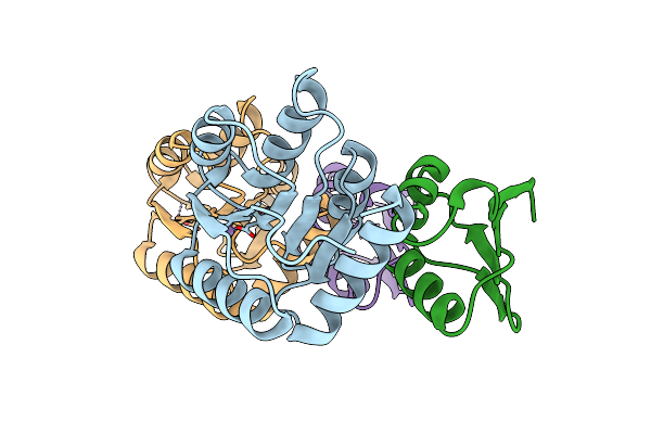
Oxa 10 Class D Beta-Lactamase At Ph 6.0
Organism: Pseudomonas aeruginosa
Method: X-RAY DIFFRACTION Resolution:1.90 Å Release Date: 2001-11-07 Classification: HYDROLASE Ligands: SO4 |
Organism: Pseudomonas aeruginosa
Method: X-RAY DIFFRACTION
Release Date: 2001-11-07
Ligands: SO4
 |
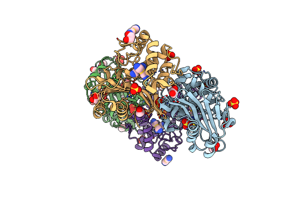
Chey-Binding Domain Of Chea In Complex With Chey At 2.1 A Resolution
Organism: Escherichia coli
Method: X-RAY DIFFRACTION Resolution:2.10 Å Release Date: 2001-01-17 Classification: TRANSFERASE/SIGNALING PROTEIN Ligands: MN |
Organism: Escherichia coli
Method: X-RAY DIFFRACTION
Release Date: 2001-01-17
Ligands: MN
 |
Chey-Binding Domain Of Chea In Complex With Chey From Crystals Soaked In Acetyl Phosphate
Organism: Escherichia coli
Method: X-RAY DIFFRACTION Resolution:2.40 Å Release Date: 2001-01-17 Classification: TRANSFERASE/SIGNALING PROTEIN Ligands: MN |
Organism: Escherichia coli
Method: X-RAY DIFFRACTION
Release Date: 2001-01-17
Ligands: MN
 |
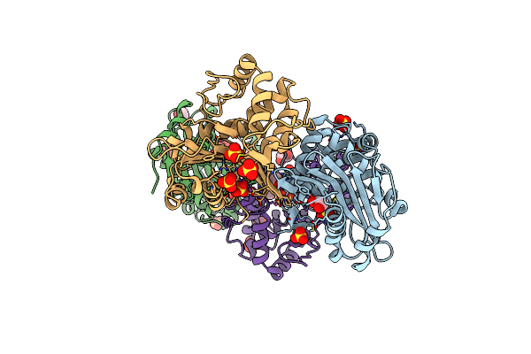
Mad Structure Of Oxa10 Class D Beta-Lactamase
Organism: Pseudomonas aeruginosa
Method: X-RAY DIFFRACTION Resolution:1.66 Å Release Date: 2001-01-12 Classification: BETA-LACTAMASE Ligands: AUC, SO4, EDO |
Organism: Pseudomonas aeruginosa
Method: X-RAY DIFFRACTION
Release Date: 2001-01-12
Ligands: AUC, SO4, EDO
 |
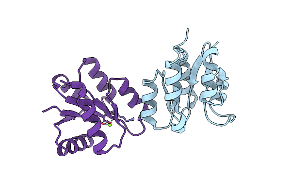
Structure Of Oxa10 Beta-Lactamase At Ph 8.3
Organism: Pseudomonas aeruginosa
Method: X-RAY DIFFRACTION Resolution:1.80 Å Release Date: 2001-01-12 Classification: BETA-LACTAMASE Ligands: SO4, EDO |
Organism: Pseudomonas aeruginosa
Method: X-RAY DIFFRACTION
Release Date: 2001-01-12
Ligands: SO4, EDO
 |
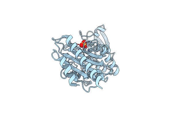
Structure Of Unphosphorylated Fixj-N With An Atypical Conformer (Monomer A)
Organism: Sinorhizobium meliloti
Method: X-RAY DIFFRACTION Resolution:3.00 Å Release Date: 2000-11-08 Classification: TRANSCRIPTION Ligands: MG |
Organism: Sinorhizobium meliloti
Method: X-RAY DIFFRACTION
Release Date: 2000-11-08
Ligands: MG
 |
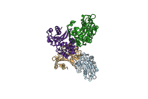
The High Resolution Structure Of Per-1 Class A Beta-Lactamase
Organism: Pseudomonas aeruginosa
Method: X-RAY DIFFRACTION Resolution:1.90 Å Release Date: 2000-11-06 Classification: HYDROLASE Ligands: SO4 |
Organism: Pseudomonas aeruginosa
Method: X-RAY DIFFRACTION
Release Date: 2000-11-06
Ligands: SO4
 |
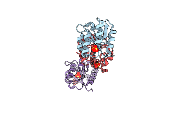
Crystal Structure Of The Oxa-10 Beta-Lactamase From Pseudomonas Aeruginosa
Organism: Pseudomonas aeruginosa
Method: X-RAY DIFFRACTION Resolution:2.40 Å Release Date: 2000-11-01 Classification: HYDROLASE |
Organism: Pseudomonas aeruginosa
Method: X-RAY DIFFRACTION
Release Date: 2000-11-01
 |
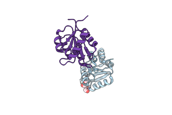
Phosphorylated Fixj Receiver Domain
Organism: Sinorhizobium meliloti
Method: X-RAY DIFFRACTION Resolution:2.30 Å Release Date: 2000-10-11 Classification: GENE REGULATION Ligands: SO4 |
Organism: Sinorhizobium meliloti
Method: X-RAY DIFFRACTION
Release Date: 2000-10-11
Ligands: SO4
 |
Crystal Structure Of Fixj-N
Organism: Sinorhizobium meliloti
Method: X-RAY DIFFRACTION Resolution:1.60 Å Release Date: 1999-11-26 Classification: TRANSCRIPTION Ligands: 15P |
Organism: Sinorhizobium meliloti
Method: X-RAY DIFFRACTION
Release Date: 1999-11-26
Ligands: 15P