

Search Count: 11
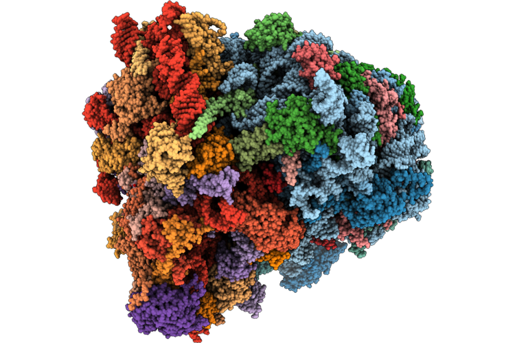 |
Rps26Dc Hek Mutant 80S Ribosome Bound To Kozak Mrna (Rps26Dc-Kozak)
Organism: Homo sapiens
Method: ELECTRON MICROSCOPY Release Date: 2026-06-03 Classification: RIBOSOME Ligands: K, SAC, MG, ZN, AME, MET, HYG, ANM, NA |
Organism: Homo sapiens
Method: ELECTRON MICROSCOPY
Release Date: 2026-06-03
Ligands: K, SAC, MG, ZN, AME, MET, HYG, ANM, NA
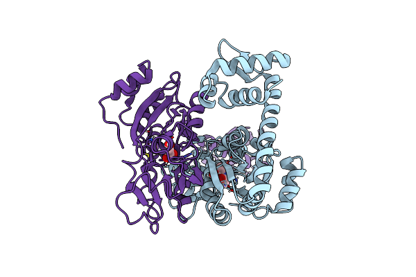 |
The Crystal Structure Of Full Length Tetramer Cysb From Klebsiella Aerogenes In Complex With N-Acetylserine
Organism: Klebsiella aerogenes
Method: X-RAY DIFFRACTION Resolution:2.30 Å Release Date: 2024-07-24 Classification: TRANSCRIPTION Ligands: SAC |
Organism: Klebsiella aerogenes
Method: X-RAY DIFFRACTION
Release Date: 2024-07-24
Ligands: SAC
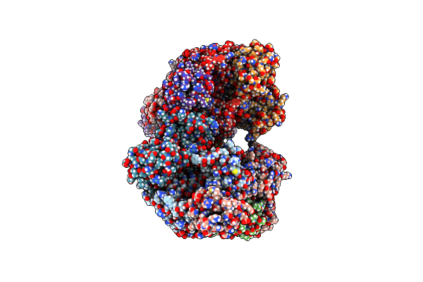 |
The Crystal Structure Of Full Length Tetramer Cysb From Klebsiella Aerogenes In Complex With N-Acetylserine
Organism: Klebsiella
Method: X-RAY DIFFRACTION Resolution:2.80 Å Release Date: 2024-07-03 Classification: TRANSCRIPTION Ligands: SAC |
Organism: Klebsiella
Method: X-RAY DIFFRACTION
Release Date: 2024-07-03
Ligands: SAC
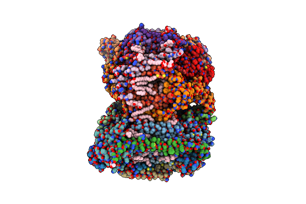 |
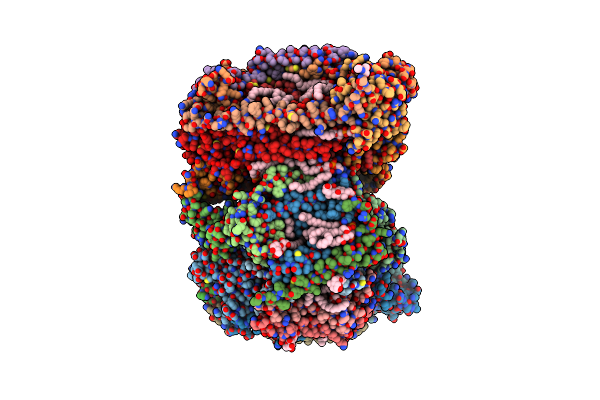
Sfx Structure Of Oxidized Cytochrome C Oxidase At 2.38 Angstrom Resolution
Organism: Bos taurus
Method: X-RAY DIFFRACTION Resolution:2.38 Å Release Date: 2023-09-27 Classification: MEMBRANE PROTEIN Ligands: CU, MG, NA, PGV, HEA, OH, CUA, TGL, PSC, CHD, PEK, CDL, DMU, ZN, SAC |
 |
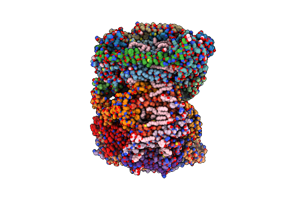
Time-Resolve Sfx Structure Of A Photoproduct Of Carbon Monoxide Complex Of Bovine Cytochrome C Oxidase
Organism: Bos taurus
Method: X-RAY DIFFRACTION Resolution:2.80 Å Release Date: 2023-09-20 Classification: MEMBRANE PROTEIN Ligands: HEA, CU, MG, NA, TGL, PGV, CMO, CUA, CHD, OH, CDL, PSC, ZN, PEK, DMU, SAC |
 |
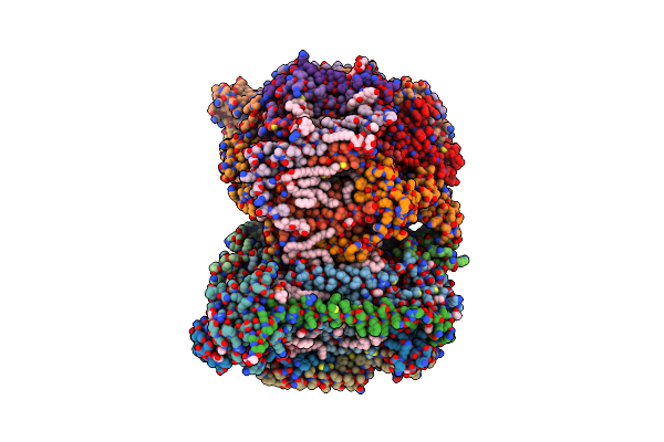
Structure Of Reduced Bovine Cytochrome C Oxidase At 1.93 Angstrom Resolution Obtained By Synchrotron X-Rays
Organism: Bos taurus
Method: X-RAY DIFFRACTION Resolution:1.93 Å Release Date: 2022-03-30 Classification: OXIDOREDUCTASE/MEMBRANE PROTEIN Ligands: CU, MG, NA, PGV, HEA, EDO, TGL, CUA, CHD, DMU, PEK, CDL, PSC, ZN, SAC |
 |
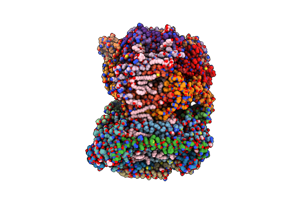
Structure Of Oxidized Bovine Cytochrome C Oxidase At 1.90 Angstrom Resolution Obtained By Synchrotron X-Rays
Organism: Bos taurus
Method: X-RAY DIFFRACTION Resolution:1.90 Å Release Date: 2022-03-30 Classification: OXIDOREDUCTASE/MEMBRANE PROTEIN Ligands: CU, MG, NA, PGV, HEA, EDO, TGL, OH, CUA, PSC, CHD, PEK, CDL, DMU, ZN, SAC |
 |
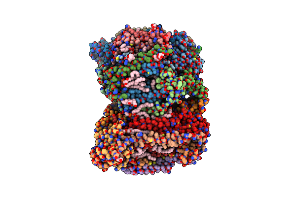
Structure Of Oxidized Bovine Cytochrome C Oxidase With Reduced Metal Centers Induced By Synchrotron X-Ray Exposure
Organism: Bos taurus
Method: X-RAY DIFFRACTION Resolution:2.35 Å Release Date: 2022-03-30 Classification: MEMBRANE PROTEIN Ligands: CU, MG, NA, PGV, HEA, EDO, OH, CUA, TGL, PSC, CHD, CDL, DMU, PEK, ZN, SAC |
 |
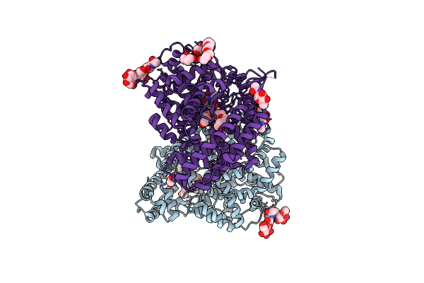
Annealed Structure Of Oxidized Bovine Cytochrome C Oxidase With Reduced Metal Centers Induced By Synchrotron X-Ray Exposure
Organism: Bos taurus
Method: X-RAY DIFFRACTION Resolution:2.45 Å Release Date: 2022-03-30 Classification: MEMBRANE PROTEIN Ligands: HEA, CU, MG, NA, PGV, EDO, OH, TGL, CUA, DMU, PEK, CDL, CHD, PSC, ZN, SAC |
 |
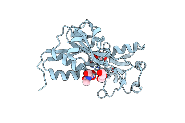
Crystal Structure Of The Angiotensin-1 Converting Enzyme N-Domain In Complex With Ac-Sd
Organism: Homo sapiens
Method: X-RAY DIFFRACTION Resolution:1.80 Å Release Date: 2015-10-07 Classification: HYDROLASE Ligands: ZN, CL, NAG, PEG, P6G, SAC, ASP |
Organism: Homo sapiens
Method: X-RAY DIFFRACTION
Release Date: 2015-10-07
Ligands: ZN, CL, NAG, PEG, P6G, SAC, ASP
 |
Crystal Structure Of Ligand Binding Domain Of Cysb, A Lysr Member From Salmonella Typhimurium Lt2 In Complex With Effector Ligand, N-Acetylserine.
Organism: Salmonella enterica subsp. enterica serovar typhimurium
Method: X-RAY DIFFRACTION Resolution:2.70 Å Release Date: 2014-08-13 Classification: GENE REGULATION Ligands: SAC, PEG |
Organism: Salmonella enterica subsp. enterica serovar typhimurium
Method: X-RAY DIFFRACTION
Release Date: 2014-08-13
Ligands: SAC, PEG