

Search Count: 20
 |
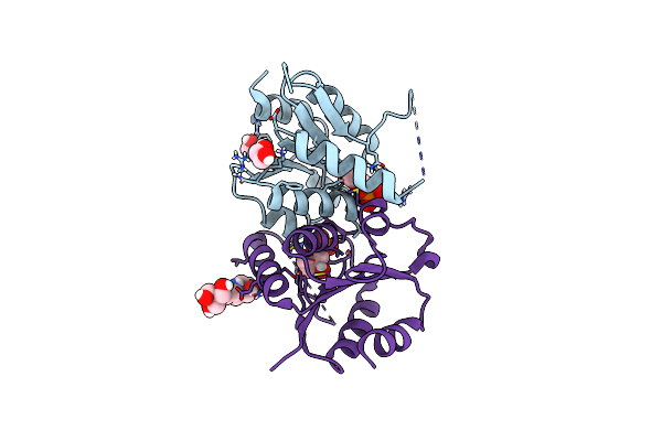
Crystal Structure Of Human Dnph1 Bound To Hmdump
Organism: Homo sapiens
Method: X-RAY DIFFRACTION Resolution:1.78 Å Release Date: 2023-11-08 Classification: HYDROLASE Ligands: 5HU, EDO |
Organism: Homo sapiens
Method: X-RAY DIFFRACTION
Release Date: 2023-11-08
Ligands: 5HU, EDO
 |
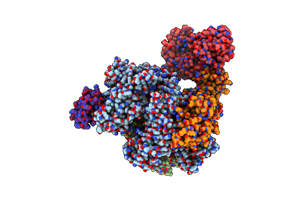
Crystal Structure Of The Human Dnph1 Glycosyl-Enzyme Intermediate
Organism: Homo sapiens
Method: X-RAY DIFFRACTION Resolution:1.65 Å Release Date: 2023-11-08 Classification: HYDROLASE Ligands: 3DR, EDO, B3P |
Organism: Homo sapiens
Method: X-RAY DIFFRACTION
Release Date: 2023-11-08
Ligands: 3DR, EDO, B3P
 |
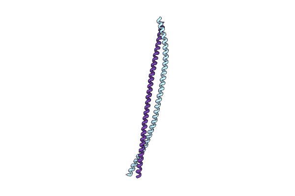
Cryoem Structure Of Dna Polymerase Alpha - Primase Bound To Sars Cov Nsp1
Organism: Homo sapiens, Severe acute respiratory syndrome coronavirus
Method: ELECTRON MICROSCOPY Release Date: 2021-11-10 Classification: DNA BINDING PROTEIN Ligands: ZN, SF4 |
Organism: Homo sapiens, Severe acute respiratory syndrome coronavirus
Method: ELECTRON MICROSCOPY
Release Date: 2021-11-10
Ligands: ZN, SF4
 |
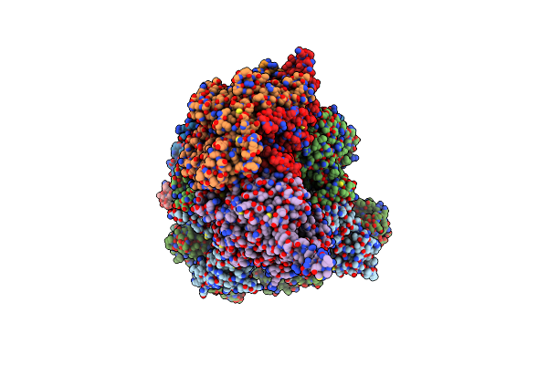
Crystal Structure Of The N-Terminal Dimeric Coiled Coil Of The Human Ctip Protein
Organism: Homo sapiens
Method: X-RAY DIFFRACTION Resolution:2.80 Å Release Date: 2021-06-30 Classification: DNA BINDING PROTEIN |
Organism: Homo sapiens
Method: X-RAY DIFFRACTION
Release Date: 2021-06-30
 |
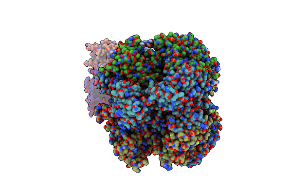
Cryoem Structure Of Human Cmg Bound To Atpgammas And Dna
Organism: Homo sapiens, Synthetic construct
Method: ELECTRON MICROSCOPY Release Date: 2020-05-27 Classification: REPLICATION Ligands: ZN, ADP, AGS, MG |
Organism: Homo sapiens, Synthetic construct
Method: ELECTRON MICROSCOPY
Release Date: 2020-05-27
Ligands: ZN, ADP, AGS, MG
 |
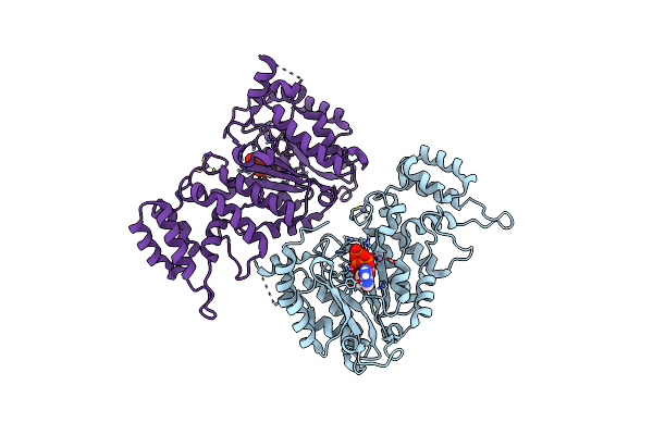
Cryoem Structure Of Human Cmg Bound To And-1 (Cmga)
Organism: Homo sapiens
Method: ELECTRON MICROSCOPY Release Date: 2020-05-27 Classification: REPLICATION Ligands: ZN |
Organism: Homo sapiens
Method: ELECTRON MICROSCOPY
Release Date: 2020-05-27
Ligands: ZN
 |
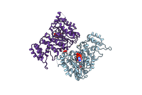
Crystal Structure Of The Pri1 Subunit Of Human Primase Bound To Atp
Organism: Homo sapiens
Method: X-RAY DIFFRACTION Resolution:2.75 Å Release Date: 2019-09-11 Classification: REPLICATION Ligands: ZN, MN, ATP |
Organism: Homo sapiens
Method: X-RAY DIFFRACTION
Release Date: 2019-09-11
Ligands: ZN, MN, ATP
 |
Crystal Structure Of The Pri1 Subunit Of Human Primase Bound To Vidarabine Triphosphate
Organism: Homo sapiens
Method: X-RAY DIFFRACTION Resolution:2.35 Å Release Date: 2019-09-11 Classification: REPLICATION Ligands: ZN, MN, HEJ, EDO |
Organism: Homo sapiens
Method: X-RAY DIFFRACTION
Release Date: 2019-09-11
Ligands: ZN, MN, HEJ, EDO
 |
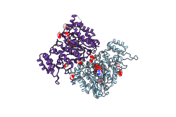
Crystal Structure Of The Pri1 Subunit Of Human Primase Bound To Fludarabine Triphosphate
Organism: Homo sapiens
Method: X-RAY DIFFRACTION Resolution:2.20 Å Release Date: 2019-09-11 Classification: REPLICATION Ligands: ZN, MN, HFD, EDO |
Organism: Homo sapiens
Method: X-RAY DIFFRACTION
Release Date: 2019-09-11
Ligands: ZN, MN, HFD, EDO
 |
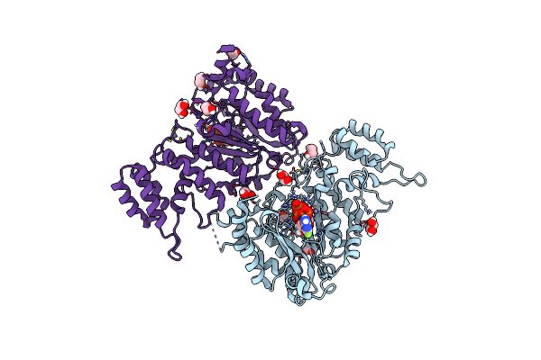
Crystal Structure Of The Pri1 Subunit Of Human Primase Bound To Datp
Organism: Homo sapiens
Method: X-RAY DIFFRACTION Resolution:1.95 Å Release Date: 2019-09-11 Classification: REPLICATION Ligands: ZN, MN, DTP, EDO |
Organism: Homo sapiens
Method: X-RAY DIFFRACTION
Release Date: 2019-09-11
Ligands: ZN, MN, DTP, EDO
 |
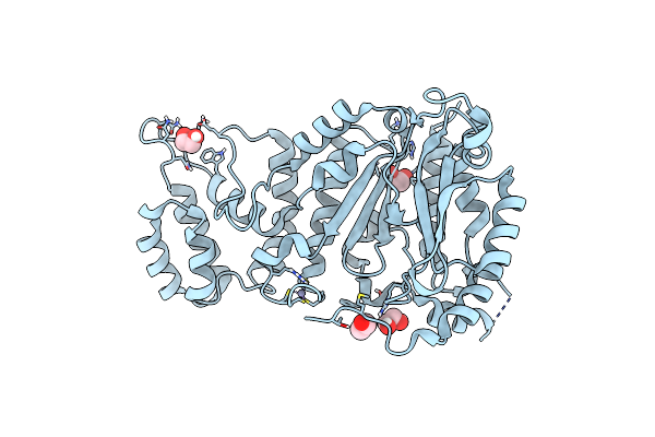
Crystal Structure Of The Pri1 Subunit Of Human Primase Bound To 2F-Atp
Organism: Homo sapiens
Method: X-RAY DIFFRACTION Resolution:1.85 Å Release Date: 2019-09-11 Classification: REPLICATION Ligands: ZN, MN, JSQ, EDO |
Organism: Homo sapiens
Method: X-RAY DIFFRACTION
Release Date: 2019-09-11
Ligands: ZN, MN, JSQ, EDO
 |
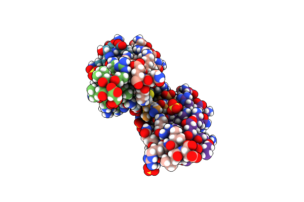
Crystal Structure Of The Pri1 Subunit Of Human Primase
Organism: Homo sapiens
Method: X-RAY DIFFRACTION Resolution:1.50 Å Release Date: 2019-09-11 Classification: REPLICATION Ligands: EDO, ZN |
Organism: Homo sapiens
Method: X-RAY DIFFRACTION
Release Date: 2019-09-11
Ligands: EDO, ZN
 |
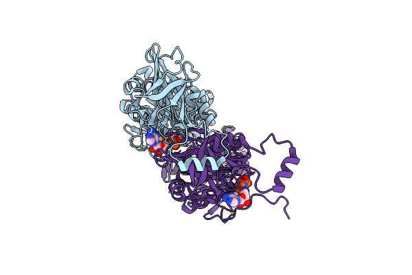
Crystal Structure Of The Tetramerisation Domain Of Human Ctip
Organism: Homo sapiens
Method: X-RAY DIFFRACTION Resolution:1.90 Å Release Date: 2015-01-14 Classification: GENE REGULATION Ligands: SO4 |
Organism: Homo sapiens
Method: X-RAY DIFFRACTION
Release Date: 2015-01-14
Ligands: SO4
 |
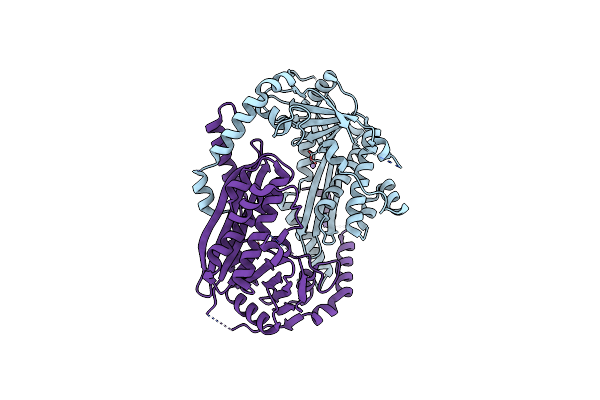
Crystal Structure Of The Hera Hexameric Dna Translocase From Sulfolobus Solfataricus Bound To Amp-Pnp
Organism: Sulfolobus solfataricus
Method: X-RAY DIFFRACTION Resolution:2.84 Å Release Date: 2014-12-03 Classification: HYDROLASE Ligands: ANP, MG |
Organism: Sulfolobus solfataricus
Method: X-RAY DIFFRACTION
Release Date: 2014-12-03
Ligands: ANP, MG
 |
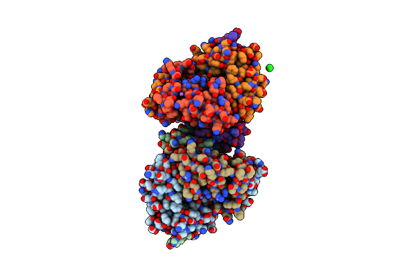
Crystal Structure Of The Nura Nuclease From Sulfolobus Solfataricus
Organism: Sulfolobus solfataricus
Method: X-RAY DIFFRACTION Resolution:2.50 Å Release Date: 2011-12-14 Classification: HYDROLASE Ligands: MN |
Organism: Sulfolobus solfataricus
Method: X-RAY DIFFRACTION
Release Date: 2011-12-14
Ligands: MN
 |
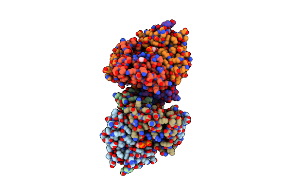
Structure Of Yncf,The Genomic Dutpase From Bacillus Subtilis
Organism: Bacillus subtilis
Method: X-RAY DIFFRACTION Resolution:1.84 Å Release Date: 2010-08-11 Classification: HYDROLASE Ligands: MG, NA, CL, GOL |
Organism: Bacillus subtilis
Method: X-RAY DIFFRACTION
Release Date: 2010-08-11
Ligands: MG, NA, CL, GOL
 |
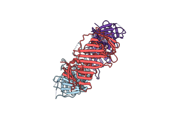
Structure Of Yncf In Complex With Dupnhpp
Organism: Bacillus subtilis
Method: X-RAY DIFFRACTION Resolution:1.85 Å Release Date: 2010-08-11 Classification: HYDROLASE Ligands: DUP, CA, GOL |
Organism: Bacillus subtilis
Method: X-RAY DIFFRACTION
Release Date: 2010-08-11
Ligands: DUP, CA, GOL
 |
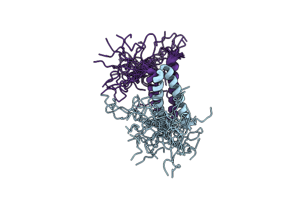
Crystal Structure Of The Human Rad9-Rad1-Hus1 Dna Damage Checkpoint Complex
Organism: Homo sapiens
Method: X-RAY DIFFRACTION Resolution:2.90 Å Release Date: 2009-05-26 Classification: CELL CYCLE |
Organism: Homo sapiens
Method: X-RAY DIFFRACTION
Release Date: 2009-05-26
 |
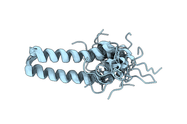
Nmr Solution Structure Of A Protein Aspartic Acid Phosphate Phosphatase From Bacillus Anthracis
Organism: Bacillus anthracis
Method: SOLUTION NMR Release Date: 2006-09-25 Classification: TRANSFERASE |
Organism: Bacillus anthracis
Method: SOLUTION NMR
Release Date: 2006-09-25
 |
Nmr Solution Structure Of A Protein Aspartic Acid Phosphate Phosphatase From Bacillus Anthracis
Organism: Bacillus anthracis str. ames
Method: SOLUTION NMR Release Date: 2006-09-25 Classification: TRANSFERASE |
Organism: Bacillus anthracis str. ames
Method: SOLUTION NMR
Release Date: 2006-09-25