

Search Count: 9
 |
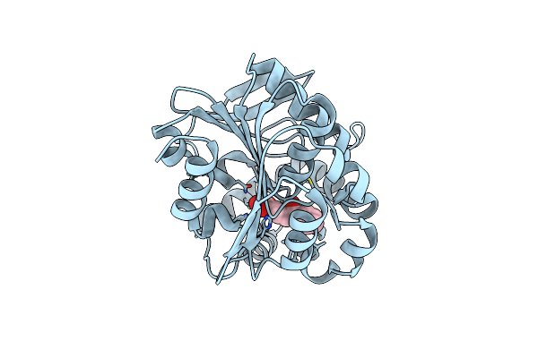
Crystal Structure Of The S105A Mutant Of A Carbon-Carbon Bond Hydrolase, Dxnb2 From Sphingomonas Wittichii Rw1, In Complex With 5,8-Dif Hopda
Organism: Sphingomonas wittichii rw1
Method: X-RAY DIFFRACTION Resolution:2.17 Å Release Date: 2013-10-09 Classification: HYDROLASE Ligands: 22J, NA |
Organism: Sphingomonas wittichii rw1
Method: X-RAY DIFFRACTION
Release Date: 2013-10-09
Ligands: 22J, NA
 |
Crystal Structure Of The S105A Mutant Of A C-C Hydrolase, Dxnb2 From Sphingomonas Wittichii Rw1, In Complex With Substrate Hopda
Organism: Sphingomonas wittichii
Method: X-RAY DIFFRACTION Resolution:2.33 Å Release Date: 2013-10-09 Classification: HYDROLASE Ligands: HPK |
Organism: Sphingomonas wittichii
Method: X-RAY DIFFRACTION
Release Date: 2013-10-09
Ligands: HPK
 |
Crystal Structure Of Dxnb2, A Carbon - Carbon Bond Hydrolase From Sphingomonas Wittichii Rw1
Organism: Sphingomonas wittichii rw1
Method: X-RAY DIFFRACTION Resolution:2.22 Å Release Date: 2013-09-18 Classification: HYDROLASE Ligands: SO4 |
Organism: Sphingomonas wittichii rw1
Method: X-RAY DIFFRACTION
Release Date: 2013-09-18
Ligands: SO4
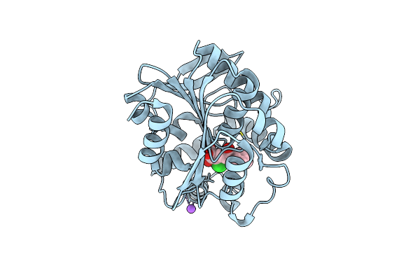 |
Crystal Structure Of The S105A Mutant Of A Carbon-Carbon Bond Hydrolase, Dxnb2 From Sphingomonas Wittichii Rw1, In Complex With 3-Cl Hopda
Organism: Sphingomonas wittichii rw1
Method: X-RAY DIFFRACTION Resolution:2.02 Å Release Date: 2013-09-18 Classification: HYDROLASE Ligands: C1E, NA |
Organism: Sphingomonas wittichii rw1
Method: X-RAY DIFFRACTION
Release Date: 2013-09-18
Ligands: C1E, NA
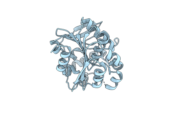 |
Crystal Structure Of The S105A Mutant Of A C-C Hydrolase, Dxnb2 From Sphingomonas Wittichii Rw1
Organism: Sphingomonas wittichii
Method: X-RAY DIFFRACTION Resolution:2.26 Å Release Date: 2013-09-18 Classification: HYDROLASE |
Organism: Sphingomonas wittichii
Method: X-RAY DIFFRACTION
Release Date: 2013-09-18
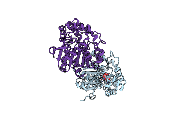 |
Crystal Structure Of The H265Q Mutant Of A C-C Hydrolase, Bphd From Burkholderia Xenovorans Lb400.
Organism: Burkholderia xenovorans
Method: X-RAY DIFFRACTION Resolution:2.13 Å Release Date: 2012-03-21 Classification: HYDROLASE Ligands: MLA |
Organism: Burkholderia xenovorans
Method: X-RAY DIFFRACTION
Release Date: 2012-03-21
Ligands: MLA
 |
Crystal Structure Of The S112A/H265Q Mutant Of A C-C Hydrolase, Bphd From Burkholderia Xenovorans Lb400
Organism: Burkholderia xenovorans
Method: X-RAY DIFFRACTION Resolution:2.11 Å Release Date: 2012-03-21 Classification: HYDROLASE Ligands: MLA |
Organism: Burkholderia xenovorans
Method: X-RAY DIFFRACTION
Release Date: 2012-03-21
Ligands: MLA
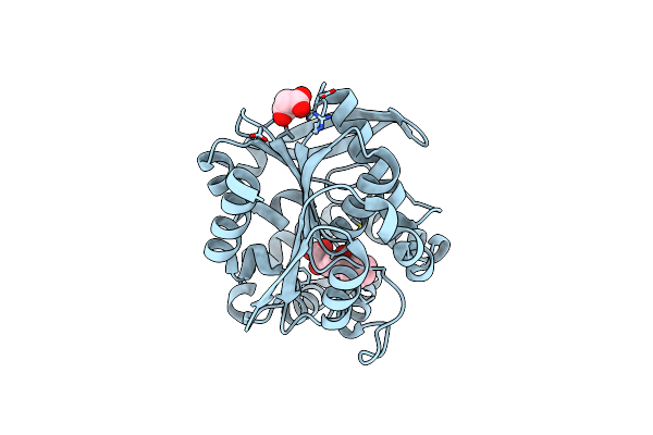 |
Crystal Structure Of The S112A/H265Q Mutant Of A C-C Hydrolase, Bphd From Burkholderia Xenovorans Lb400, After Exposure To Its Substrate Hopda
Organism: Burkholderia xenovorans
Method: X-RAY DIFFRACTION Resolution:1.92 Å Release Date: 2012-03-21 Classification: HYDROLASE Ligands: MLI, HPK |
Organism: Burkholderia xenovorans
Method: X-RAY DIFFRACTION
Release Date: 2012-03-21
Ligands: MLI, HPK
 |
Crystal Structure Of The H265Q Mutant Of A C-C Hydrolase, Bphd From Burkholderia Xenovorans Lb400, After Exposure To Its Substrate Hopda
Organism: Burkholderia xenovorans
Method: X-RAY DIFFRACTION Resolution:1.59 Å Release Date: 2012-03-21 Classification: HYDROLASE Ligands: MLA, BEZ, HPK |
Organism: Burkholderia xenovorans
Method: X-RAY DIFFRACTION
Release Date: 2012-03-21
Ligands: MLA, BEZ, HPK