

Search Count: 47
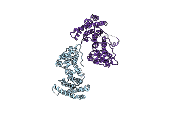 |
The Retinoblastoma Protein With Mutation M704V
Organism: Homo sapiens
Method: X-RAY DIFFRACTION Resolution:2.38 Å Release Date: 2025-02-19 Classification: CELL CYCLE |
Organism: Homo sapiens
Method: X-RAY DIFFRACTION
Release Date: 2025-02-19
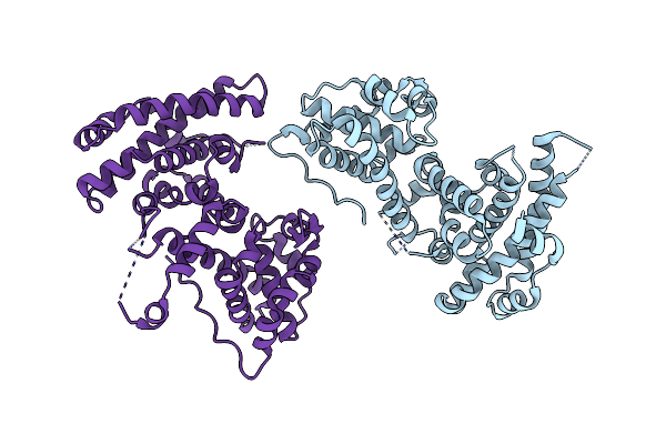 |
The Retinoblastoma Protein With Mutation S751Y
Organism: Homo sapiens
Method: X-RAY DIFFRACTION Resolution:2.32 Å Release Date: 2025-02-19 Classification: CELL CYCLE |
Organism: Homo sapiens
Method: X-RAY DIFFRACTION
Release Date: 2025-02-19
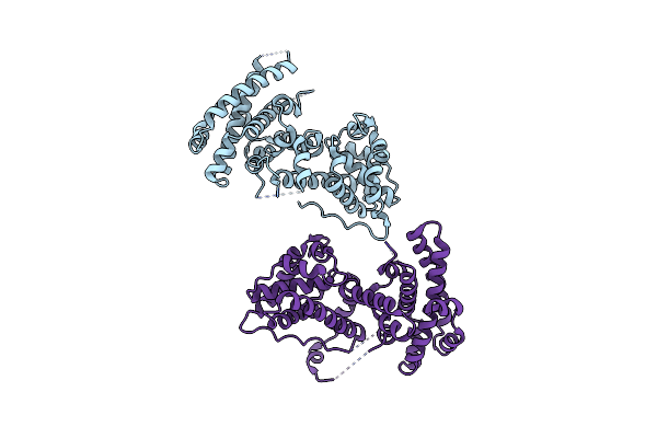 |
The Retinoblastoma Protein With Mutation E554K
Organism: Homo sapiens
Method: X-RAY DIFFRACTION Resolution:2.26 Å Release Date: 2025-02-19 Classification: CELL CYCLE |
Organism: Homo sapiens
Method: X-RAY DIFFRACTION
Release Date: 2025-02-19
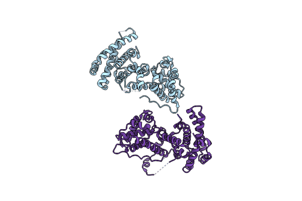 |
The Retinoblastoma Protein With Mutation E533K
Organism: Homo sapiens
Method: X-RAY DIFFRACTION Resolution:2.16 Å Release Date: 2025-02-19 Classification: CELL CYCLE |
Organism: Homo sapiens
Method: X-RAY DIFFRACTION
Release Date: 2025-02-19
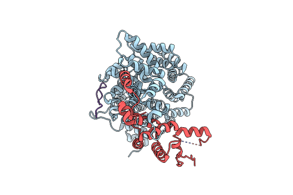 |
E2F1-Cyclin F Interface
Organism: Homo sapiens
Method: ELECTRON MICROSCOPY Release Date: 2025-02-12 Classification: CELL CYCLE |
Organism: Homo sapiens
Method: ELECTRON MICROSCOPY
Release Date: 2025-02-12
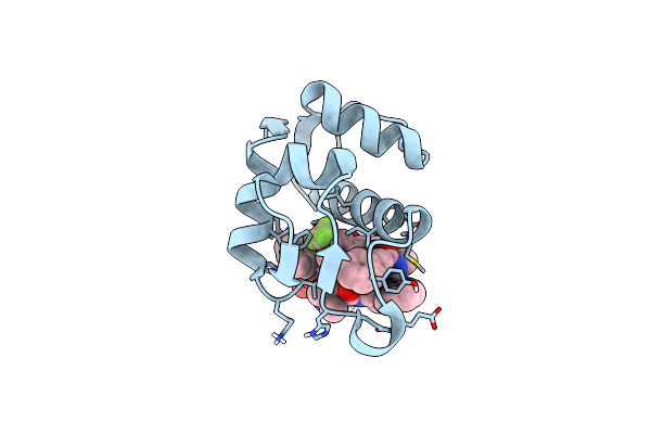 |
Mdm2 Bound To Inhibitor
Organism: Homo sapiens, Synthetic construct
Method: X-RAY DIFFRACTION Resolution:1.47 Å Release Date: 2024-10-23 Classification: TRANSFERASE/INHIBITOR |
Organism: Homo sapiens, Synthetic construct
Method: X-RAY DIFFRACTION
Release Date: 2024-10-23
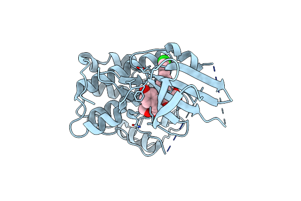 |
Crystal Structure Of Cdk2 In Complex With Cyclin A Inhibitor 6-[(E)-2-(4-Chlorophenyl)Ethenyl]-2-{[(2R)-3-(4-Hydroxyphenyl)-1-Methoxy-1-Oxopropan-2-Yl]Carbamoyl}Quinoline-4-Carboxylic Acid
Organism: Homo sapiens
Method: X-RAY DIFFRACTION Resolution:2.20 Å Release Date: 2023-04-05 Classification: TRANSFERASE/INHIBITOR Ligands: OW6 |
Organism: Homo sapiens
Method: X-RAY DIFFRACTION
Release Date: 2023-04-05
Ligands: OW6
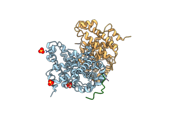 |
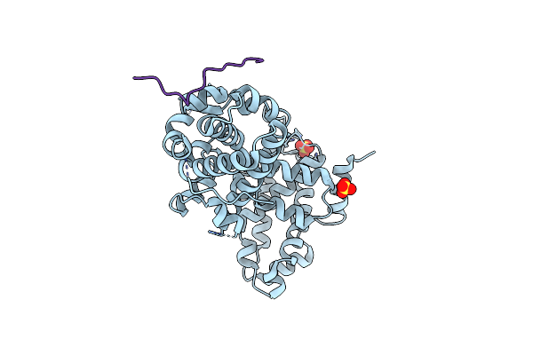
P107 Pocket Domain Complexed With Arid4A Peptide
Organism: Homo sapiens
Method: X-RAY DIFFRACTION Resolution:2.70 Å Release Date: 2022-06-29 Classification: CELL CYCLE Ligands: SO4 |
Organism: Homo sapiens
Method: X-RAY DIFFRACTION
Release Date: 2022-06-29
Ligands: SO4
 |
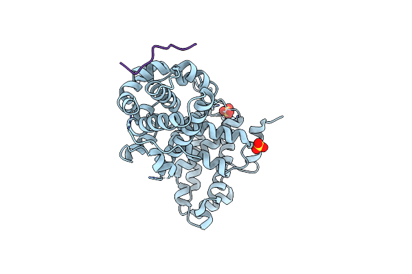
P107 Pocket Domain Complexed With Eid1 Peptide
Organism: Homo sapiens
Method: X-RAY DIFFRACTION Resolution:2.15 Å Release Date: 2022-06-29 Classification: CELL CYCLE Ligands: SO4 |
Organism: Homo sapiens
Method: X-RAY DIFFRACTION
Release Date: 2022-06-29
Ligands: SO4
 |
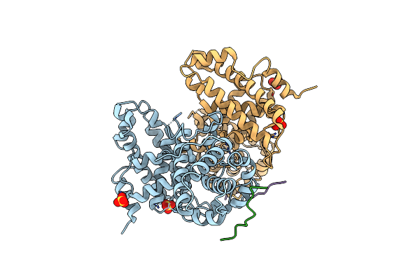
P107 Pocket Domain Complexed With Hdac1 Peptide
Organism: Homo sapiens
Method: X-RAY DIFFRACTION Resolution:2.64 Å Release Date: 2022-06-29 Classification: CELL CYCLE Ligands: SO4 |
Organism: Homo sapiens
Method: X-RAY DIFFRACTION
Release Date: 2022-06-29
Ligands: SO4
 |
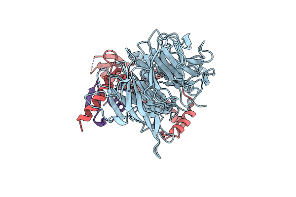
P107 Pocket Domain Complexed With Mutated Hdac1-3X Peptide
Organism: Homo sapiens
Method: X-RAY DIFFRACTION Resolution:3.00 Å Release Date: 2022-06-29 Classification: CELL CYCLE Ligands: SO4 |
Organism: Homo sapiens
Method: X-RAY DIFFRACTION
Release Date: 2022-06-29
Ligands: SO4
 |
Crystal Structure Of Lin9-Rbap48-Lin37, A Muvb Subcomplex
Organism: Homo sapiens
Method: X-RAY DIFFRACTION Resolution:2.55 Å Release Date: 2022-02-09 Classification: CELL CYCLE |
Organism: Homo sapiens
Method: X-RAY DIFFRACTION
Release Date: 2022-02-09
 |
Crystal Structure Of Vpsu (Vc0916) From Vibrio Cholerae
Organism: Vibrio cholerae
Method: X-RAY DIFFRACTION Resolution:2.20 Å Release Date: 2020-08-19 Classification: HYDROLASE Ligands: GOL |
Organism: Vibrio cholerae
Method: X-RAY DIFFRACTION
Release Date: 2020-08-19
Ligands: GOL
 |
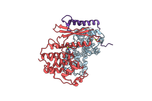
Crystal Structure Of Vpso (Vc0937) Kinase Domain
Organism: Vibrio cholerae
Method: X-RAY DIFFRACTION Resolution:2.87 Å Release Date: 2020-08-19 Classification: BIOSYNTHETIC PROTEIN Ligands: PTR |
Organism: Vibrio cholerae
Method: X-RAY DIFFRACTION
Release Date: 2020-08-19
Ligands: PTR
 |
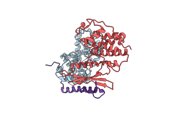
Crystal Structure Of Cdk4 In Complex With Cyclind1 And P27
Organism: Homo sapiens
Method: X-RAY DIFFRACTION Resolution:2.30 Å Release Date: 2019-12-25 Classification: cell cycle, transferase Ligands: MG |
Organism: Homo sapiens
Method: X-RAY DIFFRACTION
Release Date: 2019-12-25
Ligands: MG
 |
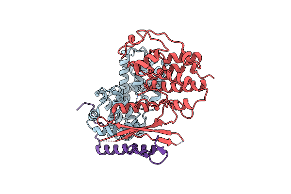
Crystal Structure Of Cdk4 In Complex With Cyclind1 And P27
Organism: Homo sapiens
Method: X-RAY DIFFRACTION Resolution:2.89 Å Release Date: 2019-12-25 Classification: cell cycle, transferase |
Organism: Homo sapiens
Method: X-RAY DIFFRACTION
Release Date: 2019-12-25
 |
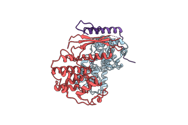
Crystal Structure Of Cdk4 In Complex With Cyclind1 And P27
Organism: Homo sapiens
Method: X-RAY DIFFRACTION Resolution:2.80 Å Release Date: 2019-12-25 Classification: cell cycle, transferase |
Organism: Homo sapiens
Method: X-RAY DIFFRACTION
Release Date: 2019-12-25
 |
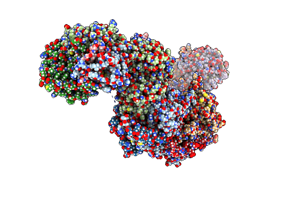
Crystal Structure Of Cdk4 In Complex With Cyclind1 And P21
Organism: Homo sapiens
Method: X-RAY DIFFRACTION Resolution:3.19 Å Release Date: 2019-12-25 Classification: cell cycle, transferase |
Organism: Homo sapiens
Method: X-RAY DIFFRACTION
Release Date: 2019-12-25
 |
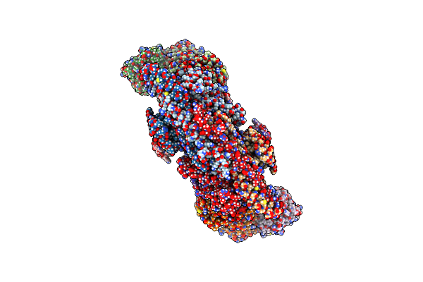
Crystal Structure Of The Brca1-A Complex
Organism: Mus musculus
Method: X-RAY DIFFRACTION Resolution:3.75 Å Release Date: 2019-07-10 Classification: SIGNALING PROTEIN Ligands: ZN |
Organism: Mus musculus
Method: X-RAY DIFFRACTION
Release Date: 2019-07-10
Ligands: ZN
 |
Cryo-Em Structure Of The Brisc Complex Bound To Shmt2
Organism: Homo sapiens
Method: ELECTRON MICROSCOPY Release Date: 2019-07-10 Classification: SIGNALING PROTEIN Ligands: ZN |
Organism: Homo sapiens
Method: ELECTRON MICROSCOPY
Release Date: 2019-07-10
Ligands: ZN