

Search Count: 48
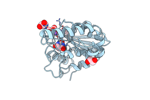 |
Crystal Structure Of Mycobacterium Tuberculosis Uracil-Dna Glycosylase In Complex With Barbituric Acid And Citric Acid, Form I
Organism: Mycobacterium tuberculosis h37rv
Method: X-RAY DIFFRACTION Resolution:1.24 Å Release Date: 2023-07-12 Classification: HYDROLASE Ligands: BR8, CIT, EDO, NA |
Organism: Mycobacterium tuberculosis h37rv
Method: X-RAY DIFFRACTION
Release Date: 2023-07-12
Ligands: BR8, CIT, EDO, NA
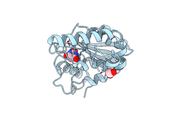 |
Crystal Structure Of Mycobacterium Tuberculosis Uracil-Dna Glycosylase In Complex With Barbituric Acid, Form I
Organism: Mycobacterium tuberculosis h37rv
Method: X-RAY DIFFRACTION Resolution:1.26 Å Release Date: 2023-07-12 Classification: HYDROLASE/INHIBITOR Ligands: BR8, EDO, CL |
Organism: Mycobacterium tuberculosis h37rv
Method: X-RAY DIFFRACTION
Release Date: 2023-07-12
Ligands: BR8, EDO, CL
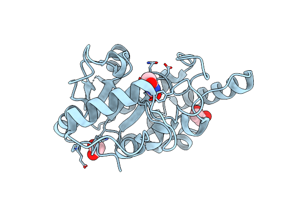 |
Crystal Structure Of Mycobacterium Tuberculosis Uracil-Dna Glycosylase In Complex With Barbituric Acid, Form Iii
Organism: Mycobacterium tuberculosis h37rv
Method: X-RAY DIFFRACTION Resolution:1.95 Å Release Date: 2023-07-12 Classification: HYDROLASE/INHIBITOR Ligands: BR8, EDO |
Organism: Mycobacterium tuberculosis h37rv
Method: X-RAY DIFFRACTION
Release Date: 2023-07-12
Ligands: BR8, EDO
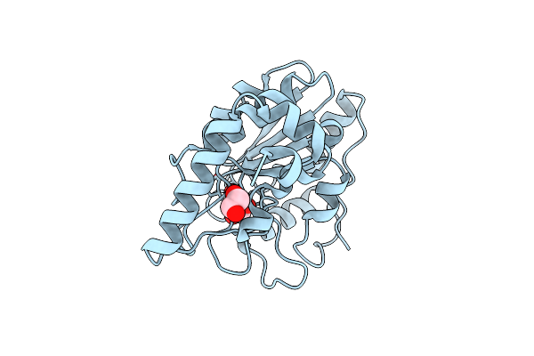 |
Crystal Structure Of Mycobacterium Tuberculosis Uracil-Dna Glycosylase In Complex With Barbituric Acid, Form Ii
Organism: Mycobacterium tuberculosis h37rv
Method: X-RAY DIFFRACTION Resolution:2.26 Å Release Date: 2023-07-12 Classification: HYDROLASE/INHIBITOR Ligands: BR8, EDO |
Organism: Mycobacterium tuberculosis h37rv
Method: X-RAY DIFFRACTION
Release Date: 2023-07-12
Ligands: BR8, EDO
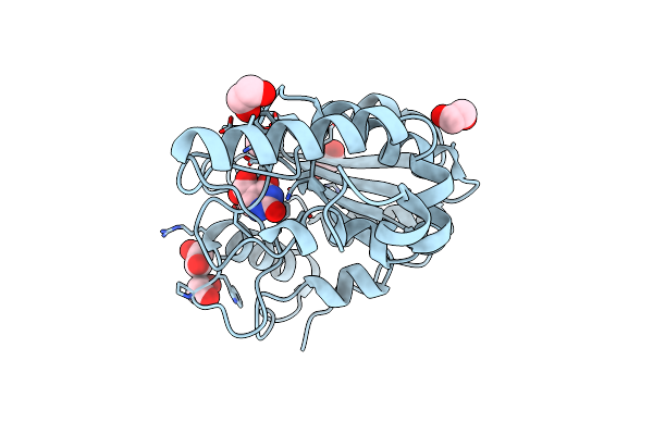 |
Crystal Structure Of Mycobacterium Tuberculosis Uracil-Dna Glycosylase In Complex With Isoorotic Acid (2,4-Dihydroxypyrimidine-5-Carboxylic Acid), Form I
Organism: Mycobacterium tuberculosis h37rv
Method: X-RAY DIFFRACTION Resolution:1.72 Å Release Date: 2023-07-12 Classification: HYDROLASE/INHIBITOR Ligands: 5CU, EDO |
Organism: Mycobacterium tuberculosis h37rv
Method: X-RAY DIFFRACTION
Release Date: 2023-07-12
Ligands: 5CU, EDO
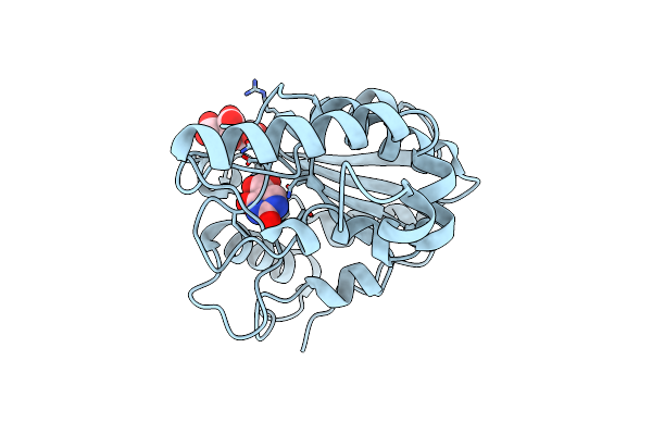 |
Crystal Structure Of Mycobacterium Tuberculosis Uracil-Dna Glycosylase In Complex With Isoorotic Acid (2,4-Dihydroxypyrimidine-5-Carboxylic Acid) And Citric Acid, Form I
Organism: Mycobacterium tuberculosis h37rv
Method: X-RAY DIFFRACTION Resolution:2.60 Å Release Date: 2023-07-12 Classification: HYDROLASE/INHIBITOR Ligands: 5CU, CIT |
Organism: Mycobacterium tuberculosis h37rv
Method: X-RAY DIFFRACTION
Release Date: 2023-07-12
Ligands: 5CU, CIT
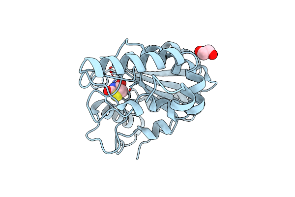 |
Crystal Structure Of Mycobacterium Tuberculosis Uracil-Dna Glycosylase In Complex With 2,4-Thiazolidinedione, Form I
Organism: Mycobacterium tuberculosis h37rv
Method: X-RAY DIFFRACTION Resolution:1.72 Å Release Date: 2023-07-12 Classification: HYDROLASE/INHIBITOR Ligands: OD3, EDO |
Organism: Mycobacterium tuberculosis h37rv
Method: X-RAY DIFFRACTION
Release Date: 2023-07-12
Ligands: OD3, EDO
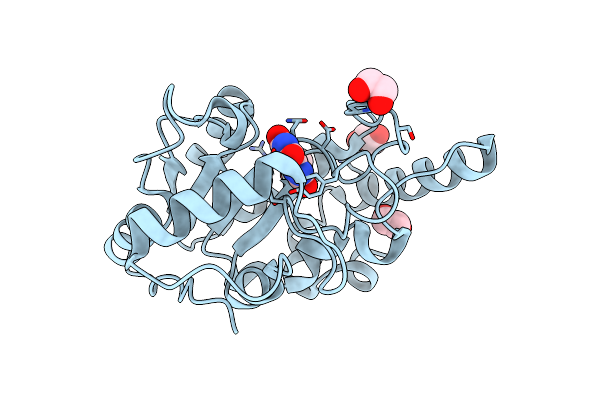 |
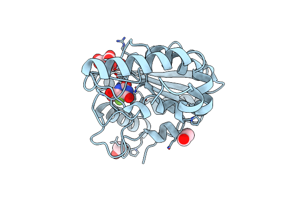
Crystal Structure Of Mycobacterium Tuberculosis Uracil-Dna Glycosylase In Complex With Uric Acid, Form Iii
Organism: Mycobacterium tuberculosis h37rv
Method: X-RAY DIFFRACTION Resolution:1.88 Å Release Date: 2023-07-12 Classification: HYDROLASE/INHIBITOR Ligands: URC, EDO |
Organism: Mycobacterium tuberculosis h37rv
Method: X-RAY DIFFRACTION
Release Date: 2023-07-12
Ligands: URC, EDO
 |
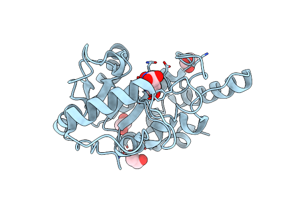
Crystal Structure Of Mycobacterium Tuberculosis Uracil-Dna Glycosylase In Complex With 5-Fluoroorotic Acid And Citric Acid, Form I
Organism: Mycobacterium tuberculosis h37rv
Method: X-RAY DIFFRACTION Resolution:2.00 Å Release Date: 2023-07-12 Classification: HYDROLASE/INHIBITOR Ligands: FOT, CIT, EDO |
Organism: Mycobacterium tuberculosis h37rv
Method: X-RAY DIFFRACTION
Release Date: 2023-07-12
Ligands: FOT, CIT, EDO
 |
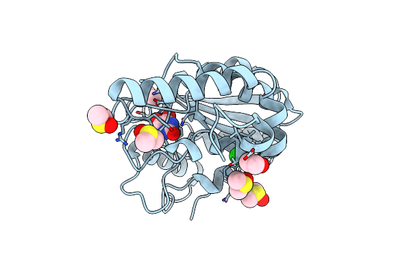
Crystal Structure Of Mycobacterium Tuberculosis Uracil-Dna Glycosylase In Complex With Orotic Acid, Form Iii
Organism: Mycobacterium tuberculosis h37rv
Method: X-RAY DIFFRACTION Resolution:2.00 Å Release Date: 2023-07-12 Classification: HYDROLASE/INHIBITOR Ligands: ORO, EDO |
Organism: Mycobacterium tuberculosis h37rv
Method: X-RAY DIFFRACTION
Release Date: 2023-07-12
Ligands: ORO, EDO
 |
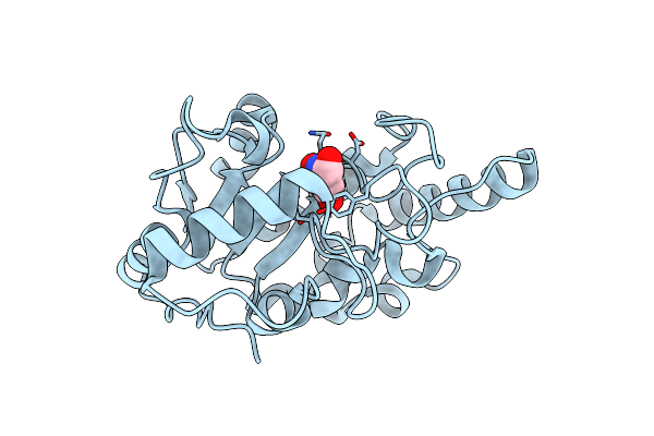
Crystal Structure Of Mycobacterium Tuberculosis Uracil-Dna Glycosylase In Complex With 5-Hydroxy-2,4(1H,3H)-Pyrimidinedione, Form I
Organism: Mycobacterium tuberculosis h37rv
Method: X-RAY DIFFRACTION Resolution:1.60 Å Release Date: 2023-07-12 Classification: HYDROLASE/INHIBITOR Ligands: ODF, EDO, DMS, NA, CL |
Organism: Mycobacterium tuberculosis h37rv
Method: X-RAY DIFFRACTION
Release Date: 2023-07-12
Ligands: ODF, EDO, DMS, NA, CL
 |
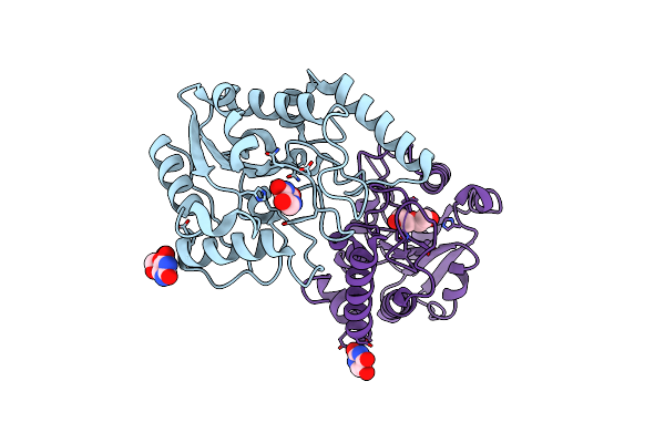
Crystal Structure Of Mycobacterium Tuberculosis Uracil-Dna Glycosylase In Complex With 6-Formyl-Uracil, Form Iii
Organism: Mycobacterium tuberculosis h37rv
Method: X-RAY DIFFRACTION Resolution:2.28 Å Release Date: 2023-07-12 Classification: HYDROLASE/INHIBITOR Ligands: OG6 |
Organism: Mycobacterium tuberculosis h37rv
Method: X-RAY DIFFRACTION
Release Date: 2023-07-12
Ligands: OG6
 |
Crystal Structure Of Mycobacterium Tuberculosis Uracil-Dna Glycosylase In Complex With 5-Hydroxy-2,4(1H,3H)-Pyrimidinedione, Form Vi
Organism: Mycobacterium tuberculosis h37rv
Method: X-RAY DIFFRACTION Resolution:2.40 Å Release Date: 2023-07-12 Classification: HYDROLASE/INHIBITOR Ligands: ODF, EDO, PEG |
Organism: Mycobacterium tuberculosis h37rv
Method: X-RAY DIFFRACTION
Release Date: 2023-07-12
Ligands: ODF, EDO, PEG
 |
Structure Of Human Serotonin Transporter Bound To Small Molecule '8090 In Lipid Nanodisc And Nacl
Organism: Mus musculus, Homo sapiens
Method: ELECTRON MICROSCOPY Release Date: 2023-03-15 Classification: TRANSPORT PROTEIN Ligands: KWC |
Organism: Mus musculus, Homo sapiens
Method: ELECTRON MICROSCOPY
Release Date: 2023-03-15
Ligands: KWC
 |
Cryo-Em Structure Of Human Nkcc1 K289Na492El671C Bound With Bumetanide
Organism: Homo sapiens
Method: ELECTRON MICROSCOPY Release Date: 2022-05-25 Classification: TRANSPORT PROTEIN Ligands: 82U, K, CL |
Organism: Homo sapiens
Method: ELECTRON MICROSCOPY
Release Date: 2022-05-25
Ligands: 82U, K, CL
 |
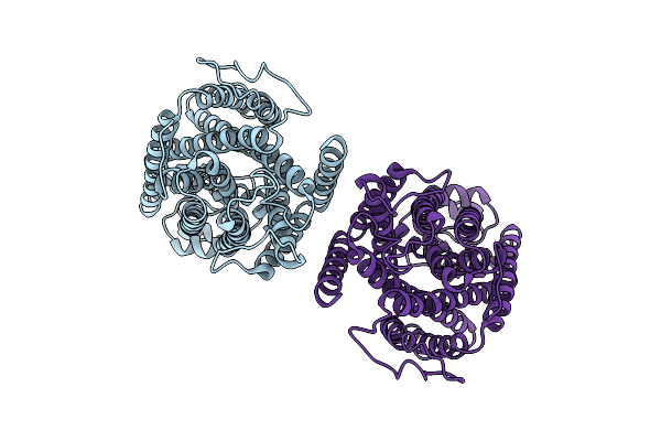
Cryo-Em Structure Of Human Nkcc1 K289Na492E Bound With Bumetanide
Organism: Homo sapiens
Method: ELECTRON MICROSCOPY Release Date: 2022-05-25 Classification: TRANSPORT PROTEIN Ligands: K, CL, 82U |
Organism: Homo sapiens
Method: ELECTRON MICROSCOPY
Release Date: 2022-05-25
Ligands: K, CL, 82U
 |
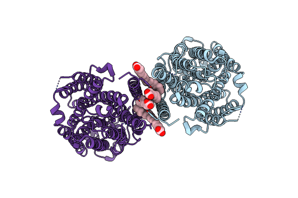
Cryo-Em Structure Of Human Nkcc1 K289Na492El671C
Organism: Homo sapiens
Method: ELECTRON MICROSCOPY Release Date: 2022-05-25 Classification: TRANSPORT PROTEIN |
Organism: Homo sapiens
Method: ELECTRON MICROSCOPY
Release Date: 2022-05-25
 |
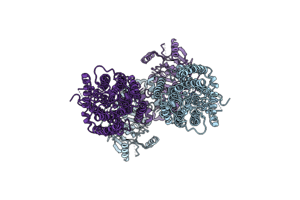
Human Nkcc1
Organism: Homo sapiens
Method: ELECTRON MICROSCOPY Release Date: 2021-04-14 Classification: MEMBRANE PROTEIN Ligands: PLM |
Organism: Homo sapiens
Method: ELECTRON MICROSCOPY
Release Date: 2021-04-14
Ligands: PLM
 |
Mouse Kcc2
Organism: Mus musculus
Method: ELECTRON MICROSCOPY Release Date: 2021-04-14 Classification: MEMBRANE PROTEIN |
Organism: Mus musculus
Method: ELECTRON MICROSCOPY
Release Date: 2021-04-14
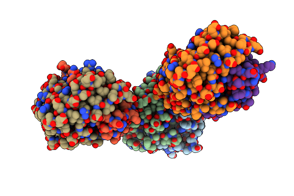 |
A Resting State Structure Of L-Dopa Dioxygenase From Streptomyces Sclerotialus
Organism: Streptomyces sclerotialus
Method: X-RAY DIFFRACTION Resolution:1.98 Å Release Date: 2019-06-26 Classification: OXIDOREDUCTASE Ligands: FE, TRS |
Organism: Streptomyces sclerotialus
Method: X-RAY DIFFRACTION
Release Date: 2019-06-26
Ligands: FE, TRS