

Search Count: 19
 |
Amine Dehydrogenase From Cystobacter Fuscus (Cfusamdh) W145A Mutant With Nadp+ And Pentylamine
Organism: Cystobacter fuscus
Method: X-RAY DIFFRACTION Resolution:1.50 Å Release Date: 2022-12-07 Classification: OXIDOREDUCTASE Ligands: NAP, TRS, AML |
Organism: Cystobacter fuscus
Method: X-RAY DIFFRACTION
Release Date: 2022-12-07
Ligands: NAP, TRS, AML
 |
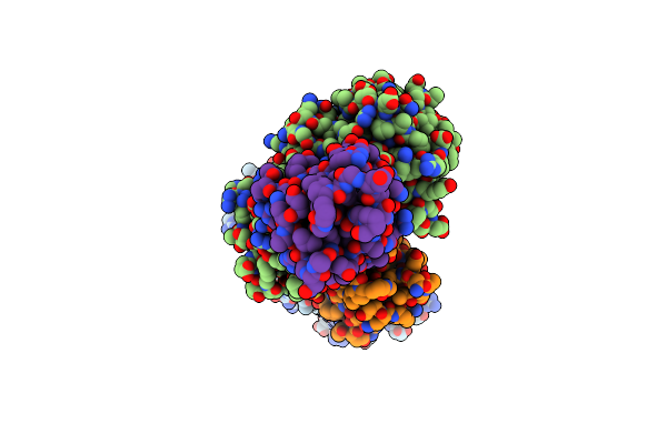
Amine Dehydrogenase From Cystobacter Fuscus (Cfusamdh) W145A Mutant With Nad+
Organism: Cystobacter fuscus
Method: X-RAY DIFFRACTION Resolution:1.64 Å Release Date: 2022-12-07 Classification: OXIDOREDUCTASE Ligands: NAD, TRS |
Organism: Cystobacter fuscus
Method: X-RAY DIFFRACTION
Release Date: 2022-12-07
Ligands: NAD, TRS
 |
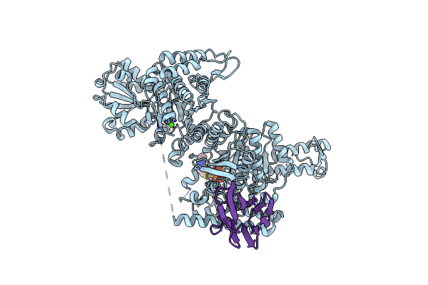
Crystal Structure Of The Mycobacterium Tuberculosis Pknb Kinase Domain (L33E Mutant) In Complex With Its Substrate Gara
Organism: Mycobacterium tuberculosis (strain atcc 25618 / h37rv)
Method: X-RAY DIFFRACTION Resolution:2.37 Å Release Date: 2019-05-22 Classification: SIGNALING PROTEIN Ligands: ACP, MG, SO4 |
Organism: Mycobacterium tuberculosis (strain atcc 25618 / h37rv)
Method: X-RAY DIFFRACTION
Release Date: 2019-05-22
Ligands: ACP, MG, SO4
 |
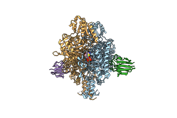
Crystal Structure Of The Wild-Type Suca Domain Of Mycobacterium Smegmatis Kgd (Alpha-Ketoglutarate Decarboxylase), In Complex With Gara
Organism: Mycobacterium smegmatis (strain atcc 700084 / mc(2)155)
Method: X-RAY DIFFRACTION Resolution:2.15 Å Release Date: 2019-05-22 Classification: OXIDOREDUCTASE Ligands: TPP, MG, CA |
Organism: Mycobacterium smegmatis (strain atcc 700084 / mc(2)155)
Method: X-RAY DIFFRACTION
Release Date: 2019-05-22
Ligands: TPP, MG, CA
 |
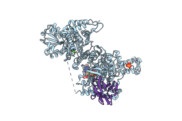
Crystal Structure Of The Suca Domain Of Mycobacterium Smegmatis Kgd (Alpha-Ketoglutarate Decarboxylase), Mutant R802A, In Complex With Gara
Organism: Mycobacterium smegmatis (strain atcc 700084 / mc(2)155)
Method: X-RAY DIFFRACTION Resolution:2.20 Å Release Date: 2019-05-22 Classification: OXIDOREDUCTASE Ligands: TPP, MG, CA |
Organism: Mycobacterium smegmatis (strain atcc 700084 / mc(2)155)
Method: X-RAY DIFFRACTION
Release Date: 2019-05-22
Ligands: TPP, MG, CA
 |
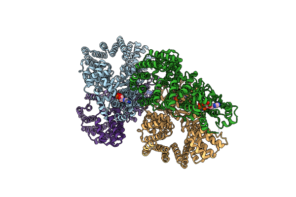
Crystal Structure Of The Suca Domain Of Mycobacterium Smegmatis Kgd (R802A) In Complex With Gara, Following 2-Oxoglutarate Soak
Organism: Mycobacterium smegmatis (strain atcc 700084 / mc(2)155)
Method: X-RAY DIFFRACTION Resolution:2.40 Å Release Date: 2019-05-22 Classification: OXIDOREDUCTASE Ligands: MG, CA, PO4, TD6 |
Organism: Mycobacterium smegmatis (strain atcc 700084 / mc(2)155)
Method: X-RAY DIFFRACTION
Release Date: 2019-05-22
Ligands: MG, CA, PO4, TD6
 |
Crystal Structure Of The Signal Transduction Atpase With Numerous Domains (Stand) Protein With A Tetratricopeptide Repeat Sensor Ph0952 From Pyrococcus Horikoshii
Organism: Pyrococcus horikoshii (strain atcc 700860 / dsm 12428 / jcm 9974 / nbrc 100139 / ot-3)
Method: X-RAY DIFFRACTION Resolution:3.40 Å Release Date: 2019-02-20 Classification: SIGNALING PROTEIN Ligands: ADP |
Organism: Pyrococcus horikoshii (strain atcc 700860 / dsm 12428 / jcm 9974 / nbrc 100139 / ot-3)
Method: X-RAY DIFFRACTION
Release Date: 2019-02-20
Ligands: ADP
 |
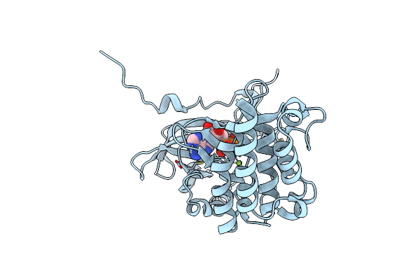
Oh1 From The Orf Virus: A Tyrosine Phosphatase That Displays Distinct Structural Features And Triple Substrate Specificity
Organism: Orf virus
Method: X-RAY DIFFRACTION Resolution:1.89 Å Release Date: 2017-08-09 Classification: HYDROLASE Ligands: PO4, SO4 |
Organism: Orf virus
Method: X-RAY DIFFRACTION
Release Date: 2017-08-09
Ligands: PO4, SO4
 |
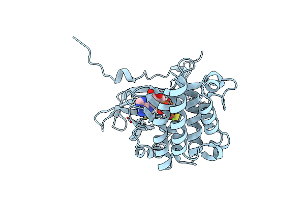
Crystal Structure Of The S/T Protein Kinase Pkng From Mycobacterium Tuberculosis In Complex With Adp
Organism: Mycobacterium tuberculosis (strain atcc 25618 / h37rv)
Method: X-RAY DIFFRACTION Resolution:1.74 Å Release Date: 2015-05-20 Classification: TRANSFERASE Ligands: ADP, ZN, MG |
Organism: Mycobacterium tuberculosis (strain atcc 25618 / h37rv)
Method: X-RAY DIFFRACTION
Release Date: 2015-05-20
Ligands: ADP, ZN, MG
 |
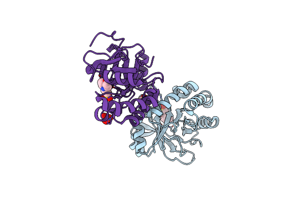
Crystal Structure Of The S/T Protein Kinase Pkng From Mycobacterium Tuberculosis In Complex With Ags
Organism: Mycobacterium tuberculosis (strain atcc 25618 / h37rv)
Method: X-RAY DIFFRACTION Resolution:1.90 Å Release Date: 2015-05-20 Classification: TRANSFERASE Ligands: AGS, ZN, MG |
Organism: Mycobacterium tuberculosis (strain atcc 25618 / h37rv)
Method: X-RAY DIFFRACTION
Release Date: 2015-05-20
Ligands: AGS, ZN, MG
 |
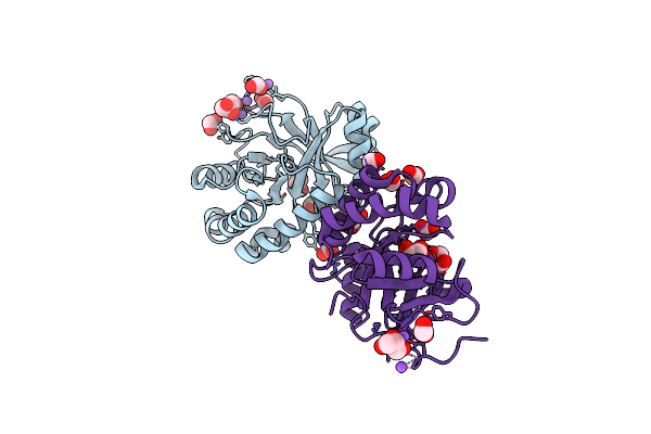
Crystal Structure Of The Mycobacterium Tuberculosis Tat-Secreted Protein Rv2525C In Complex With Mes (Monoclinic Crystal Form I)
Organism: Mycobacterium tuberculosis
Method: X-RAY DIFFRACTION Resolution:1.44 Å Release Date: 2014-10-08 Classification: UNKNOWN FUNCTION Ligands: MES, GOL, SO4 |
Organism: Mycobacterium tuberculosis
Method: X-RAY DIFFRACTION
Release Date: 2014-10-08
Ligands: MES, GOL, SO4
 |
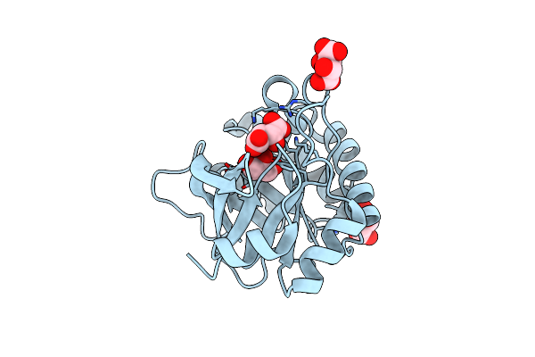
Crystal Structure Of The Mycobacterium Tuberculosis Tat-Secreted Protein Rv2525C, Monoclinic Crystal Form I
Organism: Mycobacterium tuberculosis
Method: X-RAY DIFFRACTION Resolution:1.33 Å Release Date: 2014-10-08 Classification: UNKNOWN FUNCTION Ligands: GOL, FMT, NA |
Organism: Mycobacterium tuberculosis
Method: X-RAY DIFFRACTION
Release Date: 2014-10-08
Ligands: GOL, FMT, NA
 |
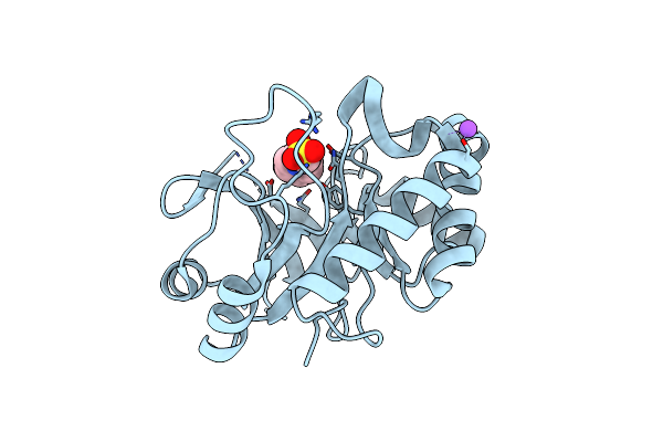
Crystal Structure Of The Mycobacterium Tuberculosis Tat-Secreted Protein Rv2525C In Complex With L-Tartrate (Orthorhombic Crystal Form)
Organism: Mycobacterium tuberculosis
Method: X-RAY DIFFRACTION Resolution:1.61 Å Release Date: 2014-10-08 Classification: UNKNOWN FUNCTION Ligands: TLA, GOL |
Organism: Mycobacterium tuberculosis
Method: X-RAY DIFFRACTION
Release Date: 2014-10-08
Ligands: TLA, GOL
 |
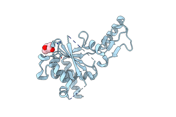
Crystal Structure Of The Mycobacterium Tuberculosis Tat-Secreted Protein Rv2525C In Complex With Hepes (Monoclinic Crystal Form Ii)
Organism: Mycobacterium tuberculosis
Method: X-RAY DIFFRACTION Resolution:1.81 Å Release Date: 2014-10-08 Classification: UNKNOWN FUNCTION Ligands: EPE, NA |
Organism: Mycobacterium tuberculosis
Method: X-RAY DIFFRACTION
Release Date: 2014-10-08
Ligands: EPE, NA
 |
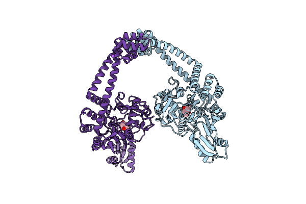
Crystal Structure Of The Second Part Of The Mycobacterium Tuberculosis Dna Gyrase Reaction Core: The Toprim Domain At 1.95 A Resolution
Organism: Mycobacterium tuberculosis
Method: X-RAY DIFFRACTION Resolution:1.95 Å Release Date: 2010-09-29 Classification: ISOMERASE Ligands: MPD |
Organism: Mycobacterium tuberculosis
Method: X-RAY DIFFRACTION
Release Date: 2010-09-29
Ligands: MPD
 |
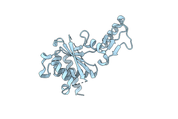
Crystal Structure Of The First Part Of The Mycobacterium Tuberculosis Dna Gyrase Reaction Core: The Breakage And Reunion Domain At 2.7 A Resolution
Organism: Mycobacterium tuberculosis
Method: X-RAY DIFFRACTION Resolution:2.70 Å Release Date: 2010-07-28 Classification: ISOMERASE Ligands: MPD, NA |
Organism: Mycobacterium tuberculosis
Method: X-RAY DIFFRACTION
Release Date: 2010-07-28
Ligands: MPD, NA
 |
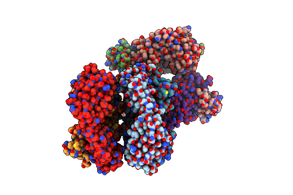
Crystal Structure Of The Second Part Of The Mycobacterium Tuberculosis Dna Gyrase Reaction Core: The Toprim Domain At 2.1 A Resolution
Organism: Mycobacterium tuberculosis
Method: X-RAY DIFFRACTION Resolution:2.10 Å Release Date: 2010-07-28 Classification: ISOMERASE |
Organism: Mycobacterium tuberculosis
Method: X-RAY DIFFRACTION
Release Date: 2010-07-28
 |
Crystal Structure Of A Monoclonal Antibody Directed Against An Antigenic Determinant Common To Ogawa And Inaba Serotypes Of Vibrio Cholerae O1
Organism: Mus musculus
Method: X-RAY DIFFRACTION Resolution:2.50 Å Release Date: 2008-05-13 Classification: IMMUNE SYSTEM |
Organism: Mus musculus
Method: X-RAY DIFFRACTION
Release Date: 2008-05-13
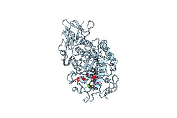 |
Structure Of Human Pancreatic Alpha-Amylase In Complex With The Carbohydrate Inhibitor Acarbose
Organism: Homo sapiens
Method: X-RAY DIFFRACTION Resolution:3.20 Å Release Date: 2000-02-16 Classification: HYDROLASE Ligands: CL, CA |
Organism: Homo sapiens
Method: X-RAY DIFFRACTION
Release Date: 2000-02-16
Ligands: CL, CA