

Search Count: 12
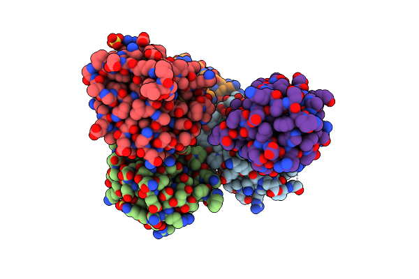 |
Crystal Structure Of The Third Catalytic Domain Of Erp46
Organism: Homo sapiens
Method: X-RAY DIFFRACTION Resolution:2.00 Å Release Date: 2012-04-11 Classification: ISOMERASE Ligands: SO4 |
Organism: Homo sapiens
Method: X-RAY DIFFRACTION
Release Date: 2012-04-11
Ligands: SO4
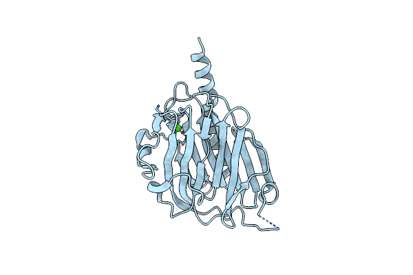 |
Crystal Structure Of The Calreticulin Lectin Domain
Organism: Mus musculus
Method: X-RAY DIFFRACTION Resolution:2.30 Å Release Date: 2010-09-29 Classification: CHAPERONE Ligands: CA |
Organism: Mus musculus
Method: X-RAY DIFFRACTION
Release Date: 2010-09-29
Ligands: CA
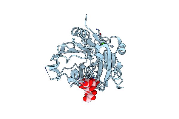 |
Structural Basis Of Carbohydrate Recognition By Calreticulin
Organism: Mus musculus
Method: X-RAY DIFFRACTION Resolution:1.95 Å Release Date: 2010-09-29 Classification: CHAPERONE Ligands: CA |
Organism: Mus musculus
Method: X-RAY DIFFRACTION
Release Date: 2010-09-29
Ligands: CA
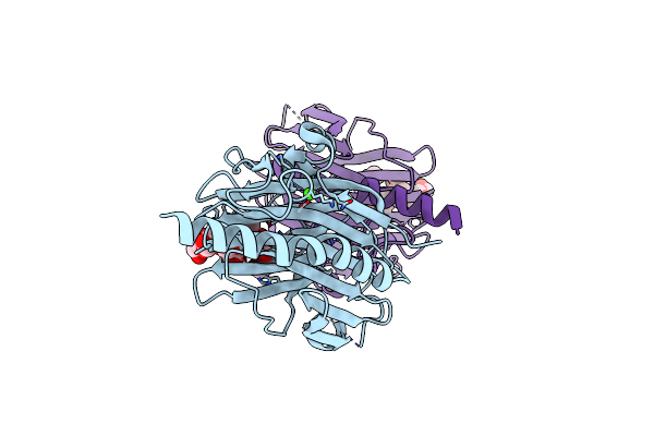 |
Structural Basis Of Carbohydrate Recognition By Calreticulin
Organism: Mus musculus
Method: X-RAY DIFFRACTION Resolution:2.01 Å Release Date: 2010-09-29 Classification: CHAPERONE Ligands: CA |
Organism: Mus musculus
Method: X-RAY DIFFRACTION
Release Date: 2010-09-29
Ligands: CA
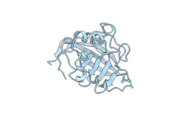 |
Crystal Structure Of Cyclophilin B At 1.2 A Resolution
Organism: Homo sapiens
Method: X-RAY DIFFRACTION Resolution:1.20 Å Release Date: 2010-07-21 Classification: ISOMERASE |
Organism: Homo sapiens
Method: X-RAY DIFFRACTION
Release Date: 2010-07-21
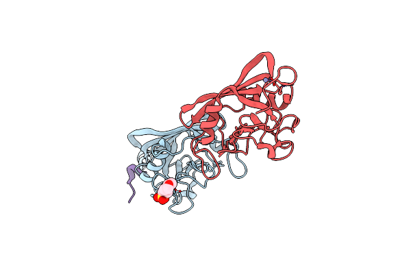 |
Crystal Structure Of Cyclophilin B In Complex With Calmegin Fragment
Organism: Homo sapiens, Canis lupus familiaris
Method: X-RAY DIFFRACTION Resolution:1.70 Å Release Date: 2010-07-21 Classification: ISOMERASE Ligands: ZN, MES |
Organism: Homo sapiens, Canis lupus familiaris
Method: X-RAY DIFFRACTION
Release Date: 2010-07-21
Ligands: ZN, MES
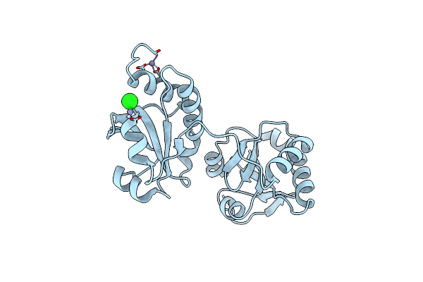 |
Crystal Structure Of The A0A Fragment Of Erp72
Organism: Homo sapiens
Method: X-RAY DIFFRACTION Resolution:1.95 Å Release Date: 2010-07-07 Classification: ISOMERASE Ligands: ZN, CL |
Organism: Homo sapiens
Method: X-RAY DIFFRACTION
Release Date: 2010-07-07
Ligands: ZN, CL
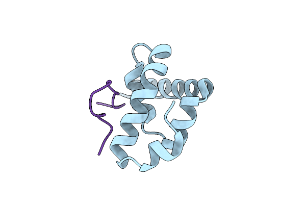 |
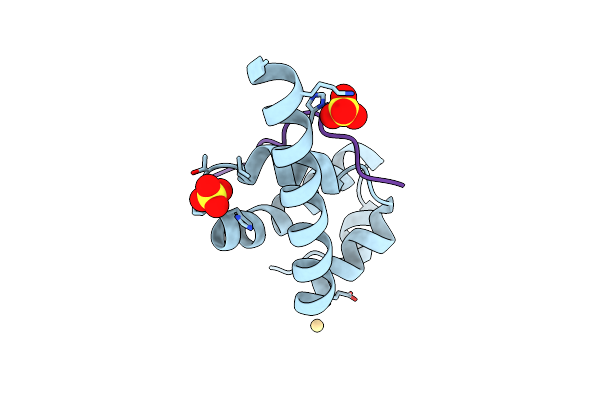
Structural Basis Of Gw182 Recognition By Poly(A)-Binding Protein
Organism: Homo sapiens
Method: X-RAY DIFFRACTION Resolution:1.50 Å Release Date: 2010-02-23 Classification: PROTEIN BINDING |
Organism: Homo sapiens
Method: X-RAY DIFFRACTION
Release Date: 2010-02-23
 |
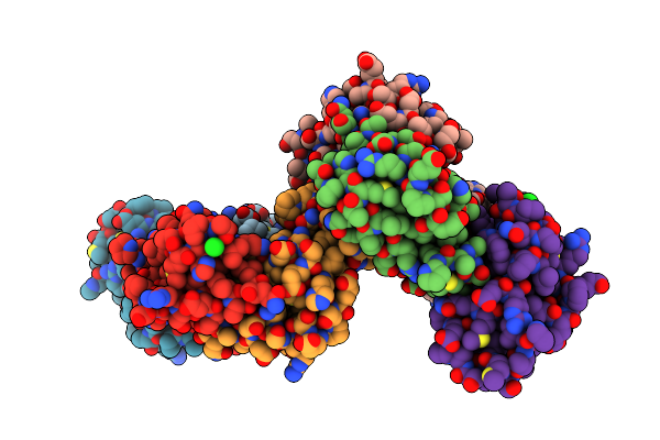
Structural Basis Of Ataxin-2 Recognition By Poly(A)-Binding Protein
Organism: Homo sapiens
Method: X-RAY DIFFRACTION Resolution:1.70 Å Release Date: 2010-02-23 Classification: PROTEIN BINDING Ligands: CD, SO4 |
Organism: Homo sapiens
Method: X-RAY DIFFRACTION
Release Date: 2010-02-23
Ligands: CD, SO4
 |
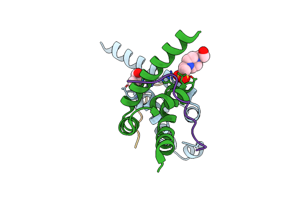
Crystal Structure Of The Mlle Domain Of Poly(A)-Binding Protein
Organism: Homo sapiens
Method: X-RAY DIFFRACTION Resolution:2.50 Å Release Date: 2010-02-09 Classification: RNA BINDING PROTEIN Ligands: CL |
Organism: Homo sapiens
Method: X-RAY DIFFRACTION
Release Date: 2010-02-09
Ligands: CL
 |
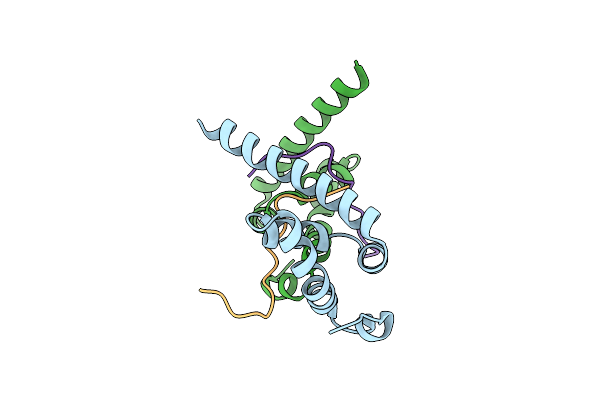
Crystal Structure Of The Mlle Domain Of Poly(A)-Binding Protein In Complex With The Binding Region Of Paip2
Organism: Homo sapiens
Method: X-RAY DIFFRACTION Resolution:1.40 Å Release Date: 2010-02-09 Classification: PROTEIN BINDING Ligands: GOL, EPE |
Organism: Homo sapiens
Method: X-RAY DIFFRACTION
Release Date: 2010-02-09
Ligands: GOL, EPE
 |
Crystal Structure Of The Mlle Domain Of Poly(A)-Binding Protein In Complex With The Binding Region Of Paip2
Organism: Homo sapiens
Method: X-RAY DIFFRACTION Resolution:1.50 Å Release Date: 2010-02-09 Classification: PROTEIN BINDING Ligands: CL |
Organism: Homo sapiens
Method: X-RAY DIFFRACTION
Release Date: 2010-02-09
Ligands: CL