

Search Count: 30
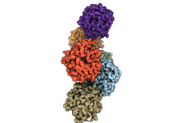 |
Isobutylene Epoxide Hydrolase From Mycolicibacterium
Organism: Mycolicibacterium
Method: X-RAY DIFFRACTION Resolution:2.29 Å Release Date: 2025-10-01 Classification: HYDROLASE Ligands: GOL, MG, 8K6, SO4 |
Organism: Mycolicibacterium
Method: X-RAY DIFFRACTION
Release Date: 2025-10-01
Ligands: GOL, MG, 8K6, SO4
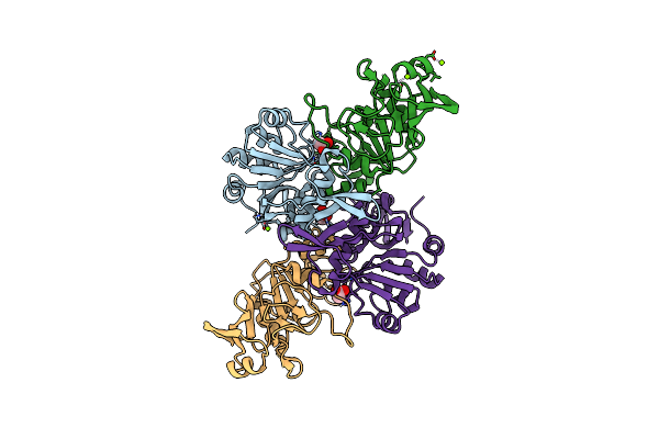 |
Ctfab E46D Active Site Mutant Hydrolase
Organism: Thermosipho melanesiensis
Method: X-RAY DIFFRACTION Resolution:2.20 Å Release Date: 2025-02-05 Classification: TRANSFERASE Ligands: MG, CL, ACT |
Organism: Thermosipho melanesiensis
Method: X-RAY DIFFRACTION
Release Date: 2025-02-05
Ligands: MG, CL, ACT
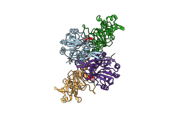 |
Ctfab E46S Active Site Mutant Inactive
Organism: Thermosipho melanesiensis
Method: X-RAY DIFFRACTION Resolution:2.60 Å Release Date: 2025-02-05 Classification: TRANSFERASE Ligands: ACT, CL, MG |
Organism: Thermosipho melanesiensis
Method: X-RAY DIFFRACTION
Release Date: 2025-02-05
Ligands: ACT, CL, MG
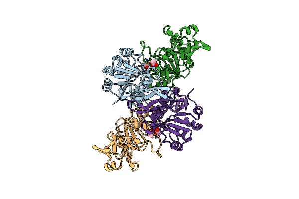 |
Ctfab Native Acetoacetate-Coa Transferase Protein
Organism: Thermosipho melanesiensis
Method: X-RAY DIFFRACTION Resolution:2.00 Å Release Date: 2025-02-05 Classification: TRANSFERASE Ligands: ACT |
Organism: Thermosipho melanesiensis
Method: X-RAY DIFFRACTION
Release Date: 2025-02-05
Ligands: ACT
 |
Ctfab F42Ts45C Mutant Co-Crystallized With Acetyl-Coa
Organism: Thermosipho melanesiensis
Method: X-RAY DIFFRACTION Resolution:1.90 Å Release Date: 2025-02-05 Classification: TRANSFERASE Ligands: ACT, PEG, COA |
Organism: Thermosipho melanesiensis
Method: X-RAY DIFFRACTION
Release Date: 2025-02-05
Ligands: ACT, PEG, COA
 |
Caenorhabditis Elegans Swsn-4 (Smarca4-Brg1) Atpase Bromodomain In Complex With A Modified Histone H3, N6-Epsilon-Acetyl-L-Lysine 14 (H3K14Ac) Polypeptide
Organism: Caenorhabditis elegans
Method: X-RAY DIFFRACTION Resolution:1.30 Å Release Date: 2021-09-08 Classification: PEPTIDE BINDING PROTEIN |
Organism: Caenorhabditis elegans
Method: X-RAY DIFFRACTION
Release Date: 2021-09-08
 |
Biotin Carboxylase Domain Of Thermophilic 2-Oxoglutarate Carboxylase Bound To Bicarbonate
Organism: Hydrogenobacter thermophilus
Method: X-RAY DIFFRACTION Resolution:2.30 Å Release Date: 2021-01-27 Classification: LIGASE Ligands: BCT |
Organism: Hydrogenobacter thermophilus
Method: X-RAY DIFFRACTION
Release Date: 2021-01-27
Ligands: BCT
 |
Biotin Carboxylase Domain Of Thermophilic 2-Oxoglutarate Carboxylase Bound To Adp Without Magnesium With Disordered Phosphate Tail
Organism: Hydrogenobacter thermophilus
Method: X-RAY DIFFRACTION Resolution:2.20 Å Release Date: 2021-01-27 Classification: LIGASE Ligands: PO4, ADP |
Organism: Hydrogenobacter thermophilus
Method: X-RAY DIFFRACTION
Release Date: 2021-01-27
Ligands: PO4, ADP
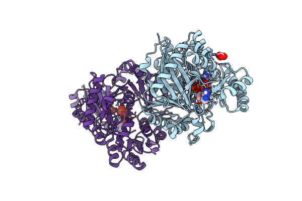 |
Crystal Structure Of The Hydrogenobacter Thermophilus 2-Oxoglutarate Carboxylase (Ogc) Biotin Carboxylase (Bc) Domain Dimer In Complex With Adenosine 5'-Diphosphate Magnesium Salt (Mgadp), Adenosine 5'-Diphosphate (Adp, And Bicarbonate Anion (Hydrogen Carbonate/Hco3-)
Organism: Hydrogenobacter thermophilus
Method: X-RAY DIFFRACTION Resolution:2.02 Å Release Date: 2021-01-27 Classification: LIGASE Ligands: ADP, MG, BCT |
Organism: Hydrogenobacter thermophilus
Method: X-RAY DIFFRACTION
Release Date: 2021-01-27
Ligands: ADP, MG, BCT
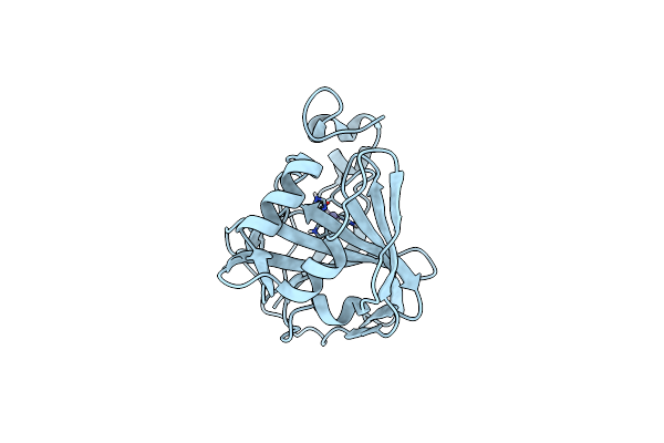 |
Photobacterium Profundum Alpha-Carbonic Anhydrase
Organism: Photobacterium profundum ss9
Method: X-RAY DIFFRACTION Resolution:1.50 Å Release Date: 2016-12-07 Classification: LYASE Ligands: ZN, CL |
Organism: Photobacterium profundum ss9
Method: X-RAY DIFFRACTION
Release Date: 2016-12-07
Ligands: ZN, CL
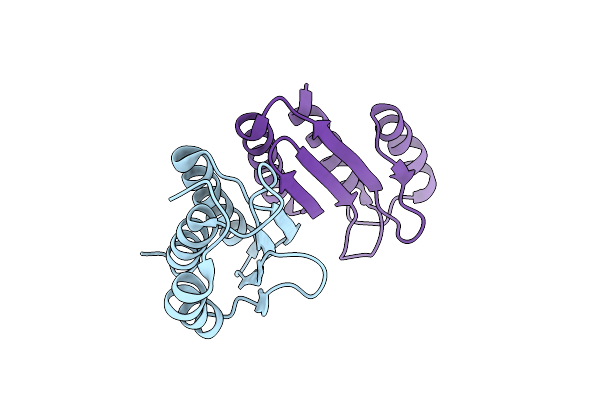 |
Crystal Structure Of Dcoh2 S51T
Organism: Mus musculus
Method: X-RAY DIFFRACTION Resolution:1.36 Å Release Date: 2014-12-31 Classification: LYASE |
Organism: Mus musculus
Method: X-RAY DIFFRACTION
Release Date: 2014-12-31
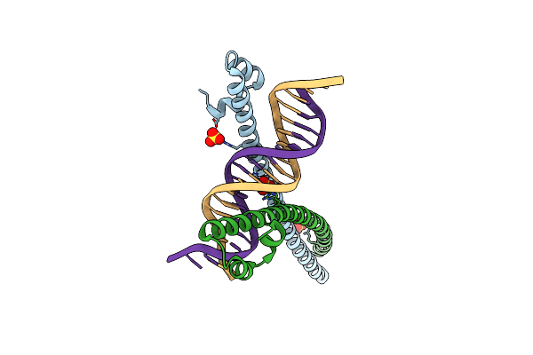 |
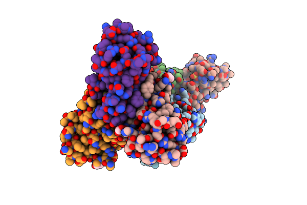
Crystal Structure Of The Mafa Homodimer Bound To The Consensus Mare
Organism: Homo sapiens
Method: X-RAY DIFFRACTION Resolution:2.86 Å Release Date: 2012-11-28 Classification: TRANSCRIPTION/DNA Ligands: SO4 |
Organism: Homo sapiens
Method: X-RAY DIFFRACTION
Release Date: 2012-11-28
Ligands: SO4
 |
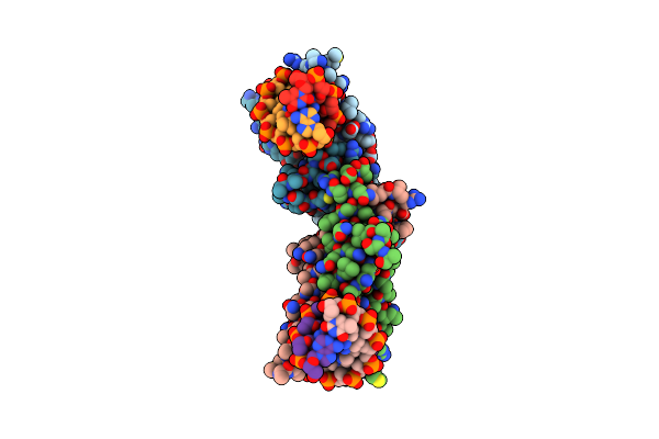
Crystal Structure Of Dcoh1Thr51Ser
Organism: Rattus norvegicus
Method: X-RAY DIFFRACTION Resolution:1.80 Å Release Date: 2010-08-25 Classification: LYASE Ligands: SO4, GOL |
Organism: Rattus norvegicus
Method: X-RAY DIFFRACTION
Release Date: 2010-08-25
Ligands: SO4, GOL
 |
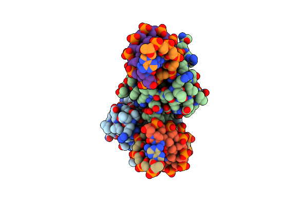
Crystal Structure Of The Basic-Helix-Loop-Helix Domains Of The Heterodimer E47/Neurod1 Bound To Dna
Organism: Mus musculus
Method: X-RAY DIFFRACTION Resolution:2.50 Å Release Date: 2008-11-04 Classification: transcription/DNA |
Organism: Mus musculus
Method: X-RAY DIFFRACTION
Release Date: 2008-11-04
 |
Crystal Structure Of The Pdx1 Homeodomain In Complex With Dna
Organism: Mesocricetus auratus
Method: X-RAY DIFFRACTION Resolution:2.42 Å Release Date: 2007-03-20 Classification: Transcription/DNA |
Organism: Mesocricetus auratus
Method: X-RAY DIFFRACTION
Release Date: 2007-03-20
 |
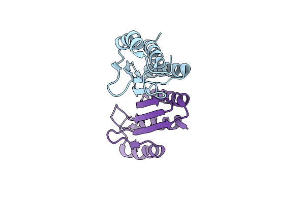
Structure Of Erh (Enhencer Of Rudimentary Gene)
Organism: Homo sapiens
Method: X-RAY DIFFRACTION Resolution:2.00 Å Release Date: 2005-04-06 Classification: ERH(ENHANCER OF RUDIMENTARY HOMOLOG) |
Organism: Homo sapiens
Method: X-RAY DIFFRACTION
Release Date: 2005-04-06
 |
Crystal Structure Of Dcoh2, A Paralog Of Dcoh, The Dimerization Cofactor Of Hnf-1
Organism: Mus musculus
Method: X-RAY DIFFRACTION Resolution:1.60 Å Release Date: 2004-10-12 Classification: LYASE |
Organism: Mus musculus
Method: X-RAY DIFFRACTION
Release Date: 2004-10-12
 |
X-Ray Structure Of An Early-M Intermediate Of Bacteriorhodopsin
Organism: Halobacterium salinarum
Method: X-RAY DIFFRACTION Resolution:2.00 Å Release Date: 2001-12-05 Classification: PROTON TRANSPORT Ligands: RET, LI1 |
Organism: Halobacterium salinarum
Method: X-RAY DIFFRACTION
Release Date: 2001-12-05
Ligands: RET, LI1
 |
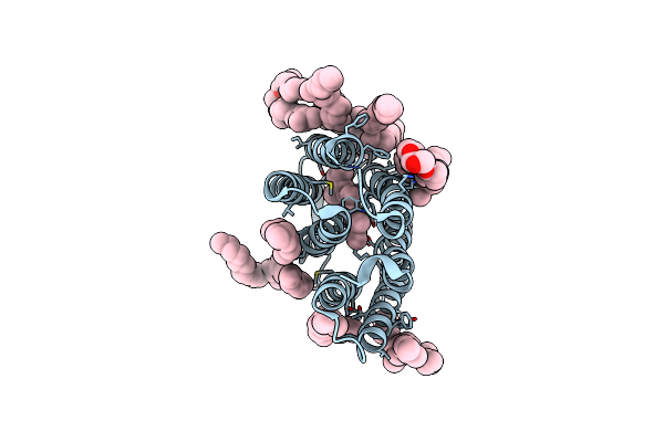
Structure Of A "Mock-Trapped" Early-M Intermediate Of Bacteriorhosopsin
Organism: Halobacterium salinarum
Method: X-RAY DIFFRACTION Resolution:1.81 Å Release Date: 2001-12-05 Classification: PROTON TRANSPORT Ligands: RET, LI1 |
Organism: Halobacterium salinarum
Method: X-RAY DIFFRACTION
Release Date: 2001-12-05
Ligands: RET, LI1
 |
Structure Of Ground-State Bacteriorhodopsin
Organism: Halobacterium salinarum
Method: X-RAY DIFFRACTION Resolution:1.65 Å Release Date: 2001-12-05 Classification: PROTON TRANSPORT Ligands: RET, LI1 |
Organism: Halobacterium salinarum
Method: X-RAY DIFFRACTION
Release Date: 2001-12-05
Ligands: RET, LI1