
Search Count: 73
 |
Organism: Streptococcus pneumoniae
Method: X-RAY DIFFRACTION Resolution:1.70 Å Release Date: 2026-05-27 Classification: HYDROLASE Ligands: EDO, CA |
 |
Organism: Streptococcus pneumoniae
Method: X-RAY DIFFRACTION Resolution:1.80 Å Release Date: 2026-05-27 Classification: HYDROLASE Ligands: EDO |
 |
Organism: Streptococcus pneumoniae
Method: X-RAY DIFFRACTION Resolution:1.57 Å Release Date: 2026-02-18 Classification: TRANSFERASE Ligands: A1JNO, CL, TAU |
 |
Organism: Streptococcus pneumoniae
Method: X-RAY DIFFRACTION Resolution:1.74 Å Release Date: 2026-02-18 Classification: TRANSFERASE Ligands: A1JNV, CL |
 |
Organism: Streptococcus pneumoniae
Method: X-RAY DIFFRACTION Resolution:1.53 Å Release Date: 2026-02-18 Classification: TRANSFERASE Ligands: A1JNN, CL, TAU |
 |
Organism: Streptococcus pneumoniae
Method: X-RAY DIFFRACTION Resolution:1.81 Å Release Date: 2026-02-18 Classification: TRANSFERASE Ligands: A1JNR, CL |
 |
Organism: Streptococcus pneumoniae
Method: X-RAY DIFFRACTION Resolution:1.88 Å Release Date: 2026-02-18 Classification: TRANSFERASE Ligands: A1JNW, SO3, CL |
 |
Organism: Streptococcus pneumoniae
Method: X-RAY DIFFRACTION Resolution:1.50 Å Release Date: 2026-02-18 Classification: TRANSFERASE Ligands: A1JNU, CL, TAU |
 |
Organism: Streptococcus pneumoniae
Method: X-RAY DIFFRACTION Resolution:1.52 Å Release Date: 2026-02-18 Classification: TRANSFERASE Ligands: A1JNQ, CL, TAU |
 |
Organism: Streptococcus pneumoniae
Method: X-RAY DIFFRACTION Resolution:1.51 Å Release Date: 2026-02-18 Classification: TRANSFERASE Ligands: A1JNP, CL, TAU |
 |
Organism: Streptococcus pneumoniae
Method: X-RAY DIFFRACTION Resolution:1.57 Å Release Date: 2026-02-18 Classification: TRANSFERASE Ligands: A1JNT, SO3, CL, TAU |
 |
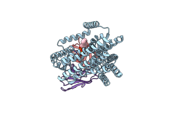
Penicillin-Binding Protein 1B (Pbp-1B) In Complex With A Monobactam (Aztreonam)
Organism: Streptococcus pneumoniae
Method: X-RAY DIFFRACTION Resolution:1.65 Å Release Date: 2026-02-18 Classification: TRANSFERASE Ligands: AZR, CL, TAU |
 |
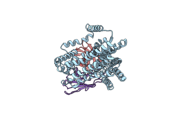
The Structure Of E. Coli Penicillin Binding Protein 3 (Pbp3) In Complex With A Bicyclic Peptide Inhibitor
Organism: Escherichia coli, Synthetic construct
Method: X-RAY DIFFRACTION Resolution:1.52 Å Release Date: 2024-04-03 Classification: HYDROLASE Ligands: 29N |
 |
Organism: Escherichia coli
Method: ELECTRON MICROSCOPY Release Date: 2023-08-30 Classification: MEMBRANE PROTEIN |
 |
Organism: Citrobacter rodentium
Method: X-RAY DIFFRACTION Resolution:3.38 Å Release Date: 2023-06-14 Classification: HYDROLASE Ligands: ZN, PO4 |
 |
Organism: Escherichia coli
Method: X-RAY DIFFRACTION Resolution:2.35 Å Release Date: 2023-06-14 Classification: HYDROLASE Ligands: ZN |
 |
Organism: Acinetobacter baumannii
Method: X-RAY DIFFRACTION Resolution:2.65 Å Release Date: 2023-02-22 Classification: HYDROLASE Ligands: ZN |
 |
Single-Particle Cryo-Em Structure Of The Waal O-Antigen Ligase In Its Ligand Bound State
Organism: Cupriavidus metallidurans, Homo sapiens
Method: ELECTRON MICROSCOPY Release Date: 2022-04-06 Classification: MEMBRANE PROTEIN Ligands: GPP |
 |
Single-Particle Cryo-Em Structure Of The Waal O-Antigen Ligase In Its Apo State
Organism: Cupriavidus metallidurans, Homo sapiens
Method: ELECTRON MICROSCOPY Release Date: 2022-04-06 Classification: MEMBRANE PROTEIN |
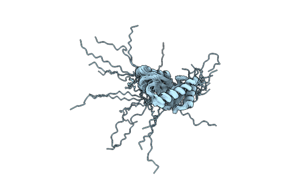 |
Structure-Function Analyses Of Dual-Bon Domain Protein Dolp Identifies Phospholipid Binding As A New Mechanism For Protein Localisation To The Cell Division Site
Organism: Escherichia coli (strain k12)
Method: SOLUTION NMR Release Date: 2020-12-30 Classification: PROTEIN BINDING |