

Search Count: 16
 |
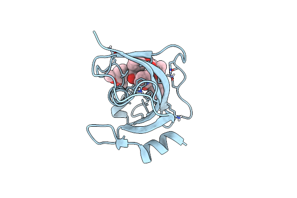
Structure Of The Fk1 Domain Of The Fkbp51 G64S Variant In Complex With Safit1
Organism: Homo sapiens
Method: X-RAY DIFFRACTION Resolution:1.10 Å Release Date: 2022-11-16 Classification: ISOMERASE Ligands: GY1, EPE, GOL |
Organism: Homo sapiens
Method: X-RAY DIFFRACTION
Release Date: 2022-11-16
Ligands: GY1, EPE, GOL
 |
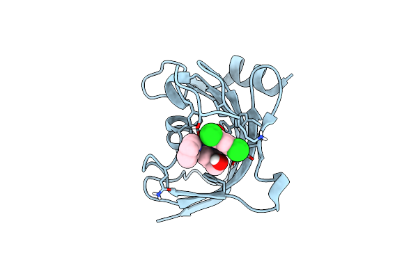
Structure Of The Fk1 Domain Of The Fkbp51 G64S Variant In Complex With (2R,5S,12R)-12-Cyclohexyl-2-[2-(3,4-Dimethoxyphenyl)Ethyl]-15,15,16-Trimethyl-3,19-Dioxa-10,13,16-Triazatricyclo[18.3.1.0^5,^10]Tetracosa-1(24),20,22-Triene-4,11,14,17-Tetrone
Organism: Homo sapiens
Method: X-RAY DIFFRACTION Resolution:1.10 Å Release Date: 2022-11-16 Classification: ISOMERASE Ligands: S8N |
Organism: Homo sapiens
Method: X-RAY DIFFRACTION
Release Date: 2022-11-16
Ligands: S8N
 |
Structure Of The Fk1 Domain Of The Fkbp51 G64S Variant In Complex With (1S,5S,6R)-10-((3,5-Dichlorophenyl)Sulfonyl)-5-(Hydroxymethyl)-3-(Pyridin-2-Ylmethyl)-3,10-Diazabicyclo[4.3.1]Decan-2-One
Organism: Homo sapiens
Method: X-RAY DIFFRACTION Resolution:1.20 Å Release Date: 2022-11-16 Classification: ISOMERASE Ligands: 9QN |
Organism: Homo sapiens
Method: X-RAY DIFFRACTION
Release Date: 2022-11-16
Ligands: 9QN
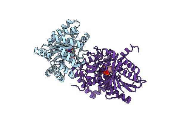 |
Crystal Structure Of Tryptophan Synthase In Complex With F9, Nh4+, Ph7.8 - Alpha Aminoacrylate Form - E(A-A)
Organism: Salmonella enterica subsp. enterica serovar typhimurium
Method: X-RAY DIFFRACTION Resolution:1.40 Å Release Date: 2021-12-29 Classification: LYASE/INHIBITOR Ligands: F9F, 0JO, NH4 |
Organism: Salmonella enterica subsp. enterica serovar typhimurium
Method: X-RAY DIFFRACTION
Release Date: 2021-12-29
Ligands: F9F, 0JO, NH4
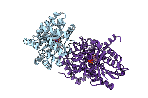 |
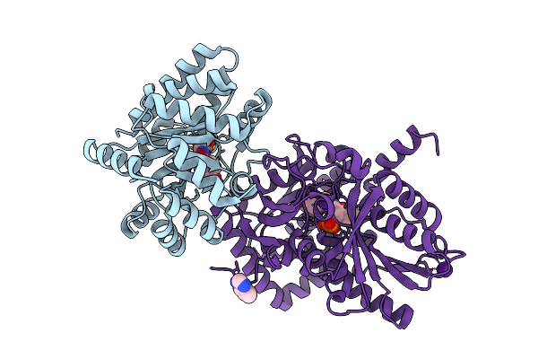
Crystal Structure Of Tryptophan Synthase In Complex With F9, Cs+, Ph7.8 - Alpha Aminoacrylate Form - E(A-A)
Organism: Salmonella typhimurium
Method: X-RAY DIFFRACTION Resolution:1.50 Å Release Date: 2021-12-29 Classification: LYASE/INHIBITOR Ligands: F9F, 0JO, CS |
Organism: Salmonella typhimurium
Method: X-RAY DIFFRACTION
Release Date: 2021-12-29
Ligands: F9F, 0JO, CS
 |
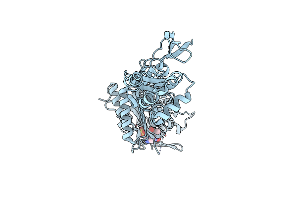
Crystal Structure Of Tryptophan Synthase In Complex With F9, Cs+, Benzimidazole, Ph7.8 - Alpha Aminoacrylate Form - E(A-A)(Bzi)
Organism: Salmonella typhimurium, Salmonella enterica subsp. enterica serovar typhimurium
Method: X-RAY DIFFRACTION Resolution:1.70 Å Release Date: 2021-12-29 Classification: LYASE/INHIBITOR Ligands: F9F, 0JO, CS, BZI |
Organism: Salmonella typhimurium, Salmonella enterica subsp. enterica serovar typhimurium
Method: X-RAY DIFFRACTION
Release Date: 2021-12-29
Ligands: F9F, 0JO, CS, BZI
 |
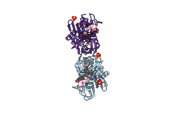
Crystal Structure Of Pseudomonas Aeruginosa Penicillin Binding Protein 3 (Pae-Pbp3) Bound To Etx0462
Organism: Pseudomonas aeruginosa
Method: X-RAY DIFFRACTION Resolution:2.20 Å Release Date: 2021-05-26 Classification: ANTIBIOTIC Ligands: CL, VMM |
Organism: Pseudomonas aeruginosa
Method: X-RAY DIFFRACTION
Release Date: 2021-05-26
Ligands: CL, VMM
 |
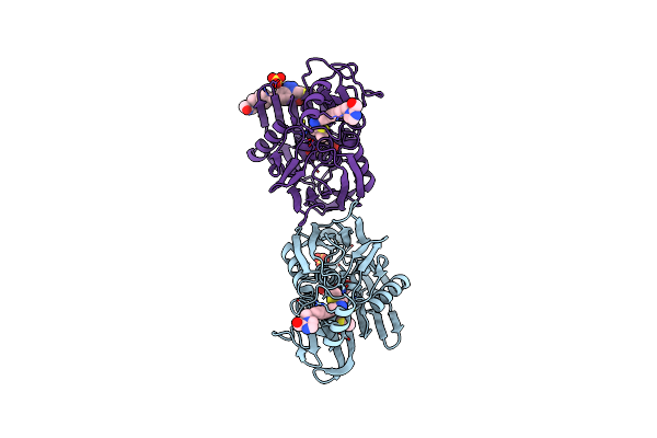
Crystal Structure Of P. Aeruginosa Lpxc With N-Hydroxyformamide Inhibitor 19
Organism: Pseudomonas aeruginosa (strain atcc 15692 / dsm 22644 / cip 104116 / jcm 14847 / lmg 12228 / 1c / prs 101 / pao1)
Method: X-RAY DIFFRACTION Resolution:1.90 Å Release Date: 2020-11-25 Classification: HYDROLASE Ligands: GOL, W4M, SO4, ZN |
Organism: Pseudomonas aeruginosa (strain atcc 15692 / dsm 22644 / cip 104116 / jcm 14847 / lmg 12228 / 1c / prs 101 / pao1)
Method: X-RAY DIFFRACTION
Release Date: 2020-11-25
Ligands: GOL, W4M, SO4, ZN
 |
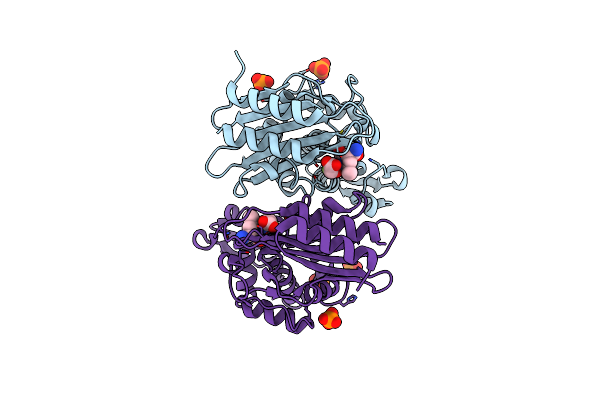
Crystal Structure Of P. Aeruginosa Lpxc With N-Hydroxyformamide Inhibitor
Organism: Pseudomonas aeruginosa (strain atcc 15692 / dsm 22644 / cip 104116 / jcm 14847 / lmg 12228 / 1c / prs 101 / pao1)
Method: X-RAY DIFFRACTION Resolution:2.00 Å Release Date: 2020-11-25 Classification: HYDROLASE Ligands: W4P, SO4, GOL, DMS, ZN, W8P |
Organism: Pseudomonas aeruginosa (strain atcc 15692 / dsm 22644 / cip 104116 / jcm 14847 / lmg 12228 / 1c / prs 101 / pao1)
Method: X-RAY DIFFRACTION
Release Date: 2020-11-25
Ligands: W4P, SO4, GOL, DMS, ZN, W8P
 |
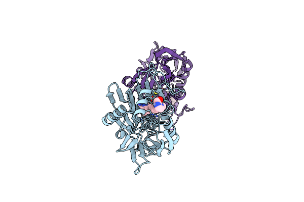
Crystal Structure Of Ctx-M-14 In Complex With Beta-Lactamase Inhibitor Etx1317
Organism: Escherichia coli
Method: X-RAY DIFFRACTION Resolution:1.28 Å Release Date: 2020-08-26 Classification: HYDROLASE Ligands: X57, PO4 |
Organism: Escherichia coli
Method: X-RAY DIFFRACTION
Release Date: 2020-08-26
Ligands: X57, PO4
 |
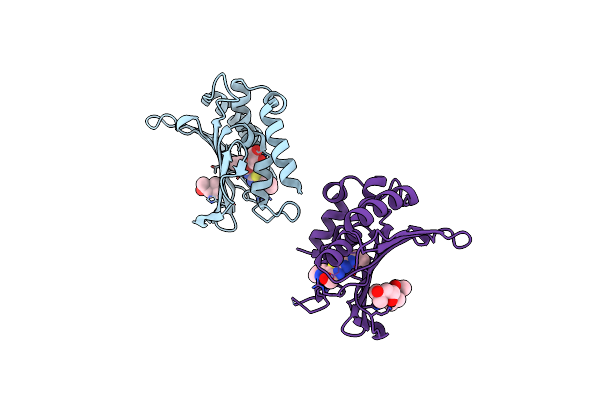
Crystal Structure Of Pseudomonas Aeruginosa Lpxc Complexed With Inhibitor
Organism: Pseudomonas aeruginosa
Method: X-RAY DIFFRACTION Resolution:1.90 Å Release Date: 2017-03-29 Classification: HYDROLASE Ligands: ZN, 8Q8, CL |
Organism: Pseudomonas aeruginosa
Method: X-RAY DIFFRACTION
Release Date: 2017-03-29
Ligands: ZN, 8Q8, CL
 |
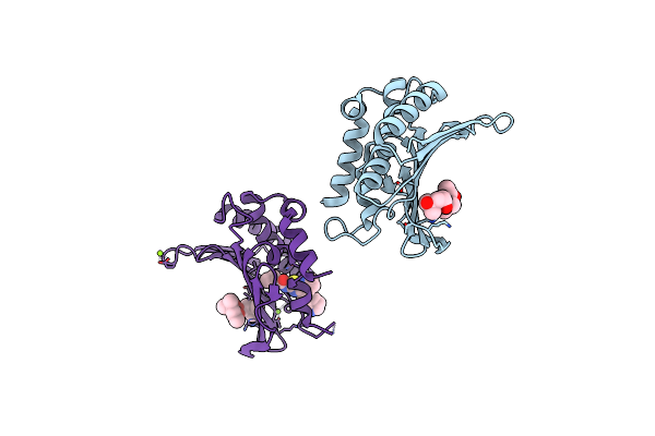
Crystal Structure Of The Atp Binding Domain Of S. Aureus Gyrb Complexed With A Ligand
Organism: Staphylococcus aureus
Method: X-RAY DIFFRACTION Resolution:2.05 Å Release Date: 2015-11-25 Classification: ISOMERASE/ISOMERASE INHIBITOR Ligands: 57U, MPD, GOL |
Organism: Staphylococcus aureus
Method: X-RAY DIFFRACTION
Release Date: 2015-11-25
Ligands: 57U, MPD, GOL
 |
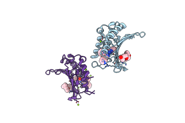
Crystal Structure Of The Atp Binding Domain Of S. Aureus Gyrb Complexed With A Ligand
Organism: Staphylococcus aureus
Method: X-RAY DIFFRACTION Resolution:1.50 Å Release Date: 2015-11-25 Classification: ISOMERASE/ISOMERASE INHIBITOR Ligands: MPD, 57V, MG |
Organism: Staphylococcus aureus
Method: X-RAY DIFFRACTION
Release Date: 2015-11-25
Ligands: MPD, 57V, MG
 |
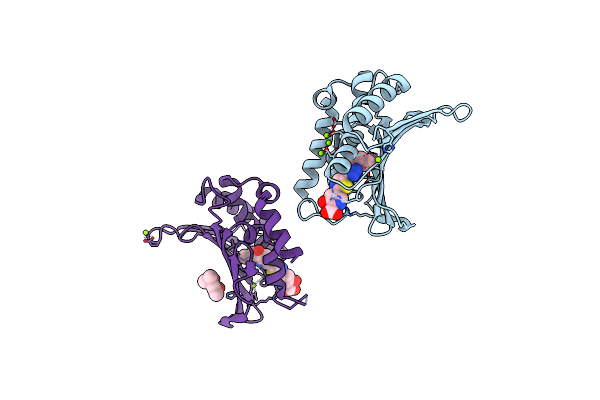
Crystal Structure Of The Atp Binding Domain Of S. Aureus Gyrb Complexed With A Ligand
Organism: Staphylococcus aureus
Method: X-RAY DIFFRACTION Resolution:1.55 Å Release Date: 2015-11-25 Classification: ISOMERASE/ISOMERASE INHIBITOR Ligands: MPD, 57W, MG |
Organism: Staphylococcus aureus
Method: X-RAY DIFFRACTION
Release Date: 2015-11-25
Ligands: MPD, 57W, MG
 |
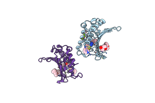
Crystal Structure Of The Atp Binding Domain Of S. Aureus Gyrb Complexed With A Ligand
Organism: Staphylococcus aureus
Method: X-RAY DIFFRACTION Resolution:1.55 Å Release Date: 2015-11-18 Classification: ISOMERASE/ISOMERASE INHIBITOR Ligands: MG, CL, 57Y, MPD |
Organism: Staphylococcus aureus
Method: X-RAY DIFFRACTION
Release Date: 2015-11-18
Ligands: MG, CL, 57Y, MPD
 |
Crystal Structure Of The Atp Binding Domain Of S. Aureus Gyrb Complexed With A Ligand
Organism: Staphylococcus aureus
Method: X-RAY DIFFRACTION Resolution:1.60 Å Release Date: 2015-11-11 Classification: ISOMERASE/ISOMERASE INHIBITOR Ligands: MPD, CL, MG, 57X |
Organism: Staphylococcus aureus
Method: X-RAY DIFFRACTION
Release Date: 2015-11-11
Ligands: MPD, CL, MG, 57X