

Search Count: 20
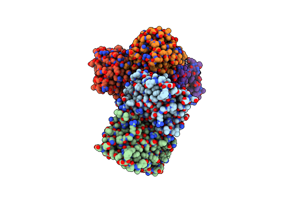 |
Structure Of Inha From Mycobacterium Tuberculosis In Complex With 5-(((4-(2-Hydroxyphenoxy)Benzyl)(Octyl)Amino)Methyl)-2-Phenoxyphenol
Organism: Mycobacterium tuberculosis
Method: X-RAY DIFFRACTION Resolution:2.11 Å Release Date: 2024-01-24 Classification: OXIDOREDUCTASE Ligands: NAD, ACT, EDO, VZE |
Organism: Mycobacterium tuberculosis
Method: X-RAY DIFFRACTION
Release Date: 2024-01-24
Ligands: NAD, ACT, EDO, VZE
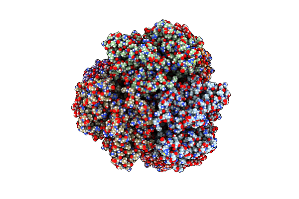 |
Cryo-Em Structure Of The Cibeles-Demetra 3:3 Heterocomplex From Galleria Mellonella Saliva
Organism: Galleria mellonella
Method: ELECTRON MICROSCOPY Release Date: 2023-10-04 Classification: UNKNOWN FUNCTION Ligands: CU |
Organism: Galleria mellonella
Method: ELECTRON MICROSCOPY
Release Date: 2023-10-04
Ligands: CU
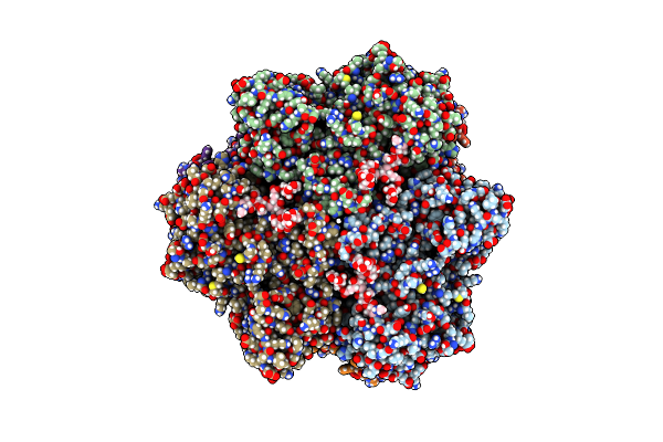 |
Cryo-Em Structure Of The Ceres Homohexamer From Galleria Mellonella Saliva
Organism: Galleria mellonella
Method: ELECTRON MICROSCOPY Release Date: 2023-10-04 Classification: UNKNOWN FUNCTION Ligands: CU |
Organism: Galleria mellonella
Method: ELECTRON MICROSCOPY
Release Date: 2023-10-04
Ligands: CU
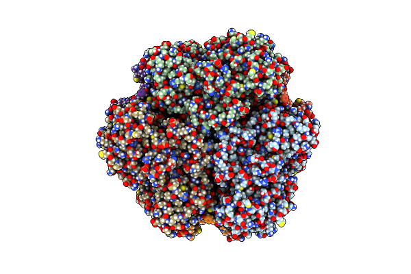 |
Cryo-Em Structure Of The Cora Homohexamer From Galleria Mellonella Saliva
Organism: Galleria mellonella
Method: ELECTRON MICROSCOPY Release Date: 2023-10-04 Classification: UNKNOWN FUNCTION Ligands: TRP, CU, NA |
Organism: Galleria mellonella
Method: ELECTRON MICROSCOPY
Release Date: 2023-10-04
Ligands: TRP, CU, NA
 |
Polyethylene Oxidation Hexamerin Pease Cibeles (Xp_026756460) From Galleria Mellonella
Organism: Galleria mellonella
Method: X-RAY DIFFRACTION Resolution:2.20 Å Release Date: 2023-10-04 Classification: OXIDOREDUCTASE Ligands: CU, MG, CL, MES, MPD, GOL, GLY, SO4 |
Organism: Galleria mellonella
Method: X-RAY DIFFRACTION
Release Date: 2023-10-04
Ligands: CU, MG, CL, MES, MPD, GOL, GLY, SO4
 |
Structure Of Inha From Mycobacterium Tuberculosis In Complex With N-((1-(3-Hydroxy-4-Phenoxybenzyl)-1H-1,2,3-Triazol-4-Yl)Methyl)-2-Oxo-2H-Chromene-3-Carboxamide
Organism: Mycobacterium tuberculosis
Method: X-RAY DIFFRACTION Resolution:1.60 Å Release Date: 2023-08-16 Classification: OXIDOREDUCTASE Ligands: NAD, VZI, EDO, NA, ACT |
Organism: Mycobacterium tuberculosis
Method: X-RAY DIFFRACTION
Release Date: 2023-08-16
Ligands: NAD, VZI, EDO, NA, ACT
 |
Structure Of Inha From Mycobacterium Tuberculosis In Complex With Inhibitor 7-((1-(3-Hydroxy-4-Phenoxybenzyl)-1H-1,2,3-Triazol-4-Yl)Methoxy)-4-Methyl-2H-Chromen-2-One
Organism: Mycobacterium tuberculosis
Method: X-RAY DIFFRACTION Resolution:1.96 Å Release Date: 2023-08-16 Classification: OXIDOREDUCTASE Ligands: NAD, VZR |
Organism: Mycobacterium tuberculosis
Method: X-RAY DIFFRACTION
Release Date: 2023-08-16
Ligands: NAD, VZR
 |
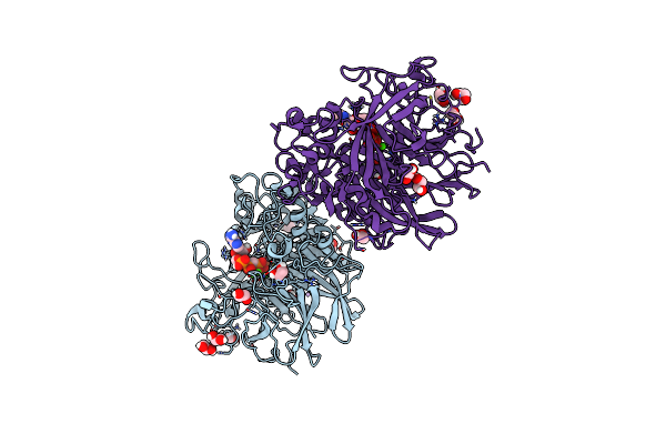
X-Ray Structure Of A Multidomain Bbgiii From Bifidobacterium Bifidum
Organism: Bifidobacterium bifidum
Method: X-RAY DIFFRACTION Resolution:2.89 Å Release Date: 2021-12-22 Classification: HYDROLASE Ligands: SO4, GOL, CA |
Organism: Bifidobacterium bifidum
Method: X-RAY DIFFRACTION
Release Date: 2021-12-22
Ligands: SO4, GOL, CA
 |
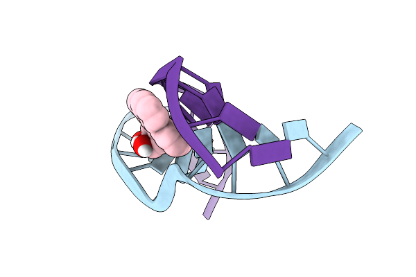
Nmr Studies Of N2-Guanine Adducts Derived From The Tumorigen Dibenzo[A,L]Pyrene In Dna: Impact Of Adduct Stereochemistry, Size, And Local Dna Structure On Solution Conformations
|
Method: SOLUTION NMR
Release Date: 2014-04-02
Ligands: 2QL
 |
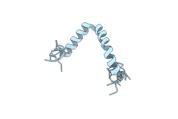
Nuclear Magnetic Resonance Studies Of N2-Guanine Adducts Derived From The Tumorigen Dibenzo[A,L]Pyrene In Dna: Impact Of Adduct Stereochemistry, Size, And Local Dna Structure On Solution Conformations
|
Method: SOLUTION NMR
Release Date: 2014-04-02
Ligands: 2QL
 |
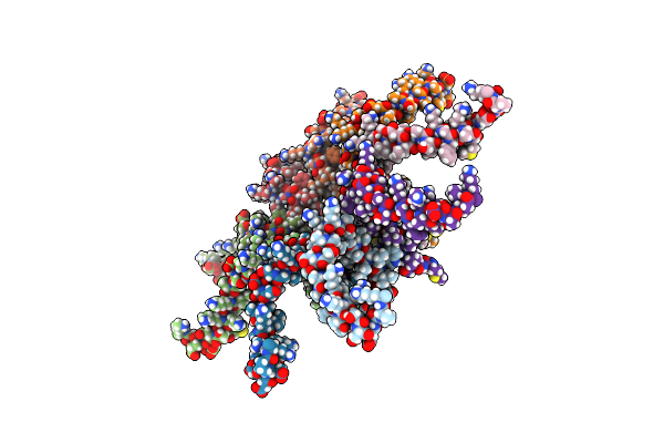
Tata T22P
Organism: Escherichia coli
Method: SOLUTION NMR Release Date: 2013-03-20 Classification: PROTEIN TRANSPORT |
Organism: Escherichia coli
Method: SOLUTION NMR
Release Date: 2013-03-20
 |
Tata Oligomer
Organism: Escherichia coli
Method: SOLUTION NMR Release Date: 2013-03-20 Classification: PROTEIN TRANSPORT |
Organism: Escherichia coli
Method: SOLUTION NMR
Release Date: 2013-03-20
 |
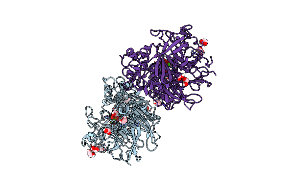
Pseudomonas Fluorescens Phox In Complex With The Substrate Analogue Appcp
Organism: Pseudomonas fluorescens
Method: X-RAY DIFFRACTION Resolution:1.52 Å Release Date: 2013-03-20 Classification: HYDROLASE Ligands: ACP, CL, LI, CA, FEO, EDO |
Organism: Pseudomonas fluorescens
Method: X-RAY DIFFRACTION
Release Date: 2013-03-20
Ligands: ACP, CL, LI, CA, FEO, EDO
 |
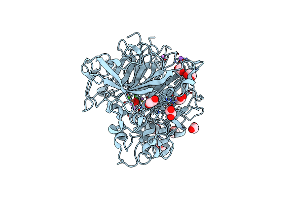
Pseudomonas Fluorescens Phox In Complex With Phosphate
Organism: Pseudomonas fluorescens
Method: X-RAY DIFFRACTION Resolution:1.25 Å Release Date: 2013-03-13 Classification: HYDROLASE Ligands: PO4, CA, FEO, EDO, CL |
Organism: Pseudomonas fluorescens
Method: X-RAY DIFFRACTION
Release Date: 2013-03-13
Ligands: PO4, CA, FEO, EDO, CL
 |
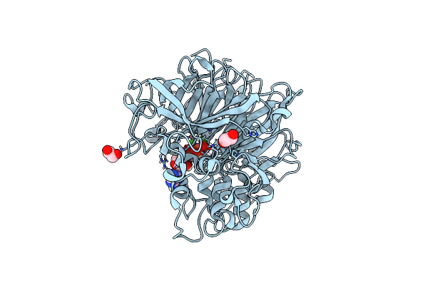
Pseudomonas Fluorescens Phox
Organism: Pseudomonas fluorescens
Method: X-RAY DIFFRACTION Resolution:1.10 Å Release Date: 2012-12-05 Classification: HYDROLASE Ligands: CL, LI, NA, EDO, CA, FEO |
Organism: Pseudomonas fluorescens
Method: X-RAY DIFFRACTION
Release Date: 2012-12-05
Ligands: CL, LI, NA, EDO, CA, FEO
 |
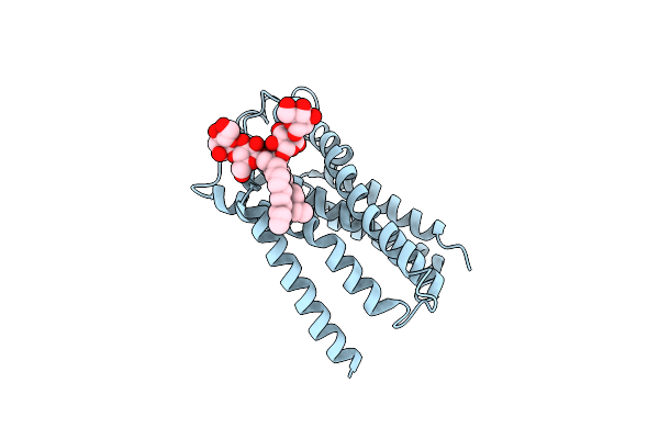
Pseudomonas Fluorescens Phox In Complex With The Substrate Analogue Appcp
Organism: Pseudomonas fluorescens
Method: X-RAY DIFFRACTION Resolution:1.79 Å Release Date: 2012-12-05 Classification: HYDROLASE Ligands: EDO, ACP, CA, FEO |
Organism: Pseudomonas fluorescens
Method: X-RAY DIFFRACTION
Release Date: 2012-12-05
Ligands: EDO, ACP, CA, FEO
 |
Structure Of The Tatc Core Of The Twin Arginine Protein Translocation System
Organism: Aquifex aeolicus
Method: X-RAY DIFFRACTION Resolution:3.50 Å Release Date: 2012-12-05 Classification: TRANSPORT PROTEIN Ligands: LMN |
Organism: Aquifex aeolicus
Method: X-RAY DIFFRACTION
Release Date: 2012-12-05
Ligands: LMN
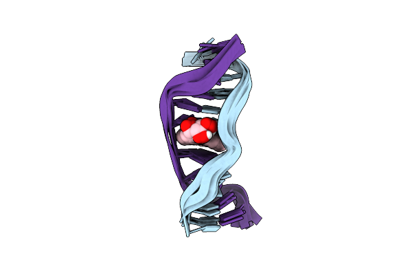 |
Nmr Solution Structure Of An N2-Guanine Dna Adduct Derived From The Potent Tumorigen Dibenzo[A,L]Pyrene: Intercalation From The Minor Groove With Ruptured Watson-Crick Base Pairing
|
Method: SOLUTION NMR
Release Date: 2012-11-21
Ligands: 14L
 |
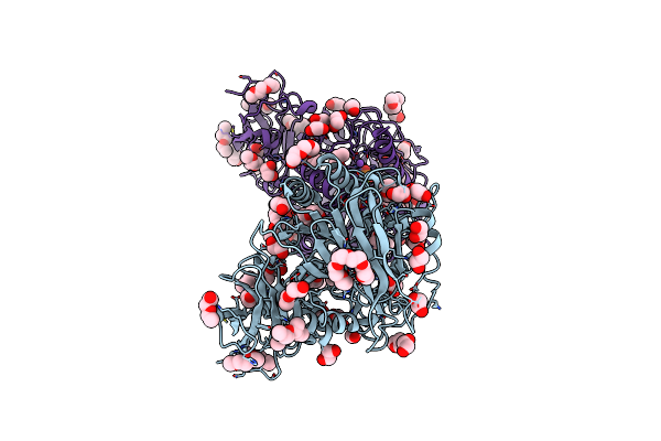
Pseudomonas Fluorescens Phox In Complex With Vanadate, A Transition State Analogue
Organism: Pseudomonas fluorescens
Method: X-RAY DIFFRACTION Resolution:1.39 Å Release Date: 2012-08-08 Classification: HYDROLASE Ligands: VO4, CL, CA, FEO |
Organism: Pseudomonas fluorescens
Method: X-RAY DIFFRACTION
Release Date: 2012-08-08
Ligands: VO4, CL, CA, FEO
 |
Structure Of Phod
Organism: Bacillus subtilis
Method: X-RAY DIFFRACTION Resolution:1.93 Å Release Date: 2012-04-18 Classification: HYDROLASE Ligands: PE5, EDO, NA, FE, CA, PO4 |
Organism: Bacillus subtilis
Method: X-RAY DIFFRACTION
Release Date: 2012-04-18
Ligands: PE5, EDO, NA, FE, CA, PO4