

Search Count: 15
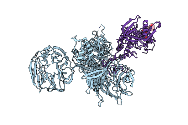 |
Cereblon In Complex With Ddb1 And Cc-220
Organism: Homo sapiens
Method: X-RAY DIFFRACTION Resolution:3.20 Å Release Date: 2017-05-03 Classification: LIGASE Ligands: 8W7, ZN |
Organism: Homo sapiens
Method: X-RAY DIFFRACTION
Release Date: 2017-05-03
Ligands: 8W7, ZN
 |
Cereblon In Complex With Ddb1, Cc-885, And Gspt1
Organism: Homo sapiens
Method: X-RAY DIFFRACTION Resolution:3.60 Å Release Date: 2016-06-29 Classification: LIGASE Ligands: ZN, 85C |
Organism: Homo sapiens
Method: X-RAY DIFFRACTION
Release Date: 2016-06-29
Ligands: ZN, 85C
 |
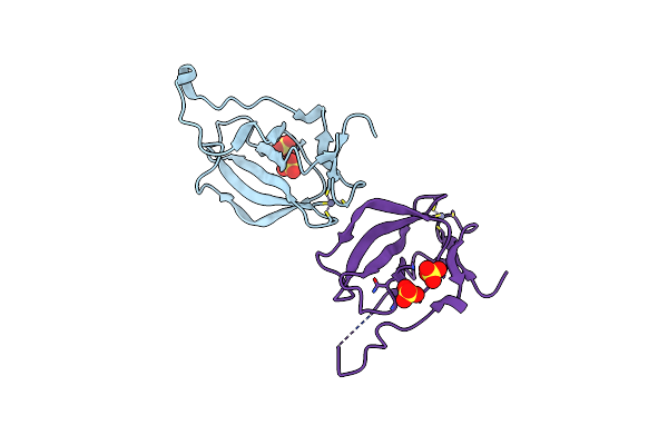
Mouse Cereblon Thalidomide Binding Domain, Selenomethionine Derivative
Organism: Mus musculus
Method: X-RAY DIFFRACTION Resolution:1.93 Å Release Date: 2014-08-06 Classification: METAL BINDING PROTEIN Ligands: ZN, SO4 |
Organism: Mus musculus
Method: X-RAY DIFFRACTION
Release Date: 2014-08-06
Ligands: ZN, SO4
 |
Mouse Cereblon Thalidomide Binding Domain, Native
Organism: Mus musculus
Method: X-RAY DIFFRACTION Resolution:2.00 Å Release Date: 2014-08-06 Classification: METAL BINDING PROTEIN Ligands: ZN, SO4 |
Organism: Mus musculus
Method: X-RAY DIFFRACTION
Release Date: 2014-08-06
Ligands: ZN, SO4
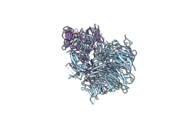 |
Crystal Structure Of Human Cereblon In Complex With Ddb1 And Lenalidomide
Organism: Homo sapiens
Method: X-RAY DIFFRACTION Resolution:3.01 Å Release Date: 2014-08-06 Classification: DNA Binding Protein/Ligase Ligands: ZN, LVY |
Organism: Homo sapiens
Method: X-RAY DIFFRACTION
Release Date: 2014-08-06
Ligands: ZN, LVY
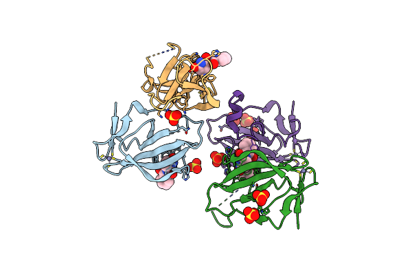 |
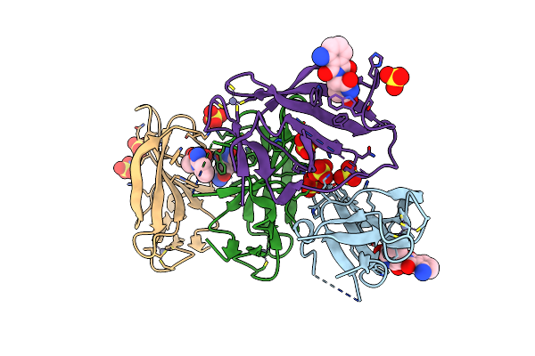
Crystal Structure Of Murine Cereblon In Complex With Thalidomide
Organism: Mus musculus
Method: X-RAY DIFFRACTION Resolution:1.88 Å Release Date: 2014-08-06 Classification: LIGASE Ligands: ZN, EF2, SO4 |
Organism: Mus musculus
Method: X-RAY DIFFRACTION
Release Date: 2014-08-06
Ligands: ZN, EF2, SO4
 |
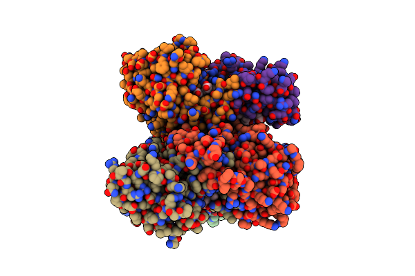
Crystal Structure Of Murine Cereblon In Complex With Pomalidomide
Organism: Mus musculus
Method: X-RAY DIFFRACTION Resolution:2.00 Å Release Date: 2014-08-06 Classification: LIGASE Ligands: SO4, ZN, Y70 |
Organism: Mus musculus
Method: X-RAY DIFFRACTION
Release Date: 2014-08-06
Ligands: SO4, ZN, Y70
 |
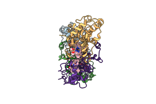
Diheme Soxax
Organism: Starkeya novella
Method: X-RAY DIFFRACTION Resolution:1.77 Å Release Date: 2011-05-18 Classification: Heme-binding protein/Heme-binding protein Ligands: HEC, SO4 |
Organism: Starkeya novella
Method: X-RAY DIFFRACTION
Release Date: 2011-05-18
Ligands: HEC, SO4
 |
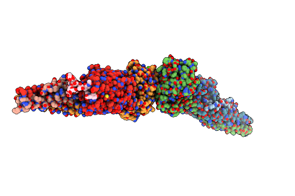
Diheme Soxax - C236M Mutant
Organism: Starkeya novella
Method: X-RAY DIFFRACTION Resolution:2.25 Å Release Date: 2011-05-18 Classification: Heme-binding protein Ligands: HEC |
Organism: Starkeya novella
Method: X-RAY DIFFRACTION
Release Date: 2011-05-18
Ligands: HEC
 |
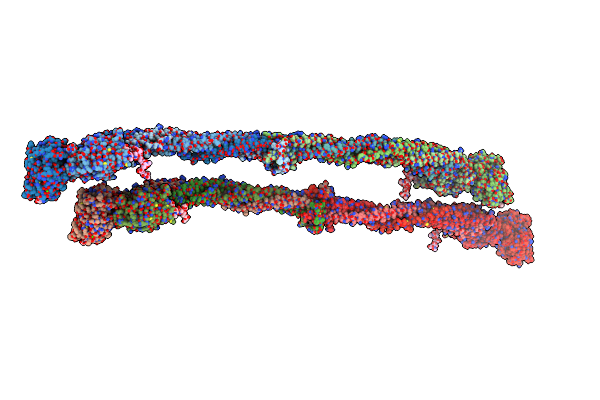
Crystal Structure Of D-Dimer From Human Fibrin Complexed With Gly-His-Arg-Pro-Tyr-Amide
Organism: Homo sapiens
Method: X-RAY DIFFRACTION Resolution:3.60 Å Release Date: 2009-07-28 Classification: BLOOD CLOTTING Ligands: CA |
Organism: Homo sapiens
Method: X-RAY DIFFRACTION
Release Date: 2009-07-28
Ligands: CA
 |
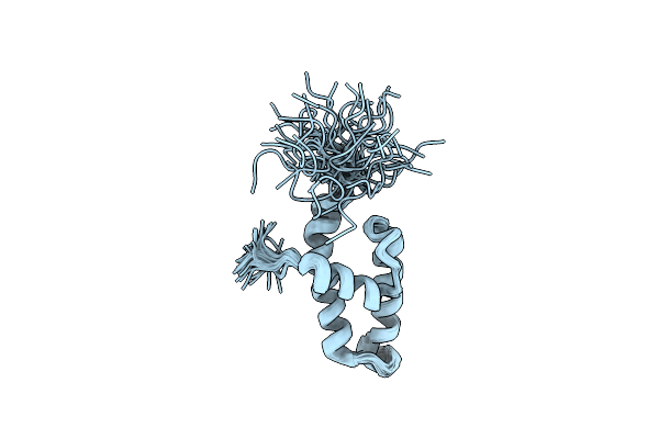
Crystal Structure Of Human Fibrinogen
Organism: Homo sapiens
Method: X-RAY DIFFRACTION Resolution:2.90 Å Release Date: 2009-05-19 Classification: BLOOD CLOTTING Ligands: CA |
Organism: Homo sapiens
Method: X-RAY DIFFRACTION
Release Date: 2009-05-19
Ligands: CA
 |
Crystal Structure Of Fragment D From Lamprey Fibrinogen Complexed With The Peptide Gly-His-Arg-Pro-Amide
Organism: Petromyzon marinus
Method: X-RAY DIFFRACTION Resolution:2.80 Å Release Date: 2002-08-23 Classification: BLOOD CLOTTING Ligands: NDG, MAN, CA, NAG, BMA, GAL |
Organism: Petromyzon marinus
Method: X-RAY DIFFRACTION
Release Date: 2002-08-23
Ligands: NDG, MAN, CA, NAG, BMA, GAL
 |
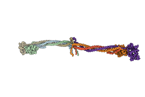
Nmr Structure Of The N-Terminal J Domain Of Murine Polyomavirus T Antigens.
Organism: Murine polyomavirus
Method: SOLUTION NMR Release Date: 2000-11-22 Classification: VIRAL PROTEIN |
Organism: Murine polyomavirus
Method: SOLUTION NMR
Release Date: 2000-11-22
 |
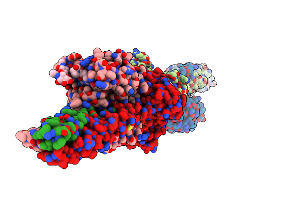
Crystal Structure Of Native Chicken Fibrinogen
Organism: Gallus gallus
Method: X-RAY DIFFRACTION Resolution:5.50 Å Release Date: 2000-05-10 Classification: BLOOD CLOTTING |
Organism: Gallus gallus
Method: X-RAY DIFFRACTION
Release Date: 2000-05-10
 |
Crystal Structure Of Fragment Double-D From Human Fibrin With Two Different Bound Ligands
Organism: Homo sapiens
Method: X-RAY DIFFRACTION Resolution:2.30 Å Release Date: 1998-10-14 Classification: BLOOD COAGULATION Ligands: MAN, CA, NAG |
Organism: Homo sapiens
Method: X-RAY DIFFRACTION
Release Date: 1998-10-14
Ligands: MAN, CA, NAG