

Search Count: 15
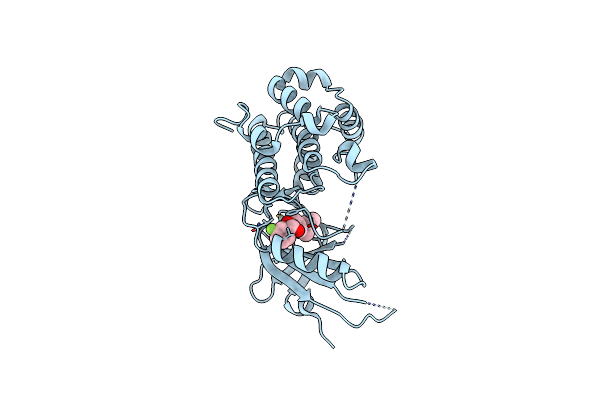 |
Co-Crystal Structure Of Perk With Compound 11
Organism: Homo sapiens
Method: X-RAY DIFFRACTION Resolution:2.86 Å Release Date: 2022-11-30 Classification: TRANSFERASE/Inhibitor Ligands: WPB |
Organism: Homo sapiens
Method: X-RAY DIFFRACTION
Release Date: 2022-11-30
Ligands: WPB
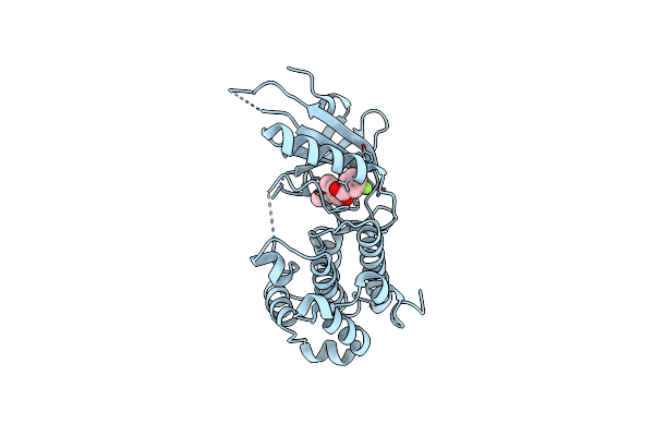 |
Co-Crystal Structure Of Perk With Compound 24
Organism: Homo sapiens
Method: X-RAY DIFFRACTION Resolution:2.92 Å Release Date: 2022-11-30 Classification: TRANSFERASE Ligands: WPX |
Organism: Homo sapiens
Method: X-RAY DIFFRACTION
Release Date: 2022-11-30
Ligands: WPX
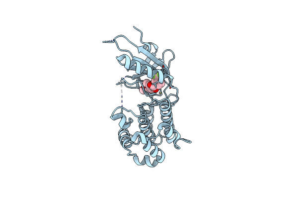 |
Co-Crystal Structure Of Perk With Compound 26
Organism: Homo sapiens
Method: X-RAY DIFFRACTION Resolution:2.56 Å Release Date: 2022-11-30 Classification: TRANSFERASE Ligands: WQ2 |
Organism: Homo sapiens
Method: X-RAY DIFFRACTION
Release Date: 2022-11-30
Ligands: WQ2
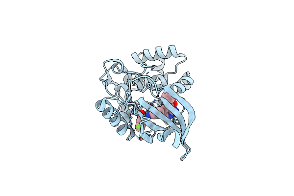 |
Co-Crystal Structure Of Perk With Inhibitor (R)-2-Amino-N-Cyclopropyl-5-(4-(2-(3,5-Difluorophenyl)-2-Hydroxyacetamido)-2-Methylphenyl)Nicotinamide
Organism: Homo sapiens
Method: X-RAY DIFFRACTION Resolution:2.81 Å Release Date: 2021-05-19 Classification: TRANSFERASE Ligands: Z6P |
Organism: Homo sapiens
Method: X-RAY DIFFRACTION
Release Date: 2021-05-19
Ligands: Z6P
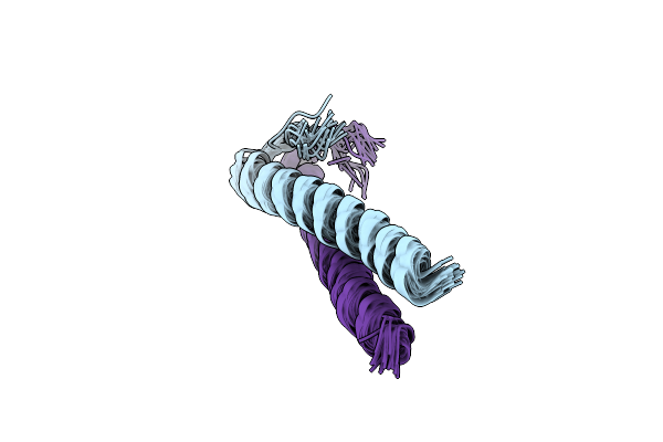 |
Solution Structure Of Coiled Coil Domain Of Myosin Binding Subunit Of Myosin Light Chain Phosphatase
Organism: Homo sapiens
Method: SOLUTION NMR Release Date: 2016-03-02 Classification: SIGNALING PROTEIN |
Organism: Homo sapiens
Method: SOLUTION NMR
Release Date: 2016-03-02
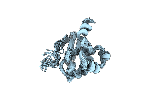 |
Solution Structure Of Stas Domain Of Rv1739C From M. Tuberculosis
Organism: Mycobacterium bovis
Method: SOLUTION NMR Release Date: 2010-12-15 Classification: TRANSPORT PROTEIN |
Organism: Mycobacterium bovis
Method: SOLUTION NMR
Release Date: 2010-12-15
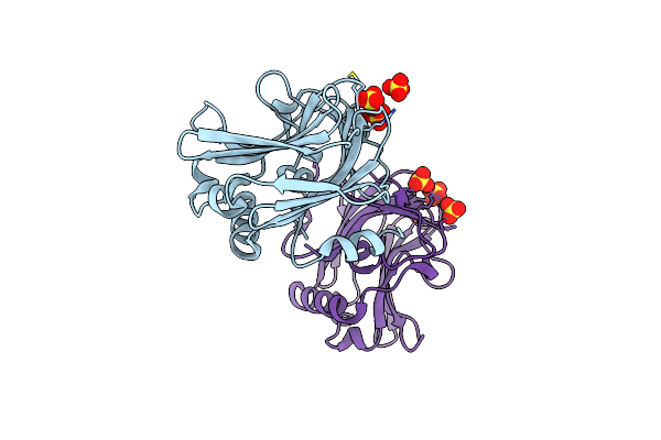 |
Crystal Structure Of The Thrombospondin-1 N-Terminal Domain In Complex With Fractionated Heparin Dp10
Organism: Homo sapiens
Method: X-RAY DIFFRACTION Resolution:2.40 Å Release Date: 2008-01-08 Classification: CELL ADHESION Ligands: SO4 |
Organism: Homo sapiens
Method: X-RAY DIFFRACTION
Release Date: 2008-01-08
Ligands: SO4
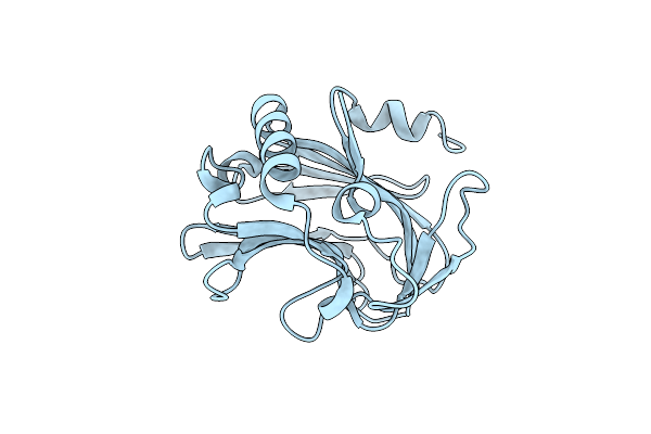 |
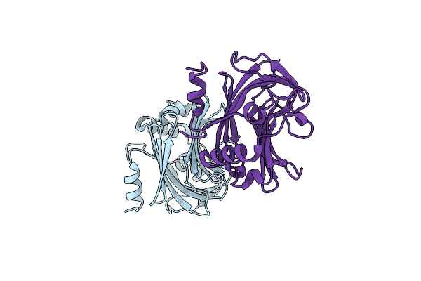
The Crystal Structure Of The Thrombospondin-1 N-Terminal Domain In Complex With Fractionated Heparin Dp8
Organism: Homo sapiens
Method: X-RAY DIFFRACTION Resolution:1.90 Å Release Date: 2008-01-08 Classification: CELL ADHESION |
Organism: Homo sapiens
Method: X-RAY DIFFRACTION
Release Date: 2008-01-08
 |
Crystal Structure Of Thrombospondin-1 N-Terminal Domain In P1 Form At 1.85A Resolution
Organism: Homo sapiens
Method: X-RAY DIFFRACTION Resolution:1.85 Å Release Date: 2006-10-31 Classification: SUGAR BINDING PROTEIN |
Organism: Homo sapiens
Method: X-RAY DIFFRACTION
Release Date: 2006-10-31
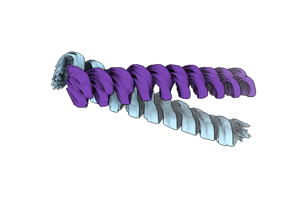 |
Solution Structure Of The Coiled-Coil Domain Of Cgmp-Dependent Protein Kinase Ia
|
Organism: Homo sapiens
Method: SOLUTION NMR
Release Date: 2005-09-13
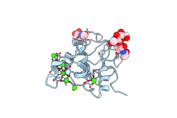 |
Bovine Prothrombin Fragment 1 In Complex With Calcium Ion
Organism: Bos taurus
Method: X-RAY DIFFRACTION Resolution:1.90 Å Release Date: 2003-09-16 Classification: HYDROLASE Ligands: NAG, CA |
Organism: Bos taurus
Method: X-RAY DIFFRACTION
Release Date: 2003-09-16
Ligands: NAG, CA
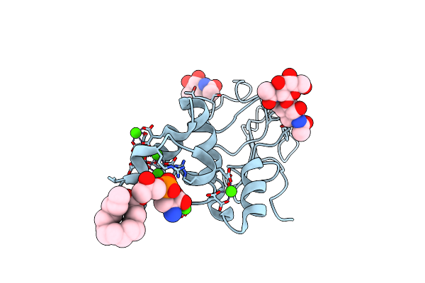 |
Bovine Prothrombin Fragment 1 In Complex With Calcium And Lysophosphotidylserine
Organism: Bos taurus
Method: X-RAY DIFFRACTION Resolution:2.30 Å Release Date: 2003-09-16 Classification: HYDROLASE Ligands: NAG, CA, CL, LPS |
Organism: Bos taurus
Method: X-RAY DIFFRACTION
Release Date: 2003-09-16
Ligands: NAG, CA, CL, LPS
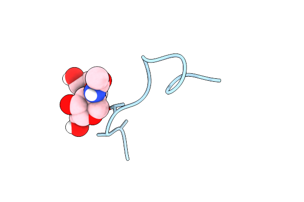 |
A Novel Conotoxin From Conus Textile With Unusual Post-Translational Modifications Reduces Presynaptic Calcium Influx, Nmr, 1 Structure, Glycosylated Protein
Organism: Conus textile
Method: SOLUTION NMR Release Date: 1999-06-08 Classification: GAMMA-CARBOXY GLUTAMIC ACID |
Organism: Conus textile
Method: SOLUTION NMR
Release Date: 1999-06-08
 |
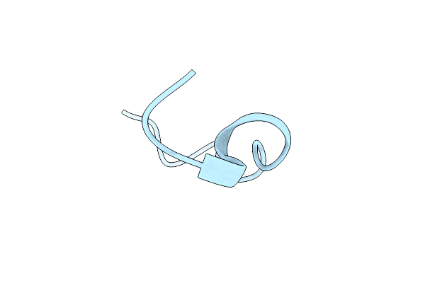
Nmr Structure Of Calcium Bound Conformer Of Conantokin G, Minimized Average Structure
|
Organism: Conus geographus
Method: SOLUTION NMR
Release Date: 1998-04-08
 |
Nmr Structure Of Metal-Free Conantokin G, 1 Structure
Organism: Conus geographus
Method: SOLUTION NMR Release Date: 1997-08-20 Classification: GAMMA-CARBOXYGLUTAMIC ACID |
Organism: Conus geographus
Method: SOLUTION NMR
Release Date: 1997-08-20