
Search Count: 18
All
Selected
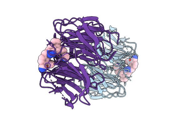 |
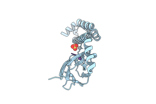
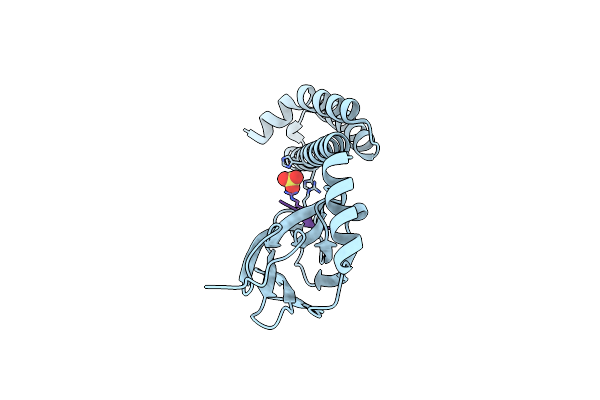
Organism: Homo sapiens
Method: X-RAY DIFFRACTION Resolution:2.11 Å Release Date: 2025-08-13 Classification: STRUCTURAL PROTEIN/INHIBITOR Ligands: A1BW7 |
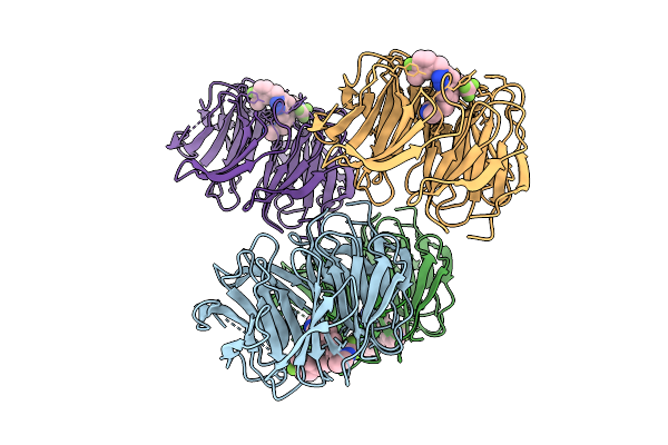 |
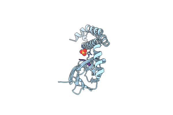
Organism: Homo sapiens
Method: X-RAY DIFFRACTION Resolution:1.58 Å Release Date: 2025-08-13 Classification: STRUCTURAL PROTEIN/INHIBITOR Ligands: A1BW8 |
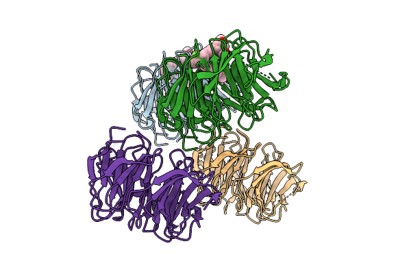 |
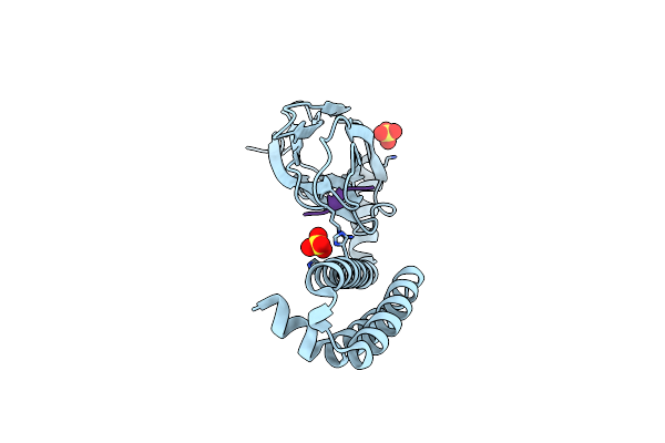
Organism: Homo sapiens
Method: X-RAY DIFFRACTION Resolution:1.58 Å Release Date: 2025-08-13 Classification: STRUCTURAL PROTEIN/INHIBITOR Ligands: A1BW9 |
 |
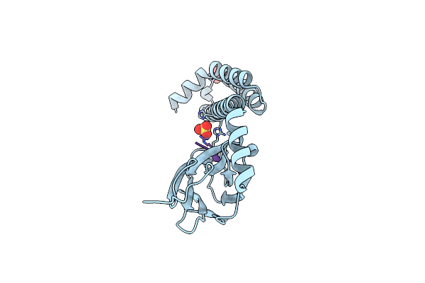
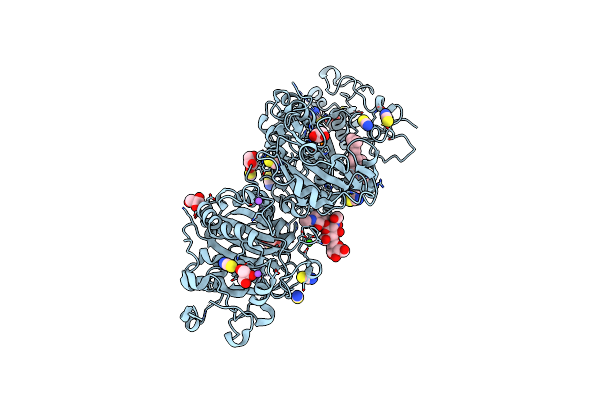
Sars-Cov-2 3Clpro In Complex With N-(4-(1H-Pyrazol-4-Yl)Phenyl)-N-(3-Chlorobenzyl)-2-(Pyridin-3-Yl)Acetamide
Organism: Severe acute respiratory syndrome coronavirus 2
Method: X-RAY DIFFRACTION Resolution:2.20 Å Release Date: 2023-01-11 Classification: VIRAL PROTEIN,HYDROLASE/INHIBITOR Ligands: I2D |
 |
Sars-Cov-2 3Clpro In Complex With N-(4-(1H-Imidazol-4-Yl)Phenyl)-N-(3-Chloro-5-Fluorobenzyl)-2-(Isoquinolin-4-Yl)Acetamide
Organism: Severe acute respiratory syndrome coronavirus 2
Method: X-RAY DIFFRACTION Resolution:2.40 Å Release Date: 2023-01-11 Classification: VIRAL PROTEIN,HYDROLASE/INHIBITOR Ligands: I2N |
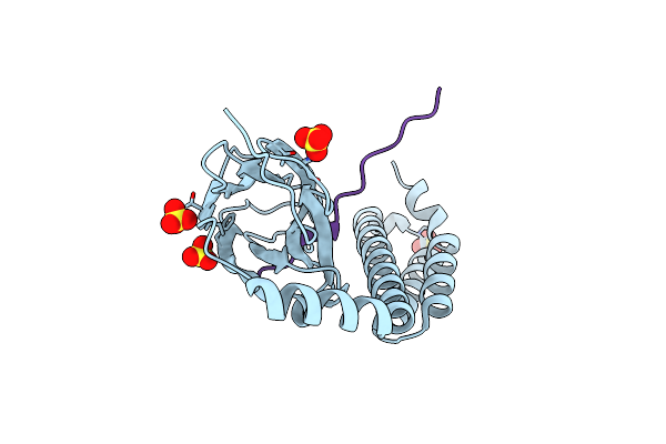 |
Crystal Structure Of The Substrate-Binding Domain Of E. Coli Dnak In Complex With The Peptide Rkqstialallpllftprr
Organism: Escherichia coli (strain k12)
Method: X-RAY DIFFRACTION Resolution:2.00 Å Release Date: 2021-06-23 Classification: CHAPERONE/HYDROLASE Ligands: SO4 |
 |
Crystal Structure Of The Substrate-Binding Domain Of E. Coli Dnak In Complex With The Peptide Ralallplsr
Organism: Escherichia coli (strain k12)
Method: X-RAY DIFFRACTION Resolution:2.55 Å Release Date: 2021-06-23 Classification: CHAPERONE/HYDROLASE Ligands: SO4 |
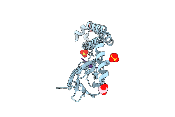 |
Crystal Structure Of The Substrate-Binding Domain Of E. Coli Dnak In Complex With The Peptide Eanqqkpllglfadg
Organism: Escherichia coli (strain k12)
Method: X-RAY DIFFRACTION Resolution:2.40 Å Release Date: 2021-06-23 Classification: CHAPERONE/HYDROLASE Ligands: GOL, SO4 |
 |
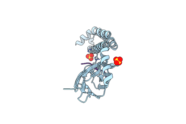
Crystal Structure Of The Substrate-Binding Domain Of E. Coli Dnak In Complex With The Peptide Rqkpllglsr
Organism: Escherichia coli (strain k12)
Method: X-RAY DIFFRACTION Resolution:1.82 Å Release Date: 2021-06-23 Classification: CHAPERONE/HYDROLASE Ligands: SO4 |
 |
Crystal Structure Of The Substrate-Binding Domain Of E. Coli Dnak In Complex With The Peptide Rakniillsr
Organism: Escherichia coli (strain k12)
Method: X-RAY DIFFRACTION Resolution:2.56 Å Release Date: 2020-08-26 Classification: CHAPERONE Ligands: SO4 |
 |
Crystal Structure Of The Substrate-Binding Domain Of E. Coli Dnak In Complex With The Peptide Rgntlvivsr
Organism: Escherichia coli (strain k12)
Method: X-RAY DIFFRACTION Resolution:3.09 Å Release Date: 2020-08-26 Classification: CHAPERONE Ligands: SO4 |
 |
Crystal Structure Of The Substrate-Binding Domain Of E. Coli Dnak In Complex With The Peptide Qehtgsqlriaaygp
Organism: Escherichia coli (strain k12)
Method: X-RAY DIFFRACTION Resolution:2.40 Å Release Date: 2020-08-26 Classification: CHAPERONE Ligands: SO4 |
 |
Crystal Structure Of The Substrate-Binding Domain Of E. Coli Dnak In Complex With The Peptide Rgsqlriasr
Organism: Escherichia coli (strain k12)
Method: X-RAY DIFFRACTION Resolution:2.54 Å Release Date: 2020-08-26 Classification: CHAPERONE Ligands: SO4 |
 |
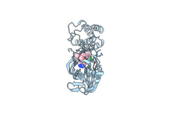
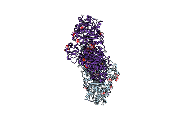
Organism: Rattus norvegicus
Method: X-RAY DIFFRACTION Resolution:1.60 Å Release Date: 2016-04-13 Classification: HYDROLASE Ligands: ZN, 5JK, CA, IOD, SCN, NA, GOL |
 |
Crystal Structure Of Autotaxin (Enpp2) With Tauroursodeoxycholic Acid (Tudca)
Organism: Rattus norvegicus
Method: X-RAY DIFFRACTION Resolution:2.00 Å Release Date: 2016-04-13 Classification: HYDROLASE Ligands: ZN, CA, IOD, PO4, NA, 5D5, SCN, GOL |
 |
Crystal Structure Of Autotaxin (Enpp2) With Tauroursodeoxycholic Acid (Tudca) And Lysophosphatidic Acid (Lpa)
Organism: Rattus norvegicus
Method: X-RAY DIFFRACTION Resolution:1.80 Å Release Date: 2016-04-13 Classification: HYDROLASE Ligands: ZN, CA, NKP, 5D5, NA, IOD, SCN, GOL |
 |
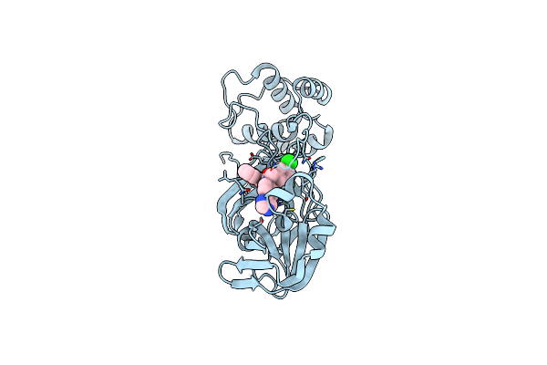
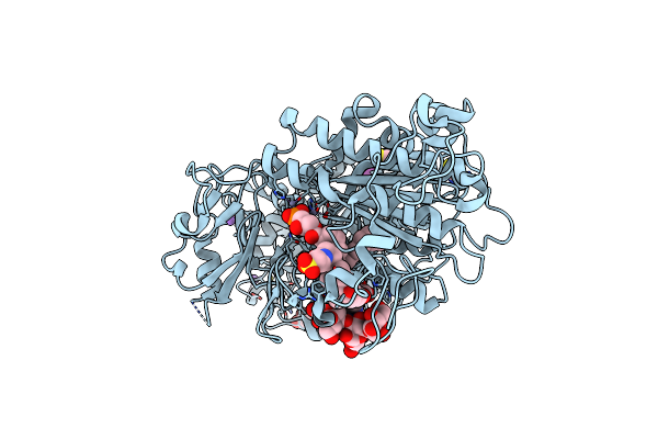
Organism: Homo sapiens
Method: X-RAY DIFFRACTION Resolution:2.70 Å Release Date: 2003-10-14 Classification: TRANSFERASE Ligands: STU, SO4 |
 |
Organism: Homo sapiens
Method: X-RAY DIFFRACTION Resolution:3.00 Å Release Date: 2003-10-14 Classification: TRANSFERASE Ligands: ADP |