

Search Count: 29
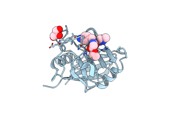 |
Cyclophilin A Complexed With The Tri-Vector Ligand 8.
Organism: Homo sapiens
Method: X-RAY DIFFRACTION Resolution:1.60 Å Release Date: 2018-11-07 Classification: ISOMERASE Ligands: F1E, GOL |
Organism: Homo sapiens
Method: X-RAY DIFFRACTION
Release Date: 2018-11-07
Ligands: F1E, GOL
 |
Cyclophilin A Complexed With Tri-Vector Ligand 2.
Organism: Homo sapiens
Method: X-RAY DIFFRACTION Resolution:1.38 Å Release Date: 2018-11-07 Classification: ISOMERASE Ligands: L36 |
Organism: Homo sapiens
Method: X-RAY DIFFRACTION
Release Date: 2018-11-07
Ligands: L36
 |
Cyclophilin A Complexed With Tri-Vector Ligand 10.
Organism: Homo sapiens
Method: X-RAY DIFFRACTION Resolution:1.16 Å Release Date: 2018-11-07 Classification: ISOMERASE Ligands: F1Q |
Organism: Homo sapiens
Method: X-RAY DIFFRACTION
Release Date: 2018-11-07
Ligands: F1Q
 |
Cyclophilin A Complexed With Tri-Vector Ligand 4.
Organism: Homo sapiens
Method: X-RAY DIFFRACTION Resolution:1.35 Å Release Date: 2018-11-07 Classification: ISOMERASE Ligands: L36 |
Organism: Homo sapiens
Method: X-RAY DIFFRACTION
Release Date: 2018-11-07
Ligands: L36
 |
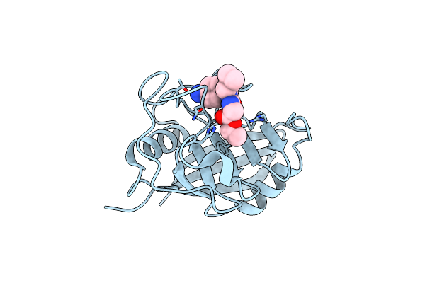
Cyclophilin A Complexed With Tri-Vector Ligand 15.
Organism: Homo sapiens
Method: X-RAY DIFFRACTION Resolution:1.70 Å Release Date: 2018-11-07 Classification: ISOMERASE Ligands: F0Q, FMT, GOL |
Organism: Homo sapiens
Method: X-RAY DIFFRACTION
Release Date: 2018-11-07
Ligands: F0Q, FMT, GOL
 |
Cyclophilin A Complexed With Tri-Vector Ligand 7.
Organism: Homo sapiens
Method: X-RAY DIFFRACTION Resolution:1.94 Å Release Date: 2018-11-07 Classification: ISOMERASE Ligands: F0T |
Organism: Homo sapiens
Method: X-RAY DIFFRACTION
Release Date: 2018-11-07
Ligands: F0T
 |
Cyclophilin A Complexed With Tri-Vector Ligand 9.
Organism: Homo sapiens
Method: X-RAY DIFFRACTION Resolution:1.69 Å Release Date: 2018-11-07 Classification: ISOMERASE Ligands: F0W |
Organism: Homo sapiens
Method: X-RAY DIFFRACTION
Release Date: 2018-11-07
Ligands: F0W
 |
Cyclophilin A Complexed With Tri-Vector Ligand 5.
Organism: Homo sapiens
Method: X-RAY DIFFRACTION Resolution:1.29 Å Release Date: 2018-11-07 Classification: ISOMERASE Ligands: F1Z |
Organism: Homo sapiens
Method: X-RAY DIFFRACTION
Release Date: 2018-11-07
Ligands: F1Z
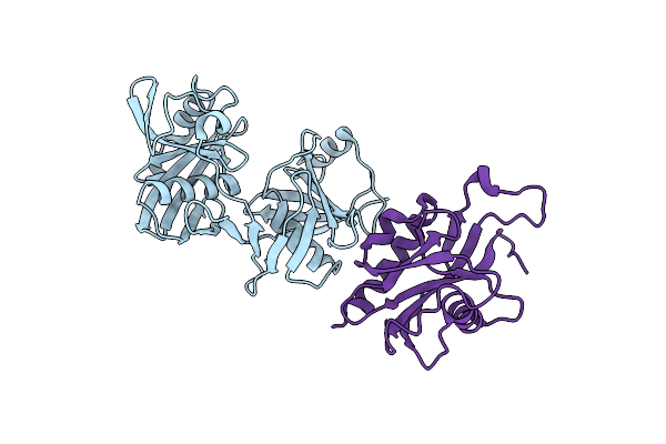 |
Crystal Structure Of E.Coli Rpph-Dapf Complex
Organism: Escherichia coli o45:k1 (strain s88 / expec)
Method: X-RAY DIFFRACTION Resolution:3.06 Å Release Date: 2018-05-23 Classification: isomerase/hydrolase Ligands: IOD, CL |
Organism: Escherichia coli o45:k1 (strain s88 / expec)
Method: X-RAY DIFFRACTION
Release Date: 2018-05-23
Ligands: IOD, CL
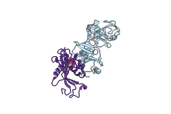 |
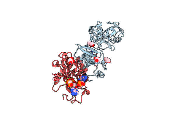
Crystal Structure Of E. Coli Rpph-Dapf Complex, Monomer
Organism: Escherichia coli (strain k12)
Method: X-RAY DIFFRACTION Resolution:2.15 Å Release Date: 2018-05-23 Classification: isomerase/hydrolase Ligands: CL, GOL |
Organism: Escherichia coli (strain k12)
Method: X-RAY DIFFRACTION
Release Date: 2018-05-23
Ligands: CL, GOL
 |
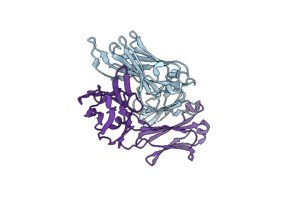
Crystal Structure Of E. Coli Rpph-Dapf Complex, Monomer Bound To Rna
Organism: Escherichia coli
Method: X-RAY DIFFRACTION Resolution:1.81 Å Release Date: 2018-05-23 Classification: isomerase/hydrolase/rna Ligands: GOL, CL, MG |
Organism: Escherichia coli
Method: X-RAY DIFFRACTION
Release Date: 2018-05-23
Ligands: GOL, CL, MG
 |
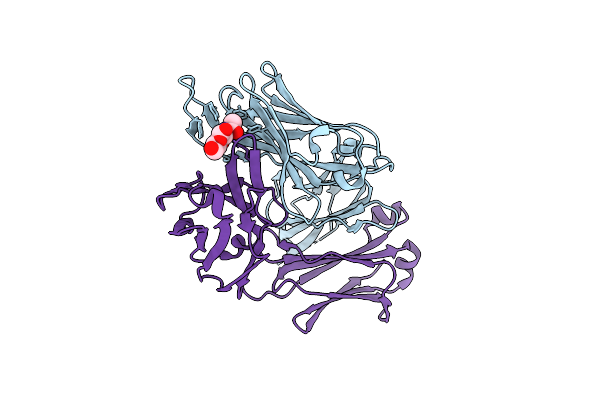
Crystal Structure Of A Polysaccharide-Binding Human Fab (F598)
Organism: Homo sapiens
Method: X-RAY DIFFRACTION Resolution:1.70 Å Release Date: 2018-02-21 Classification: IMMUNE SYSTEM |
Organism: Homo sapiens
Method: X-RAY DIFFRACTION
Release Date: 2018-02-21
 |
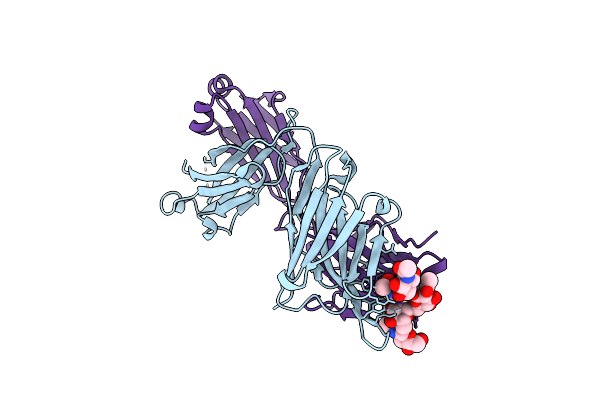
Crystal Structure Of A Polysaccharide-Binding Human Fab (F598) In Complex With N-Acetyl-D-Glucosamine (Glcnac)
Organism: Homo sapiens
Method: X-RAY DIFFRACTION Resolution:1.60 Å Release Date: 2018-02-21 Classification: IMMUNE SYSTEM Ligands: NAG |
Organism: Homo sapiens
Method: X-RAY DIFFRACTION
Release Date: 2018-02-21
Ligands: NAG
 |
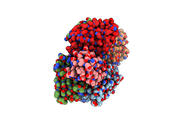
Crystal Structure Of A Polysaccharide-Binding Human Fab (F598) In Complex With Nona-N-Acetyl-D-Glucosamine (9Nac)
Organism: Homo sapiens
Method: X-RAY DIFFRACTION Resolution:1.90 Å Release Date: 2018-02-21 Classification: IMMUNE SYSTEM |
Organism: Homo sapiens
Method: X-RAY DIFFRACTION
Release Date: 2018-02-21
 |
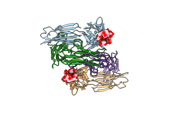
Crystal Structure Of Anti-Msp2 Fv Fragment (Mab4D11) In Complex With 3D7-Msp2 215-222
Organism: Mus musculus, Plasmodium falciparum 3d7
Method: X-RAY DIFFRACTION Resolution:2.20 Å Release Date: 2017-02-15 Classification: IMMUNE SYSTEM |
Organism: Mus musculus, Plasmodium falciparum 3d7
Method: X-RAY DIFFRACTION
Release Date: 2017-02-15
 |
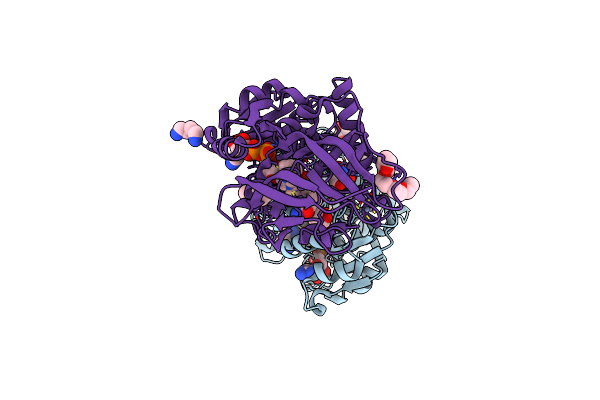
Crystal Structure Of The Igg2A Lpt3 In Complex With An 8-Sugar Inner Core Analogue Of Neisseria Meningitidis
Organism: Mus musculus
Method: X-RAY DIFFRACTION Resolution:2.69 Å Release Date: 2014-02-12 Classification: IMMUNE SYSTEM Ligands: SO4 |
Organism: Mus musculus
Method: X-RAY DIFFRACTION
Release Date: 2014-02-12
Ligands: SO4
 |
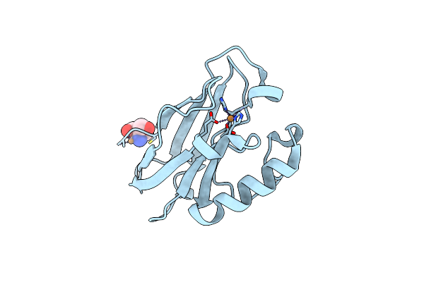
S-Nitrosoglutathione Reductase (Gsnor) In Complex With N6022
Organism: Homo sapiens
Method: X-RAY DIFFRACTION Resolution:1.90 Å Release Date: 2011-04-06 Classification: OXIDOREDUCTASE/OXIDOREDUCTASE INHIBITOR Ligands: ZN, K, NAD, 022, 1PE, N2P, TRS |
Organism: Homo sapiens
Method: X-RAY DIFFRACTION
Release Date: 2011-04-06
Ligands: ZN, K, NAD, 022, 1PE, N2P, TRS
 |
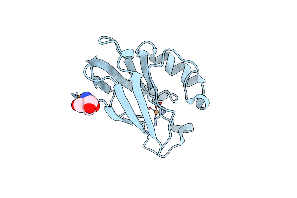
C112D/M121E Pseudomonas Aeruginosa Azurin
Organism: Pseudomonas aeruginosa
Method: X-RAY DIFFRACTION Resolution:2.10 Å Release Date: 2010-10-13 Classification: ELECTRON TRANSPORT Ligands: CU, TRS |
Organism: Pseudomonas aeruginosa
Method: X-RAY DIFFRACTION
Release Date: 2010-10-13
Ligands: CU, TRS
 |
C112D/M121E Pseudomonas Aeruginosa Azurin
Organism: Pseudomonas aeruginosa
Method: X-RAY DIFFRACTION Resolution:2.25 Å Release Date: 2010-10-13 Classification: ELECTRON TRANSPORT Ligands: CU, TRS |
Organism: Pseudomonas aeruginosa
Method: X-RAY DIFFRACTION
Release Date: 2010-10-13
Ligands: CU, TRS
 |
C112D/M121E Azurin, Ph 10.0
Organism: Pseudomonas aeruginosa
Method: X-RAY DIFFRACTION Resolution:2.40 Å Release Date: 2010-10-13 Classification: ELECTRON TRANSPORT Ligands: CU, TRS |
Organism: Pseudomonas aeruginosa
Method: X-RAY DIFFRACTION
Release Date: 2010-10-13
Ligands: CU, TRS