

Search Count: 20
 |
X-Ray Structure Of Furin (Pcsk3) In Complex With The Biphenyl-Derived Compound 26 (Mi2456)
Organism: Homo sapiens
Method: X-RAY DIFFRACTION Resolution:1.60 Å Release Date: 2025-12-24 Classification: HYDROLASE Ligands: A1JA6, CA, NA, CL, DMS, NAG |
Organism: Homo sapiens
Method: X-RAY DIFFRACTION
Release Date: 2025-12-24
Ligands: A1JA6, CA, NA, CL, DMS, NAG
 |
X-Ray Structure Of Furin (Pcsk3) In Complex With The Biphenyl-Derived Compound 42 (Mi2464)
Organism: Homo sapiens
Method: X-RAY DIFFRACTION Resolution:1.50 Å Release Date: 2025-12-24 Classification: HYDROLASE Ligands: CA, NA, CL, DMS, NAG, A1JA1 |
Organism: Homo sapiens
Method: X-RAY DIFFRACTION
Release Date: 2025-12-24
Ligands: CA, NA, CL, DMS, NAG, A1JA1
 |
X-Ray Structure Of Furin (Pcsk3) In Complex With The Biphenyl-Derived Compound 34 (Mi2470)
Organism: Homo sapiens
Method: X-RAY DIFFRACTION Resolution:1.60 Å Release Date: 2025-12-24 Classification: HYDROLASE Ligands: CA, NA, CL, DMS, NAG, A1JA2 |
Organism: Homo sapiens
Method: X-RAY DIFFRACTION
Release Date: 2025-12-24
Ligands: CA, NA, CL, DMS, NAG, A1JA2
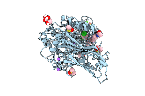 |
X-Ray Structure Of Furin (Pcsk3) In Complex With The Biphenyl-Derived Compound 27 (Mi2471)
Organism: Homo sapiens
Method: X-RAY DIFFRACTION Resolution:1.60 Å Release Date: 2025-12-24 Classification: HYDROLASE Ligands: A1JA3, CA, NA, CL, DMS, NAG |
Organism: Homo sapiens
Method: X-RAY DIFFRACTION
Release Date: 2025-12-24
Ligands: A1JA3, CA, NA, CL, DMS, NAG
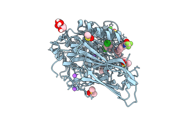 |
X-Ray Structure Of Furin (Pcsk3) In Complex With The Biphenyl-Derived Compound 13 (Mi3102)
Organism: Homo sapiens
Method: X-RAY DIFFRACTION Resolution:1.65 Å Release Date: 2025-12-24 Classification: HYDROLASE Ligands: CA, NA, CL, DMS, NAG, A1JA5 |
Organism: Homo sapiens
Method: X-RAY DIFFRACTION
Release Date: 2025-12-24
Ligands: CA, NA, CL, DMS, NAG, A1JA5
 |
X-Ray Structure Of Furin (Pcsk3) In Complex With The Biphenyl-Derived Compound 24 (Mi3140)
Organism: Homo sapiens
Method: X-RAY DIFFRACTION Resolution:1.60 Å Release Date: 2025-12-24 Classification: HYDROLASE Ligands: A1JA4, CA, NA, CL, DMS, NAG |
Organism: Homo sapiens
Method: X-RAY DIFFRACTION
Release Date: 2025-12-24
Ligands: A1JA4, CA, NA, CL, DMS, NAG
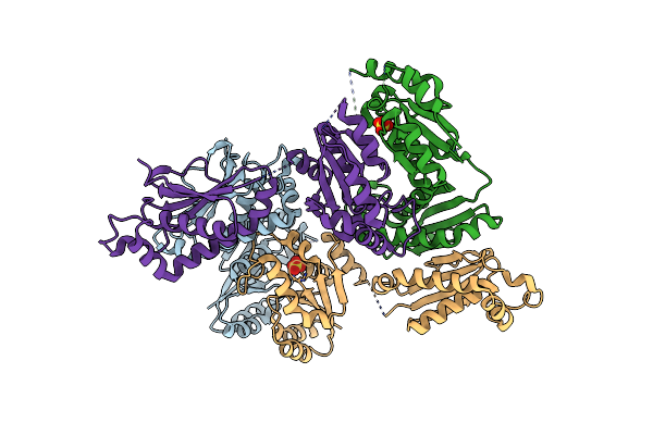 |
X-Ray Crystal Structure Of Rapz From Escherichia Coli (P32 Space Group)
Organism: Escherichia coli
Method: X-RAY DIFFRACTION Resolution:3.40 Å Release Date: 2017-08-30 Classification: CHAPERONE Ligands: SO4 |
Organism: Escherichia coli
Method: X-RAY DIFFRACTION
Release Date: 2017-08-30
Ligands: SO4
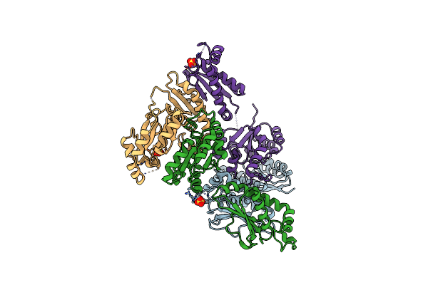 |
X-Ray Crystal Structure Of Rapz From Escherichia Coli (P3221 Space Group)
Organism: Escherichia coli
Method: X-RAY DIFFRACTION Resolution:3.25 Å Release Date: 2017-08-30 Classification: CHAPERONE Ligands: SO4 |
Organism: Escherichia coli
Method: X-RAY DIFFRACTION
Release Date: 2017-08-30
Ligands: SO4
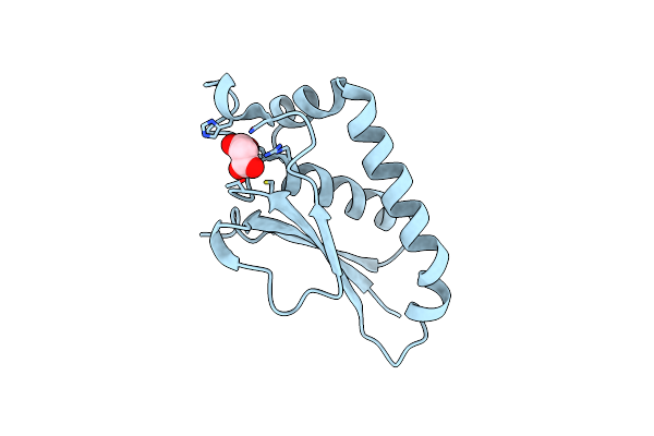 |
X-Ray Crystal Structure Of The Rapz C-Terminal Domain From Escherichia Coli
Organism: Escherichia coli
Method: X-RAY DIFFRACTION Resolution:1.17 Å Release Date: 2017-08-30 Classification: CHAPERONE Ligands: MLI |
Organism: Escherichia coli
Method: X-RAY DIFFRACTION
Release Date: 2017-08-30
Ligands: MLI
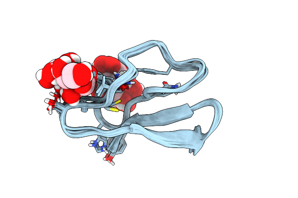 |
Solution Structure Of Family 1 Carbohydrate-Binding Module From Trichoderma Reesei Cel7A With O-Mannose Residues At Thr1 And Ser3
Organism: Trichoderma reesei
Method: SOLUTION NMR Release Date: 2015-09-02 Classification: HYDROLASE Ligands: MAN |
Organism: Trichoderma reesei
Method: SOLUTION NMR
Release Date: 2015-09-02
Ligands: MAN
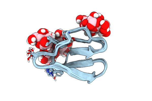 |
Family 1 Carbohydrate-Binding Module From Trichoderma Reesei Cel7A With O-Mannose Residues At Thr1, Ser3, And Ser14
Organism: Trichoderma reesei
Method: SOLUTION NMR Release Date: 2015-09-02 Classification: HYDROLASE Ligands: MAN |
Organism: Trichoderma reesei
Method: SOLUTION NMR
Release Date: 2015-09-02
Ligands: MAN
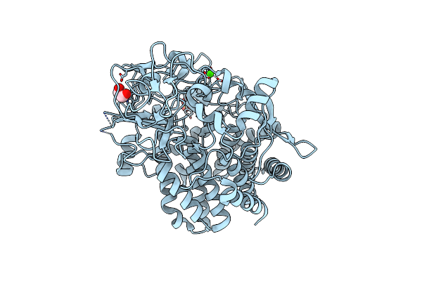 |
The Unliganded Structure Of C.Bescii Cela Gh48 Module
Organism: Caldicellulosiruptor bescii
Method: X-RAY DIFFRACTION Resolution:2.45 Å Release Date: 2013-03-20 Classification: HYDROLASE Ligands: CA, SO4, EDO |
Organism: Caldicellulosiruptor bescii
Method: X-RAY DIFFRACTION
Release Date: 2013-03-20
Ligands: CA, SO4, EDO
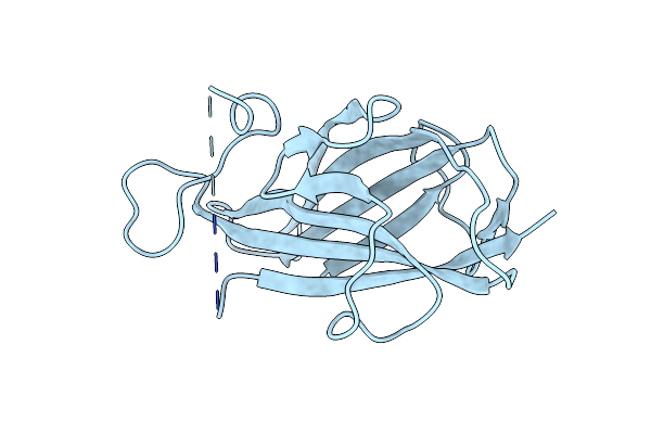 |
Structure Of Mouse Zp-C Domain Of Tgf-Beta-Receptor-3
Organism: Mus musculus
Method: X-RAY DIFFRACTION Resolution:2.70 Å Release Date: 2013-02-27 Classification: SIGNALING PROTEIN |
Organism: Mus musculus
Method: X-RAY DIFFRACTION
Release Date: 2013-02-27
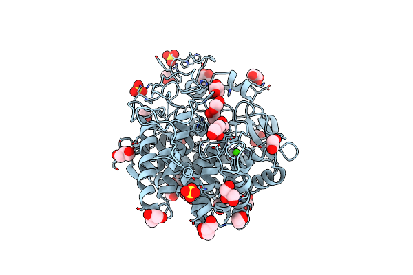 |
The Structure Of Cbescii Cela Gh9 Module
Organism: Caldicellulosiruptor bescii
Method: X-RAY DIFFRACTION Resolution:1.70 Å Release Date: 2013-02-20 Classification: HYDROLASE Ligands: DIO, CA, EDO, GOL, SO4 |
Organism: Caldicellulosiruptor bescii
Method: X-RAY DIFFRACTION
Release Date: 2013-02-20
Ligands: DIO, CA, EDO, GOL, SO4
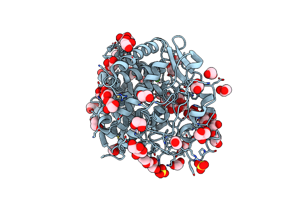 |
The Liganded Structure Of Cbescii Cela Gh9 Module
Organism: Caldicellulosiruptor bescii
Method: X-RAY DIFFRACTION Resolution:1.56 Å Release Date: 2013-02-20 Classification: HYDROLASE Ligands: DIO, CA, GOL, EDO, SO4 |
Organism: Caldicellulosiruptor bescii
Method: X-RAY DIFFRACTION
Release Date: 2013-02-20
Ligands: DIO, CA, GOL, EDO, SO4
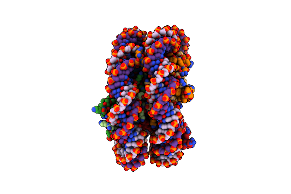 |
Structural Characterization Of H3K56Q Nucleosomes And Nucleosomal Arrays
Organism: Xenopus laevis
Method: X-RAY DIFFRACTION Resolution:3.20 Å Release Date: 2010-06-23 Classification: Transcription/DNA |
Organism: Xenopus laevis
Method: X-RAY DIFFRACTION
Release Date: 2010-06-23
 |
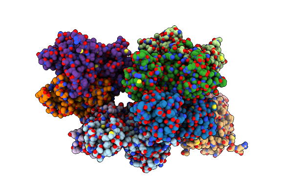
Structural Characterization Of H3K56Q Nucleosomes And Nucleosomal Arrays
Organism: Xenopus laevis
Method: X-RAY DIFFRACTION Resolution:3.50 Å Release Date: 2010-05-12 Classification: Structural Protein/DNA |
Organism: Xenopus laevis
Method: X-RAY DIFFRACTION
Release Date: 2010-05-12
 |
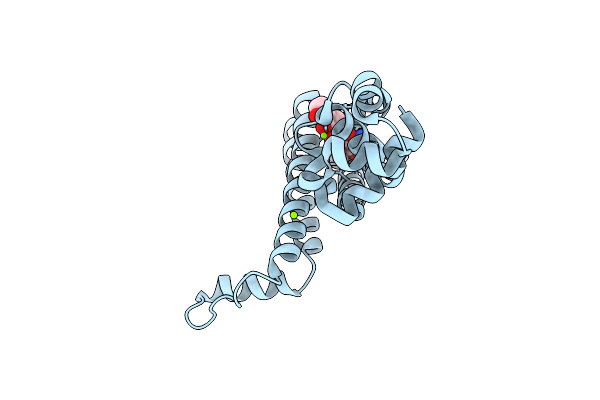
Crystal Structure Of The Gntr-Hutc Family Member Yvoa From Bacillus Subtilis
Organism: Bacillus subtilis
Method: X-RAY DIFFRACTION Resolution:2.40 Å Release Date: 2010-01-12 Classification: TRANSCRIPTION Ligands: SO4 |
Organism: Bacillus subtilis
Method: X-RAY DIFFRACTION
Release Date: 2010-01-12
Ligands: SO4
 |
Tetr (Bd) Variant L17G With Reverse Phenotype
Organism: Escherichia coli
Method: X-RAY DIFFRACTION Resolution:1.74 Å Release Date: 2008-07-08 Classification: TRANSCRIPTION Ligands: TDC, MG |
Organism: Escherichia coli
Method: X-RAY DIFFRACTION
Release Date: 2008-07-08
Ligands: TDC, MG
 |
Reconstituted Co Dehydrogenase From Oligotropha Carboxidovorans
Organism: Oligotropha carboxidovorans
Method: X-RAY DIFFRACTION Resolution:1.70 Å Release Date: 2005-10-11 Classification: OXIDOREDUCTASE Ligands: PO4, FES, CU, DW6, FAD |
Organism: Oligotropha carboxidovorans
Method: X-RAY DIFFRACTION
Release Date: 2005-10-11
Ligands: PO4, FES, CU, DW6, FAD