

Search Count: 26
 |
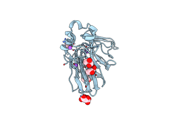
Structure Of Ribonucleotide Reductase R2 From Escherichia Coli Collected By Femtosecond Serial Crystallography On A Coc Membrane
Organism: Escherichia coli (strain k12)
Method: X-RAY DIFFRACTION Resolution:2.30 Å Release Date: 2022-01-12 Classification: OXIDOREDUCTASE Ligands: FE |
Organism: Escherichia coli (strain k12)
Method: X-RAY DIFFRACTION
Release Date: 2022-01-12
Ligands: FE
 |
Structure Of Thaumatin Collected By Femtosecond Serial Crystallography On A Coc Membrane
Organism: Thaumatococcus daniellii
Method: X-RAY DIFFRACTION Resolution:1.46 Å Release Date: 2021-11-24 Classification: PLANT PROTEIN Ligands: TLA, PGR, NA |
Organism: Thaumatococcus daniellii
Method: X-RAY DIFFRACTION
Release Date: 2021-11-24
Ligands: TLA, PGR, NA
 |
Structure Of The A2A Adenosine Receptor Determined At Swissfel Using Native-Sad At 4.57 Kev From All Available Diffraction Patterns
Organism: Homo sapiens
Method: X-RAY DIFFRACTION Resolution:2.65 Å Release Date: 2020-07-15 Classification: SIGNALING PROTEIN Ligands: ZMA, OLA, OLB, OLC, CLR |
Organism: Homo sapiens
Method: X-RAY DIFFRACTION
Release Date: 2020-07-15
Ligands: ZMA, OLA, OLB, OLC, CLR
 |
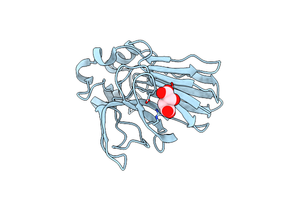
Structure Of The A2A Adenosine Receptor Determined At Swissfel Using Native-Sad At 4.57 Kev From 50,000 Diffraction Patterns
Organism: Homo sapiens, Escherichia coli
Method: X-RAY DIFFRACTION Resolution:2.65 Å Release Date: 2020-07-15 Classification: MEMBRANE PROTEIN Ligands: ZMA, OLA, OLB, OLC, CLR |
Organism: Homo sapiens, Escherichia coli
Method: X-RAY DIFFRACTION
Release Date: 2020-07-15
Ligands: ZMA, OLA, OLB, OLC, CLR
 |
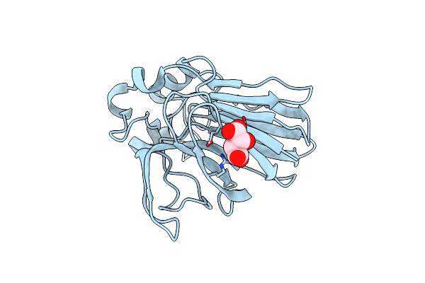
Structure Of Thaumatin Determined At Swissfel Using Native-Sad At 4.57 Kev From All Available Diffraction Patterns
Organism: Thaumatococcus daniellii
Method: X-RAY DIFFRACTION Resolution:2.65 Å Release Date: 2020-07-15 Classification: PLANT PROTEIN Ligands: TLA |
Organism: Thaumatococcus daniellii
Method: X-RAY DIFFRACTION
Release Date: 2020-07-15
Ligands: TLA
 |
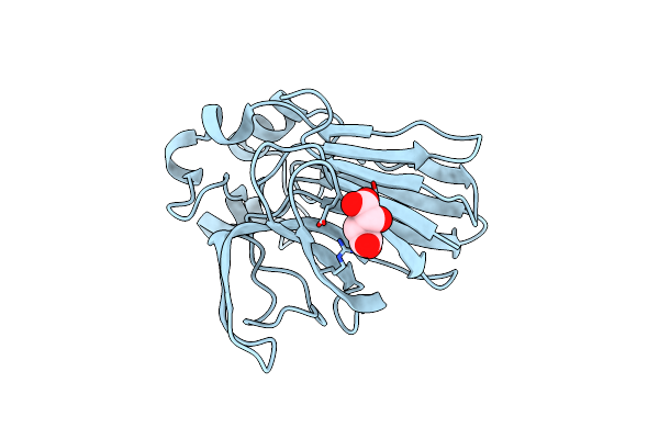
Structure Of Thaumatin Determined At Swissfel Using Native-Sad At 4.57 Kev From 20,000 Diffraction Patterns
Organism: Thaumatococcus daniellii
Method: X-RAY DIFFRACTION Resolution:2.65 Å Release Date: 2020-07-15 Classification: PLANT PROTEIN Ligands: TLA |
Organism: Thaumatococcus daniellii
Method: X-RAY DIFFRACTION
Release Date: 2020-07-15
Ligands: TLA
 |
Structure Of Thaumatin Determined At Swissfel Using Native-Sad At 6.06 Kev From All Available Diffraction Patterns
Organism: Thaumatococcus daniellii
Method: X-RAY DIFFRACTION Resolution:1.95 Å Release Date: 2020-07-15 Classification: PLANT PROTEIN Ligands: TLA |
Organism: Thaumatococcus daniellii
Method: X-RAY DIFFRACTION
Release Date: 2020-07-15
Ligands: TLA
 |
Structure Of Thaumatin Determined At Swissfel Using Native-Sad At 6.06 Kev From 50,000 Diffraction Patterns.
Organism: Thaumatococcus daniellii
Method: X-RAY DIFFRACTION Resolution:2.00 Å Release Date: 2020-07-15 Classification: PLANT PROTEIN Ligands: TLA |
Organism: Thaumatococcus daniellii
Method: X-RAY DIFFRACTION
Release Date: 2020-07-15
Ligands: TLA
 |
Pink Beam Serial Crystallography: Lysozyme, 5 Us Exposure, 14793 Patterns Merged
Organism: Gallus gallus
Method: X-RAY DIFFRACTION Resolution:1.70 Å Release Date: 2019-10-16 Classification: HYDROLASE |
Organism: Gallus gallus
Method: X-RAY DIFFRACTION
Release Date: 2019-10-16
 |
Pink Beam Serial Crystallography: Lysozyme, 5 Us Exposure, 8813 Patterns Merged (1 Chip)
Organism: Gallus gallus
Method: X-RAY DIFFRACTION Resolution:1.70 Å Release Date: 2019-10-16 Classification: HYDROLASE |
Organism: Gallus gallus
Method: X-RAY DIFFRACTION
Release Date: 2019-10-16
 |
Pink Beam Serial Crystallography: Lysozyme, 1 Us Exposure, 4448 Patterns Merged (1 Chip)
Organism: Gallus gallus
Method: X-RAY DIFFRACTION Resolution:1.84 Å Release Date: 2019-10-16 Classification: HYDROLASE |
Organism: Gallus gallus
Method: X-RAY DIFFRACTION
Release Date: 2019-10-16
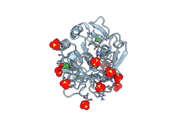 |
Pink Beam Serial Crystallography: Proteinase K, 1 Us Exposure, 1585 Patterns Merged (2 Chips)
Organism: Parengyodontium album
Method: X-RAY DIFFRACTION Resolution:1.94 Å Release Date: 2019-09-25 Classification: HYDROLASE Ligands: CA, SO4, CL |
Organism: Parengyodontium album
Method: X-RAY DIFFRACTION
Release Date: 2019-09-25
Ligands: CA, SO4, CL
 |
Pink Beam Serial Crystallography: Lysozyme, 5 Us Exposure, 24344 Patterns Merged (3 Chips)
Organism: Gallus gallus
Method: X-RAY DIFFRACTION Resolution:1.70 Å Release Date: 2019-09-25 Classification: HYDROLASE |
Organism: Gallus gallus
Method: X-RAY DIFFRACTION
Release Date: 2019-09-25
 |
Pink Beam Serial Crystallography: Lysozyme, 5 Us Exposure, 3000 Patterns Merged
Organism: Gallus gallus
Method: X-RAY DIFFRACTION Resolution:1.70 Å Release Date: 2019-09-25 Classification: HYDROLASE |
Organism: Gallus gallus
Method: X-RAY DIFFRACTION
Release Date: 2019-09-25
 |
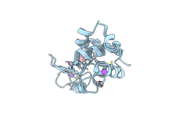
Pink Beam Serial Crystallography: Lysozyme, 5 Us Exposure, 1500 Patterns Merged
Organism: Gallus gallus
Method: X-RAY DIFFRACTION Resolution:1.70 Å Release Date: 2019-09-25 Classification: HYDROLASE |
Organism: Gallus gallus
Method: X-RAY DIFFRACTION
Release Date: 2019-09-25
 |
Pink Beam Serial Crystallography: Lysozyme, 5 Us Exposure, 750 Patterns Merged
Organism: Gallus gallus
Method: X-RAY DIFFRACTION Resolution:1.70 Å Release Date: 2019-09-25 Classification: HYDROLASE |
Organism: Gallus gallus
Method: X-RAY DIFFRACTION
Release Date: 2019-09-25
 |
Pink Beam Serial Crystallography: Lysozyme, 1 Us Exposure, 14793 Patterns Merged
Organism: Gallus gallus
Method: X-RAY DIFFRACTION Resolution:1.84 Å Release Date: 2019-09-25 Classification: HYDROLASE |
Organism: Gallus gallus
Method: X-RAY DIFFRACTION
Release Date: 2019-09-25
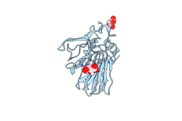 |
Thaumatin Solved By Native Sad From A Dataset Collected In 0.6 Second With Jungfrau Detector
Organism: Thaumatococcus daniellii
Method: X-RAY DIFFRACTION Resolution:2.36 Å Release Date: 2018-08-01 Classification: PLANT PROTEIN Ligands: TLA |
Organism: Thaumatococcus daniellii
Method: X-RAY DIFFRACTION
Release Date: 2018-08-01
Ligands: TLA
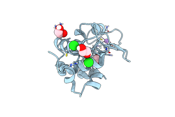 |
Lysozyme Solved By Native Sad From A Dataset Collected In 5 Seconds At 1 A Wavelength With Jungfrau Detector
Organism: Gallus gallus
Method: X-RAY DIFFRACTION Resolution:1.14 Å Release Date: 2018-08-01 Classification: HYDROLASE Ligands: CL, NA, EDO |
Organism: Gallus gallus
Method: X-RAY DIFFRACTION
Release Date: 2018-08-01
Ligands: CL, NA, EDO
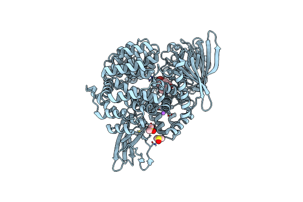 |
E. Coli Aminopeptidase N Solved By Native Sad From A Dataset Collected In 60 Second With Jungfrau Detector
Organism: Escherichia coli (strain k12)
Method: X-RAY DIFFRACTION Resolution:2.37 Å Release Date: 2018-08-01 Classification: HYDROLASE Ligands: ZN, NA, DMS, 7MF |
Organism: Escherichia coli (strain k12)
Method: X-RAY DIFFRACTION
Release Date: 2018-08-01
Ligands: ZN, NA, DMS, 7MF