

Search Count: 12
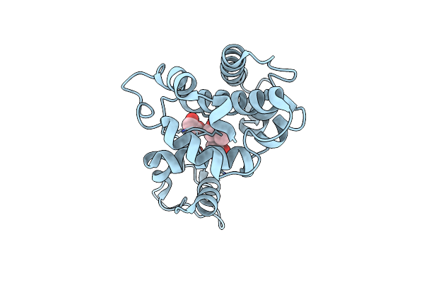 |
Class Iv Chitinase Structure From Picea Abies At 1.8A
Organism: Picea abies
Method: X-RAY DIFFRACTION Resolution:1.80 Å Release Date: 2009-08-11 Classification: HYDROLASE Ligands: MXE, ACT |
Organism: Picea abies
Method: X-RAY DIFFRACTION
Release Date: 2009-08-11
Ligands: MXE, ACT
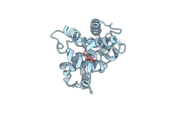 |
Class Iv Chitinase Structure From Picea Abies At 1.55A
Organism: Picea abies
Method: X-RAY DIFFRACTION Resolution:1.55 Å Release Date: 2009-08-11 Classification: HYDROLASE Ligands: MXE, ACT, FOR |
Organism: Picea abies
Method: X-RAY DIFFRACTION
Release Date: 2009-08-11
Ligands: MXE, ACT, FOR
 |
Class Iv Chitinase Structure From Picea Abies At 2.25A
Organism: Picea abies
Method: X-RAY DIFFRACTION Resolution:2.25 Å Release Date: 2009-08-11 Classification: HYDROLASE |
Organism: Picea abies
Method: X-RAY DIFFRACTION
Release Date: 2009-08-11
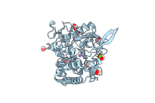 |
Crystal Structure Of Apo Form (Zinc Removed) Of The Botulinum Neurotoxin Type C Light Chain
Organism: Clostridium botulinum
Method: X-RAY DIFFRACTION Resolution:1.95 Å Release Date: 2009-04-07 Classification: HYDROLASE |
Organism: Clostridium botulinum
Method: X-RAY DIFFRACTION
Release Date: 2009-04-07
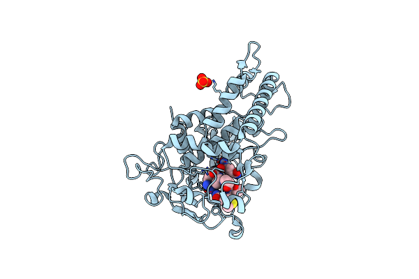 |
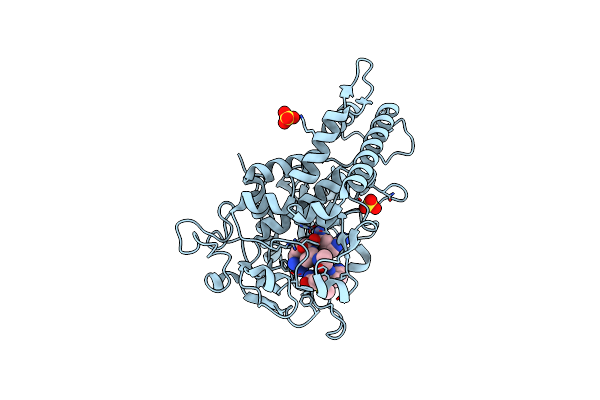
Crystal Structure Of The Catalytic Domain Of Botulinum Neurotoxin Serotype A With A Snap-25 Peptide
Organism: Clostridium botulinum, Synthetic construct
Method: X-RAY DIFFRACTION Resolution:1.50 Å Release Date: 2008-09-16 Classification: HYDROLASE Ligands: ZN, SO4 |
Organism: Clostridium botulinum, Synthetic construct
Method: X-RAY DIFFRACTION
Release Date: 2008-09-16
Ligands: ZN, SO4
 |
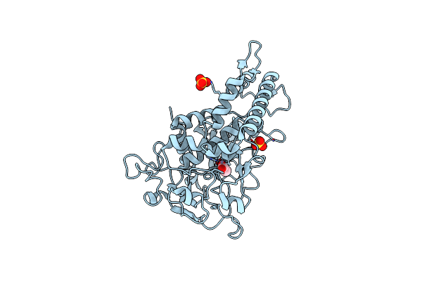
Crystal Structure Of The Catalytic Domain Of Botulinum Neurotoxin Serotype A With A Substrate Analog Peptide
Organism: Clostridium botulinum
Method: X-RAY DIFFRACTION Resolution:1.60 Å Release Date: 2008-09-16 Classification: HYDROLASE Ligands: ZN, SO4 |
Organism: Clostridium botulinum
Method: X-RAY DIFFRACTION
Release Date: 2008-09-16
Ligands: ZN, SO4
 |
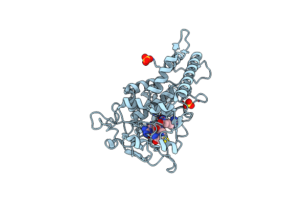
Crystal Structure Of The Catalytic Domain Of Botulinum Neurotoxin Serotype A With An Acetate Ion Bound At The Active Site
Organism: Clostridium botulinum
Method: X-RAY DIFFRACTION Resolution:1.70 Å Release Date: 2008-04-22 Classification: HYDROLASE Ligands: ZN, SO4, ACT |
Organism: Clostridium botulinum
Method: X-RAY DIFFRACTION
Release Date: 2008-04-22
Ligands: ZN, SO4, ACT
 |
Crystal Structure Of The Catalytic Domain Of Botulinum Neurotoxin Serotype A With Inhibitory Peptide Rrgc
Organism: Clostridium botulinum
Method: X-RAY DIFFRACTION Resolution:1.60 Å Release Date: 2008-04-22 Classification: HYDROLASE/HYDROLASE INHIBITOR Ligands: ZN, SO4, NA |
Organism: Clostridium botulinum
Method: X-RAY DIFFRACTION
Release Date: 2008-04-22
Ligands: ZN, SO4, NA
 |
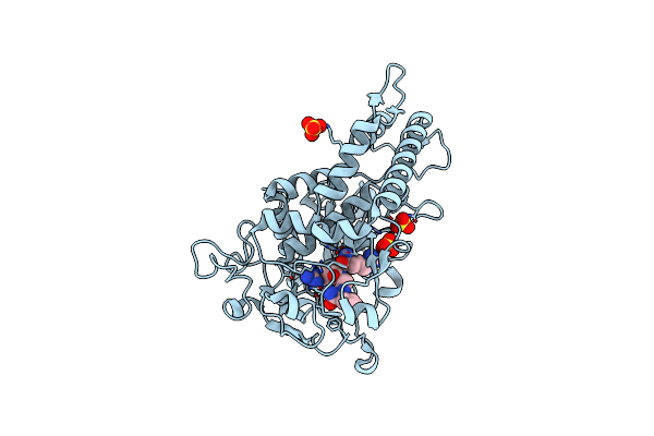
Crystal Structure Of The Catalytic Domain Of Botulinum Neurotoxin Serotype A With Inhibitory Peptide Rrgm
Organism: Clostridium botulinum
Method: X-RAY DIFFRACTION Resolution:1.58 Å Release Date: 2008-04-22 Classification: HYDROLASE/HYDROLASE INHIBITOR Ligands: ZN, SO4 |
Organism: Clostridium botulinum
Method: X-RAY DIFFRACTION
Release Date: 2008-04-22
Ligands: ZN, SO4
 |
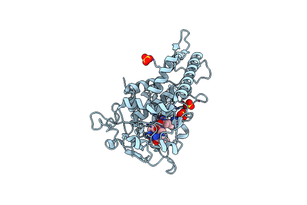
Crystal Structure Of The Catalytic Domain Of Botulinum Neurotoxin Serotype A With Inhibitory Peptide Rrgl
Organism: Clostridium botulinum, Synthetic construct
Method: X-RAY DIFFRACTION Resolution:1.52 Å Release Date: 2008-04-22 Classification: HYDROLASE/HYDROLASE INHIBITOR Ligands: ZN, SO4 |
Organism: Clostridium botulinum, Synthetic construct
Method: X-RAY DIFFRACTION
Release Date: 2008-04-22
Ligands: ZN, SO4
 |
Crystal Structure Of The Catalytic Domain Of Botulinum Neurotoxin Serotype A With Inhibitory Peptide Rrgi
Organism: Clostridium botulinum, Synthetic construct
Method: X-RAY DIFFRACTION Resolution:1.47 Å Release Date: 2008-04-22 Classification: HYDROLASE/HYDROLASE INHIBITOR Ligands: ZN, SO4 |
Organism: Clostridium botulinum, Synthetic construct
Method: X-RAY DIFFRACTION
Release Date: 2008-04-22
Ligands: ZN, SO4
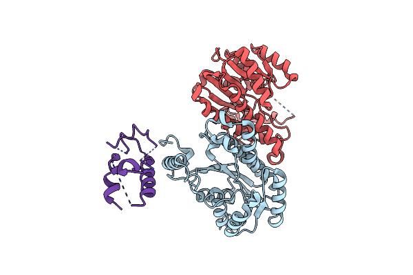 |
Structure Of Acyl Carrier Protein Bound To Fabi, The Enoyl Reductase From Escherichia Coli
Organism: Escherichia coli
Method: X-RAY DIFFRACTION Resolution:2.70 Å Release Date: 2006-10-17 Classification: OXIDOREDUCTASE/BIOSYNTHETIC PROTEIN |
Organism: Escherichia coli
Method: X-RAY DIFFRACTION
Release Date: 2006-10-17