

Search Count: 25
 |
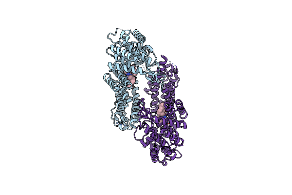
Rpl13 (El13)-Mutant 80S Ribosome From Mouse
Organism: Mus musculus
Method: ELECTRON MICROSCOPY Release Date: 2025-11-05 Classification: RIBOSOME Ligands: ZN, MG |
Organism: Mus musculus
Method: ELECTRON MICROSCOPY
Release Date: 2025-11-05
Ligands: ZN, MG
 |
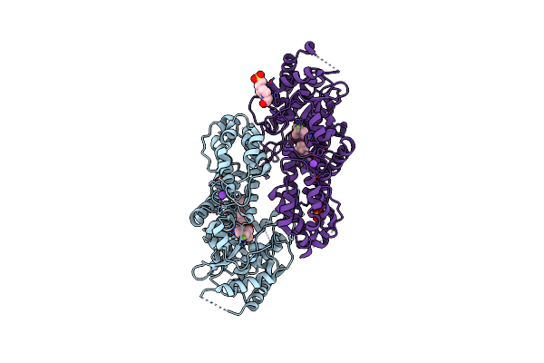
Crystal Structure Of Menin In Complex With Inhibitor Compound 15
Organism: Homo sapiens
Method: X-RAY DIFFRACTION Resolution:1.90 Å Release Date: 2025-01-15 Classification: PROTEIN BINDING/INHIBITOR Ligands: A1AVE |
Organism: Homo sapiens
Method: X-RAY DIFFRACTION
Release Date: 2025-01-15
Ligands: A1AVE
 |
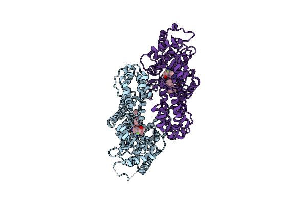
Crystal Structure Of Menin In Complex With Inhibitor Compound 26
Organism: Homo sapiens
Method: X-RAY DIFFRACTION Resolution:1.85 Å Release Date: 2025-01-15 Classification: PROTEIN BINDING/INHIBITOR Ligands: A1AVF, K, EPE, SO4 |
Organism: Homo sapiens
Method: X-RAY DIFFRACTION
Release Date: 2025-01-15
Ligands: A1AVF, K, EPE, SO4
 |
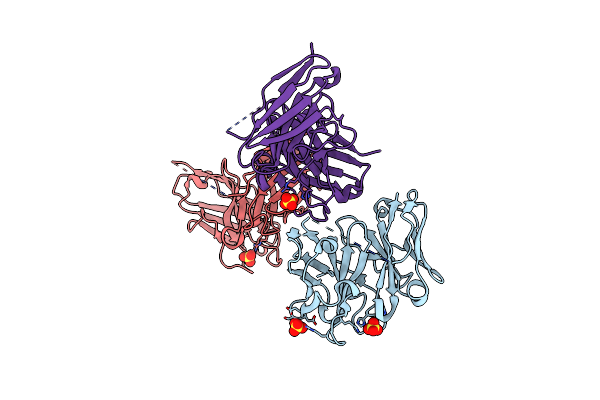
Crystal Structure Of Menin In Complex With Inhibitor Compound 20
Organism: Homo sapiens
Method: X-RAY DIFFRACTION Resolution:1.98 Å Release Date: 2025-01-15 Classification: PROTEIN BINDING/INHIBITOR Ligands: A1AVG |
Organism: Homo sapiens
Method: X-RAY DIFFRACTION
Release Date: 2025-01-15
Ligands: A1AVG
 |
Crystal Structure Of Scfv Corresponding To Human Autoantibody B96.11
Organism: Homo sapiens
Method: X-RAY DIFFRACTION Resolution:2.60 Å Release Date: 2024-11-06 Classification: IMMUNE SYSTEM Ligands: SO4 |
Organism: Homo sapiens
Method: X-RAY DIFFRACTION
Release Date: 2024-11-06
Ligands: SO4
 |
The Cryo-Em Structure Of A Complex Between Gad65 And B96.11 Fab
Organism: Homo sapiens
Method: ELECTRON MICROSCOPY Release Date: 2024-10-30 Classification: IMMUNE SYSTEM |
Organism: Homo sapiens
Method: ELECTRON MICROSCOPY
Release Date: 2024-10-30
 |
Crystal Structure Of Savinase At Cryogenic Conditions
Organism: Bacillus lentus
Method: X-RAY DIFFRACTION Resolution:0.95 Å Release Date: 2020-06-17 Classification: HYDROLASE Ligands: CA, NA |
Organism: Bacillus lentus
Method: X-RAY DIFFRACTION
Release Date: 2020-06-17
Ligands: CA, NA
 |
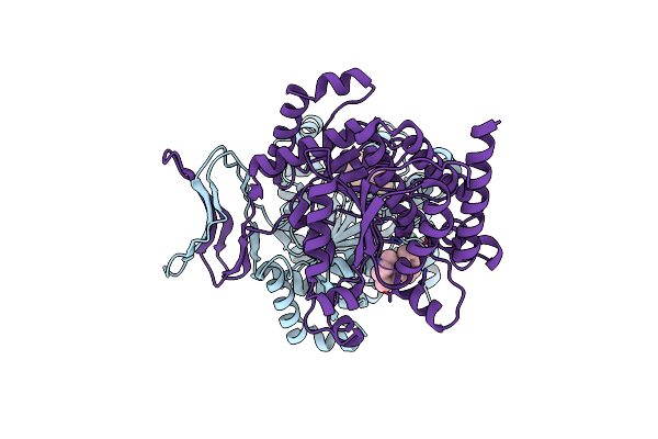
Crystal Structure Of Savinase At Room Temperature
Organism: Bacillus lentus
Method: X-RAY DIFFRACTION Resolution:1.10 Å Release Date: 2020-06-17 Classification: HYDROLASE Ligands: CA, NA |
Organism: Bacillus lentus
Method: X-RAY DIFFRACTION
Release Date: 2020-06-17
Ligands: CA, NA
 |
Crystal Structure Of Idh1 R132H In Complex With Hms101
Organism: Homo sapiens
Method: X-RAY DIFFRACTION Resolution:3.30 Å Release Date: 2019-10-16 Classification: OXIDOREDUCTASE Ligands: HJQ |
Organism: Homo sapiens
Method: X-RAY DIFFRACTION
Release Date: 2019-10-16
Ligands: HJQ
 |
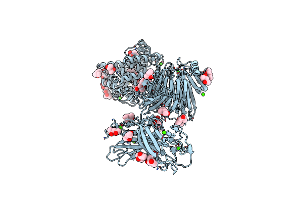
Crystal Structure Of Deaminated P Domain From Norovirus Strain Saga Gii-4 In Complex With Fuc
Organism: Norovirus hu/gii-4/saga4/2006/jp
Method: X-RAY DIFFRACTION Resolution:1.52 Å Release Date: 2019-04-03 Classification: VIRAL PROTEIN Ligands: MFU, MG |
Organism: Norovirus hu/gii-4/saga4/2006/jp
Method: X-RAY DIFFRACTION
Release Date: 2019-04-03
Ligands: MFU, MG
 |
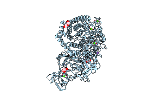
Structure Of Paenibacillus Xanthan Lyase To 2.6 A Resolution
Organism: Paenibacillus
Method: X-RAY DIFFRACTION Resolution:2.60 Å Release Date: 2018-12-05 Classification: LYASE Ligands: CA, CL, BTB, MRD, MPD |
Organism: Paenibacillus
Method: X-RAY DIFFRACTION
Release Date: 2018-12-05
Ligands: CA, CL, BTB, MRD, MPD
 |
Structural Dynamics And Catalytic Properties Of A Multi-Modular Xanthanase, Native.
Organism: Paenibacillus
Method: X-RAY DIFFRACTION Resolution:2.04 Å Release Date: 2018-08-29 Classification: HYDROLASE Ligands: CA, NA, CL, PEG, MLI |
Organism: Paenibacillus
Method: X-RAY DIFFRACTION
Release Date: 2018-08-29
Ligands: CA, NA, CL, PEG, MLI
 |
Structural Dynamics And Catalytic Properties Of A Multi-Modular Xanthanase (Pt Derivative)
Organism: Paenibacillus sp.
Method: X-RAY DIFFRACTION Resolution:2.00 Å Release Date: 2018-08-29 Classification: HYDROLASE Ligands: PT, B3P, SCN, CA, NA, CL, MLI |
Organism: Paenibacillus sp.
Method: X-RAY DIFFRACTION
Release Date: 2018-08-29
Ligands: PT, B3P, SCN, CA, NA, CL, MLI
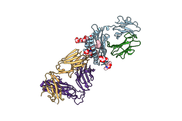 |
Structure Of Alpha-Galactosylphytosphingosine Bound By Cd1D And In Complex With The Va14Vb8.2 Tcr
Organism: Mus musculus, Homo sapiens
Method: X-RAY DIFFRACTION Resolution:3.16 Å Release Date: 2018-04-04 Classification: IMMUNE SYSTEM Ligands: N57, PLM, NAG |
Organism: Mus musculus, Homo sapiens
Method: X-RAY DIFFRACTION
Release Date: 2018-04-04
Ligands: N57, PLM, NAG
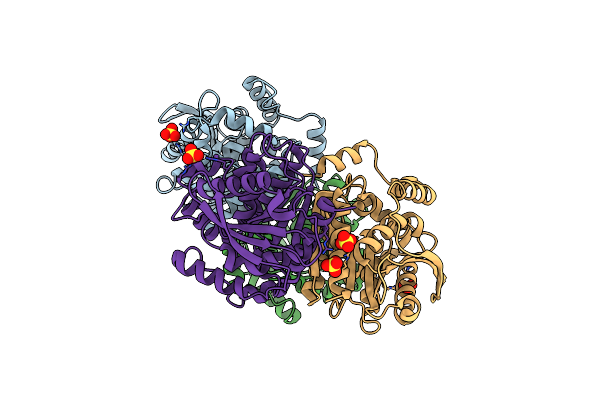 |
Crystal Structure Of A Lipase Carboxylesterase From Sulfolobus Islandicus
Organism: Sulfolobus islandicus (strain rey15a)
Method: X-RAY DIFFRACTION Resolution:2.60 Å Release Date: 2017-08-02 Classification: HYDROLASE Ligands: SO4 |
Organism: Sulfolobus islandicus (strain rey15a)
Method: X-RAY DIFFRACTION
Release Date: 2017-08-02
Ligands: SO4
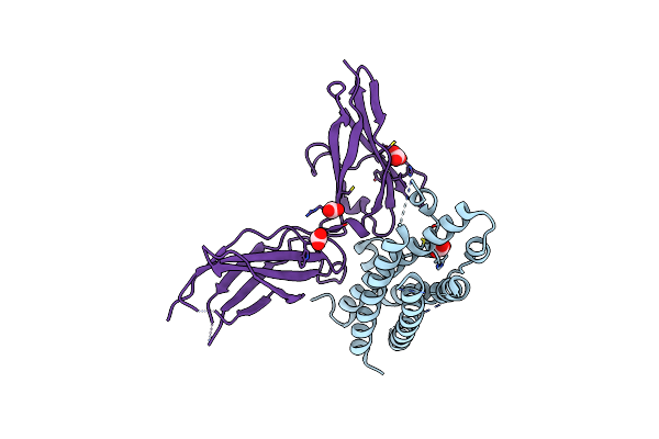 |
Crystal Structure Of A Prolactin Receptor Antagonist Bound To The Extracellular Domain Of The Prolactin Receptor
Organism: Homo sapiens
Method: X-RAY DIFFRACTION Resolution:2.50 Å Release Date: 2008-06-03 Classification: Hormone/Hormone Receptor Ligands: CO3 |
Organism: Homo sapiens
Method: X-RAY DIFFRACTION
Release Date: 2008-06-03
Ligands: CO3
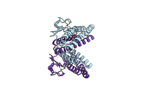 |
Pf1283- Rubrerythrin From Pyrococcus Furiosus Iron Bound Form
Organism: Pyrococcus furiosus
Method: X-RAY DIFFRACTION Resolution:2.70 Å Release Date: 2006-10-17 Classification: METAL BINDING PROTEIN Ligands: FE |
Organism: Pyrococcus furiosus
Method: X-RAY DIFFRACTION
Release Date: 2006-10-17
Ligands: FE
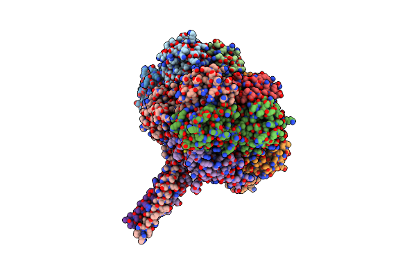 |
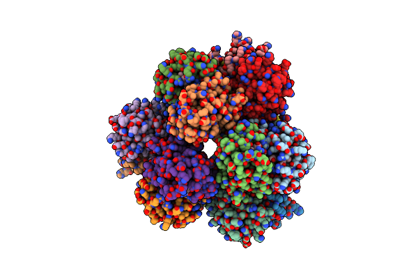
Lumbricus Erythrocruorin At 3.5A Resolution
Organism: Lumbricus terrestris
Method: X-RAY DIFFRACTION Resolution:3.50 Å Release Date: 2006-07-18 Classification: OXYGEN STORAGE/TRANSPORT Ligands: CMO, HEM, CA, ZN |
Organism: Lumbricus terrestris
Method: X-RAY DIFFRACTION
Release Date: 2006-07-18
Ligands: CMO, HEM, CA, ZN
 |
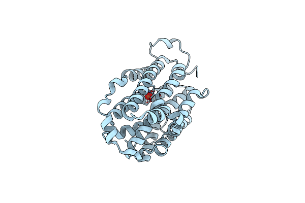
Hemoglobin Dodecamer From Lumbricus Erythrocruorin
Organism: Lumbricus terrestris
Method: X-RAY DIFFRACTION Resolution:2.60 Å Release Date: 2004-11-30 Classification: OXYGEN STORAGE/TRANSPORT Ligands: HEM, CMO, PO4 |
Organism: Lumbricus terrestris
Method: X-RAY DIFFRACTION
Release Date: 2004-11-30
Ligands: HEM, CMO, PO4
 |
Crystal Structure Of Mouse Ribonucleotide Reductase Subunit R2 Under Oxidizing Conditions. A Fully Occupied Dinuclear Iron Cluster.
Organism: Mus musculus
Method: X-RAY DIFFRACTION Resolution:2.20 Å Release Date: 2004-08-26 Classification: REDUCTASE Ligands: FEO |
Organism: Mus musculus
Method: X-RAY DIFFRACTION
Release Date: 2004-08-26
Ligands: FEO