

Search Count: 37
 |
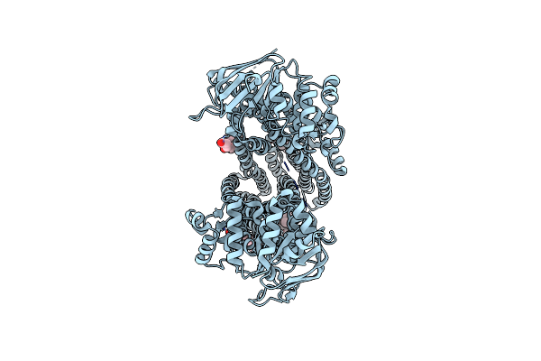
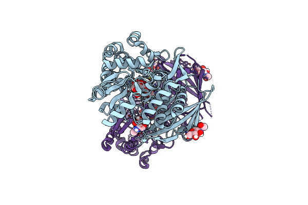
Crystal Structure Of Glutamine Synthetase-Apo
Organism: Acinetobacter baumannii ab0057
Method: X-RAY DIFFRACTION Resolution:3.40 Å Release Date: 2025-03-19 Classification: LIGASE Ligands: PGE, GOL, MG, EDO |
Organism: Acinetobacter baumannii ab0057
Method: X-RAY DIFFRACTION
Release Date: 2025-03-19
Ligands: PGE, GOL, MG, EDO
 |
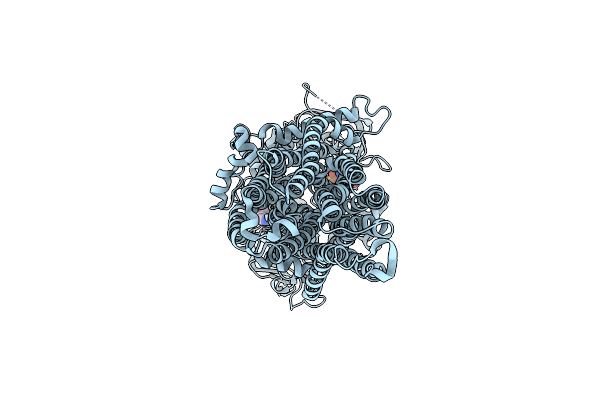
Cryo-Em Structure Of Nanodisc-Reconstituted Wildtype Human Mrp4 (Inward-Facing Conformation)
Organism: Homo sapiens
Method: ELECTRON MICROSCOPY Release Date: 2023-08-30 Classification: TRANSLOCASE Ligands: CLR, Y01 |
Organism: Homo sapiens
Method: ELECTRON MICROSCOPY
Release Date: 2023-08-30
Ligands: CLR, Y01
 |
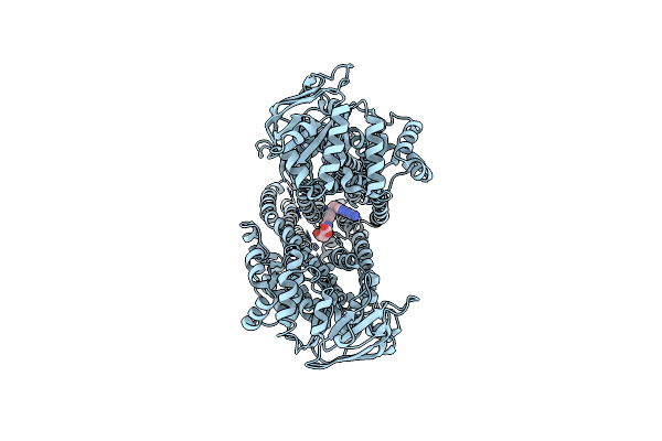
Cryo-Em Structure Of Nanodisc-Reconstituted Human Mrp4 With E1202Q Mutation (Outward-Facing Occluded Conformation)
Organism: Homo sapiens
Method: ELECTRON MICROSCOPY Release Date: 2023-08-30 Classification: TRANSLOCASE Ligands: ATP, MG |
Organism: Homo sapiens
Method: ELECTRON MICROSCOPY
Release Date: 2023-08-30
Ligands: ATP, MG
 |
Cryo-Em Structure Of Nanodisc-Reconstituted Wildtype Human Mrp4 (In Complex With Methotrexate)
Organism: Homo sapiens
Method: ELECTRON MICROSCOPY Release Date: 2023-08-30 Classification: TRANSLOCASE Ligands: MTX |
Organism: Homo sapiens
Method: ELECTRON MICROSCOPY
Release Date: 2023-08-30
Ligands: MTX
 |
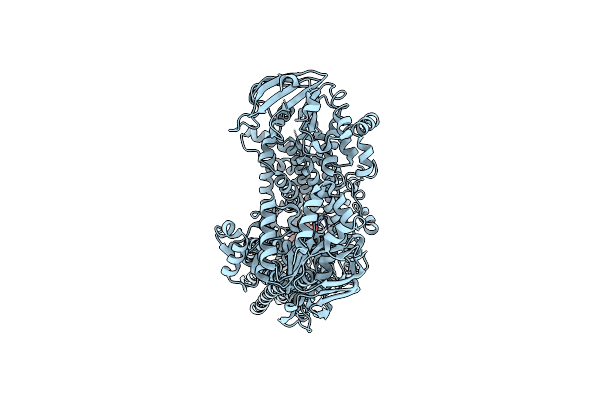
Cryo-Em Structure Of Nanodisc-Reconstituted Wildtype Human Mrp4 (In Complex With Topotecan)
Organism: Homo sapiens
Method: ELECTRON MICROSCOPY Release Date: 2023-08-30 Classification: TRANSLOCASE Ligands: TTC |
Organism: Homo sapiens
Method: ELECTRON MICROSCOPY
Release Date: 2023-08-30
Ligands: TTC
 |
Cryo-Em Structure Of Nanodisc-Reconstituted Wildtype Human Mrp4 (In Complex With Prostaglandin E2)
Organism: Homo sapiens
Method: ELECTRON MICROSCOPY Release Date: 2023-08-30 Classification: TRANSLOCASE Ligands: P2E |
Organism: Homo sapiens
Method: ELECTRON MICROSCOPY
Release Date: 2023-08-30
Ligands: P2E
 |
Structure Of Hypothetical Protein Ttha1873 With Phosphate From Thermus Thermophilus
Organism: Thermus thermophilus hb8
Method: X-RAY DIFFRACTION Resolution:2.22 Å Release Date: 2022-11-09 Classification: UNKNOWN FUNCTION Ligands: CA, PO4 |
Organism: Thermus thermophilus hb8
Method: X-RAY DIFFRACTION
Release Date: 2022-11-09
Ligands: CA, PO4
 |
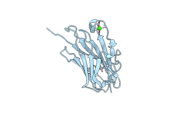
Structure Of Hypothetical Protein Ttha1873 From Thermus Thermophilus With Potassium Mercuric Iodide
Organism: Thermus thermophilus hb8
Method: X-RAY DIFFRACTION Resolution:2.05 Å Release Date: 2022-04-06 Classification: UNKNOWN FUNCTION Ligands: CA, 72I |
Organism: Thermus thermophilus hb8
Method: X-RAY DIFFRACTION
Release Date: 2022-04-06
Ligands: CA, 72I
 |
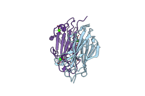
Structure Of Hypothetical Protein Ttha1873 From Thermus Thermophilus
Organism: Thermus thermophilus hb8
Method: X-RAY DIFFRACTION Resolution:1.78 Å Release Date: 2022-03-09 Classification: UNKNOWN FUNCTION Ligands: CA |
Organism: Thermus thermophilus hb8
Method: X-RAY DIFFRACTION
Release Date: 2022-03-09
Ligands: CA
 |
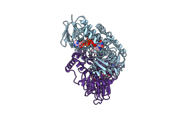
Dimer Form Of Hypothetical Protein Ttha1873 From Thermus Thermophilus
Organism: Thermus thermophilus hb8
Method: X-RAY DIFFRACTION Resolution:2.17 Å Release Date: 2022-03-09 Classification: UNKNOWN FUNCTION Ligands: CA |
Organism: Thermus thermophilus hb8
Method: X-RAY DIFFRACTION
Release Date: 2022-03-09
Ligands: CA
 |
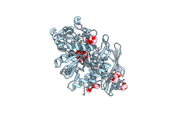
Putative Leptospira Interrogans Recombinant L-Amino Acid Oxidase
Organism: Leptospira interrogans
Method: X-RAY DIFFRACTION Resolution:1.78 Å Release Date: 2021-08-18 Classification: OXIDOREDUCTASE Ligands: FAD |
Organism: Leptospira interrogans
Method: X-RAY DIFFRACTION
Release Date: 2021-08-18
Ligands: FAD
 |
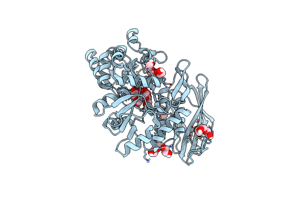
Crystal Structure Of Red Abalone Verl Repeat 1 With Linker At 2.0 A Resolution
Organism: Escherichia coli o157:h7, Haliotis rufescens, Escherichia coli
Method: X-RAY DIFFRACTION Resolution:2.00 Å Release Date: 2017-06-14 Classification: CELL ADHESION Ligands: PGE |
Organism: Escherichia coli o157:h7, Haliotis rufescens, Escherichia coli
Method: X-RAY DIFFRACTION
Release Date: 2017-06-14
Ligands: PGE
 |
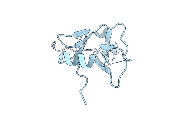
Crystal Structure Of Red Abalone Verl Repeat 1 At 1.8 A Resolution
Organism: Escherichia coli, Haliotis rufescens
Method: X-RAY DIFFRACTION Resolution:1.80 Å Release Date: 2017-06-14 Classification: CELL ADHESION Ligands: PGE |
Organism: Escherichia coli, Haliotis rufescens
Method: X-RAY DIFFRACTION
Release Date: 2017-06-14
Ligands: PGE
 |
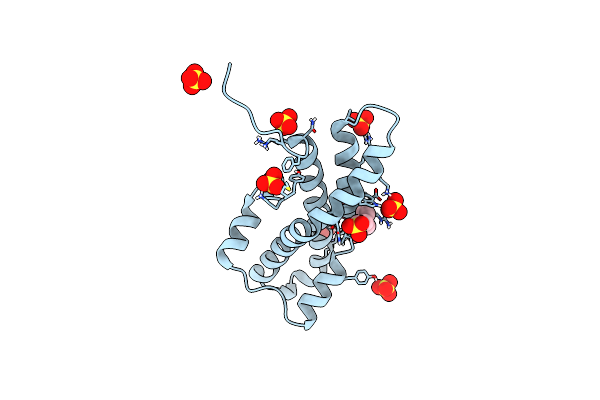
Crystal Structure Of The Zp-N1 Domain Of Mouse Sperm Receptor Zp2 At 0.95 A Resolution
Organism: Mus musculus
Method: X-RAY DIFFRACTION Resolution:0.95 Å Release Date: 2017-06-14 Classification: CELL ADHESION |
Organism: Mus musculus
Method: X-RAY DIFFRACTION
Release Date: 2017-06-14
 |
In-House Sulfur-Sad Structure Of Orthorhombic Red Abalone Lysin At 1.66 A Resolution
Organism: Haliotis rufescens
Method: X-RAY DIFFRACTION Resolution:1.66 Å Release Date: 2017-06-14 Classification: CELL ADHESION Ligands: SO4, MES |
Organism: Haliotis rufescens
Method: X-RAY DIFFRACTION
Release Date: 2017-06-14
Ligands: SO4, MES
 |
Orthorhombic Crystal Structure Of Red Abalone Lysin At 0.99 A Resolution
Organism: Haliotis rufescens
Method: X-RAY DIFFRACTION Resolution:0.99 Å Release Date: 2017-06-14 Classification: CELL ADHESION Ligands: SO4, MES |
Organism: Haliotis rufescens
Method: X-RAY DIFFRACTION
Release Date: 2017-06-14
Ligands: SO4, MES
 |
Monoclinic Crystal Structure Of Red Abalone Lysin At 2.11 A Resolution
Organism: Haliotis rufescens
Method: X-RAY DIFFRACTION Resolution:2.11 Å Release Date: 2017-06-14 Classification: CELL ADHESION |
Organism: Haliotis rufescens
Method: X-RAY DIFFRACTION
Release Date: 2017-06-14
 |
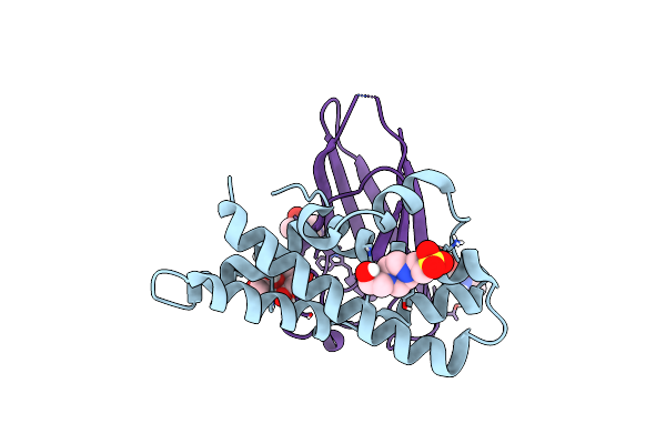
Crystal Structure Of Red Abalone Egg Verl Repeat 3 In Complex With Sperm Lysin At 1.7 A Resolution (Crystal Form I)
Organism: Haliotis rufescens
Method: X-RAY DIFFRACTION Resolution:1.70 Å Release Date: 2017-06-14 Classification: CELL ADHESION Ligands: NAG |
Organism: Haliotis rufescens
Method: X-RAY DIFFRACTION
Release Date: 2017-06-14
Ligands: NAG
 |
Crystal Structure Of Red Abalone Egg Verl Repeat 3 In Complex With Sperm Lysin At 1.64 A Resolution (Crystal Form Ii)
Organism: Haliotis rufescens
Method: X-RAY DIFFRACTION Resolution:1.64 Å Release Date: 2017-06-14 Classification: CELL ADHESION Ligands: EPE, NAG |
Organism: Haliotis rufescens
Method: X-RAY DIFFRACTION
Release Date: 2017-06-14
Ligands: EPE, NAG
 |
Crystal Structure Of Red Abalone Verl Repeat 3 At 2.9 A Resolution
Organism: Escherichia coli, Haliotis rufescens
Method: X-RAY DIFFRACTION Resolution:2.90 Å Release Date: 2017-06-14 Classification: CELL ADHESION Ligands: NAG |
Organism: Escherichia coli, Haliotis rufescens
Method: X-RAY DIFFRACTION
Release Date: 2017-06-14
Ligands: NAG