

Search Count: 71
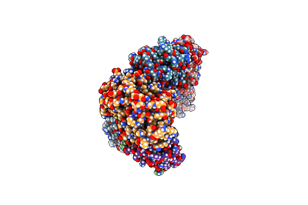 |
Mouse Cnpase Catalytic Domain With Nanobody 7E
Organism: Mus musculus, Vicugna pacos
Method: X-RAY DIFFRACTION Resolution:2.50 Å Release Date: 2024-12-25 Classification: PROTEIN BINDING |
Organism: Mus musculus, Vicugna pacos
Method: X-RAY DIFFRACTION
Release Date: 2024-12-25
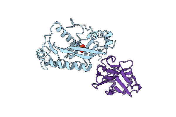 |
Mouse Cnpase Catalytic Domain With Nano Body 5E
Organism: Mus musculus, Vicugna pacos
Method: X-RAY DIFFRACTION Resolution:2.75 Å Release Date: 2024-12-11 Classification: PROTEIN BINDING Ligands: PO4 |
Organism: Mus musculus, Vicugna pacos
Method: X-RAY DIFFRACTION
Release Date: 2024-12-11
Ligands: PO4
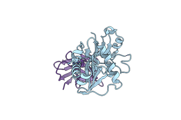 |
Mouse Cnpase Catalytic Domain With Nanobody 8C
Organism: Vicugna pacos, Mus musculus
Method: X-RAY DIFFRACTION Resolution:1.73 Å Release Date: 2024-12-11 Classification: PROTEIN BINDING |
Organism: Vicugna pacos, Mus musculus
Method: X-RAY DIFFRACTION
Release Date: 2024-12-11
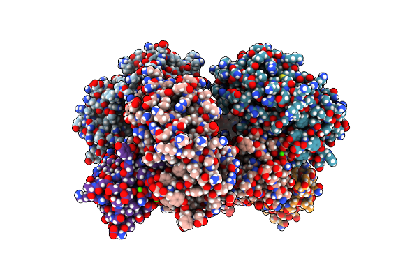 |
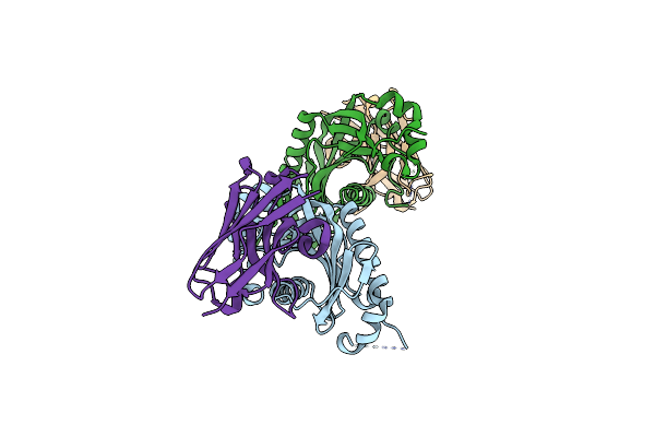
Mouse Cnpase Catalytic Domain With Nanobody 10E
Organism: Mus musculus, Vicugna pacos
Method: X-RAY DIFFRACTION Resolution:2.55 Å Release Date: 2024-12-11 Classification: PROTEIN BINDING Ligands: CA |
Organism: Mus musculus, Vicugna pacos
Method: X-RAY DIFFRACTION
Release Date: 2024-12-11
Ligands: CA
 |
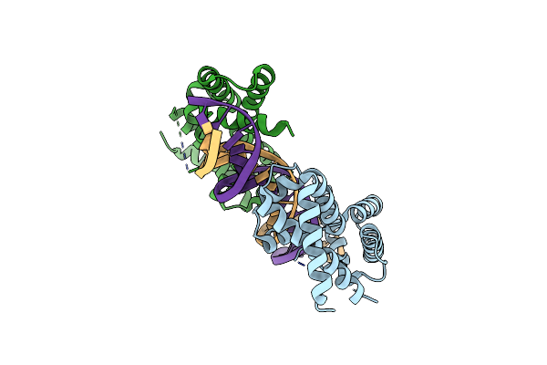
Mouse Cnpase Catalytic Domain With Nanobody 8D
Organism: Vicugna pacos, Mus musculus
Method: X-RAY DIFFRACTION Resolution:1.50 Å Release Date: 2024-12-11 Classification: PROTEIN BINDING |
Organism: Vicugna pacos, Mus musculus
Method: X-RAY DIFFRACTION
Release Date: 2024-12-11
 |
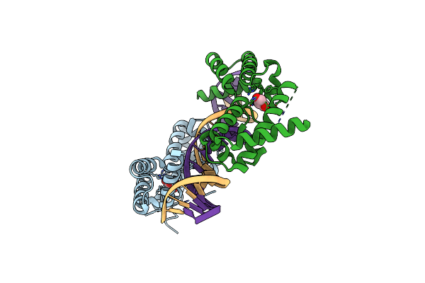
Dna Binding Domain Of Hnf-1A Bound To P2-Hnf4A Promoter Dna Variant (P2 -169C>T)
Organism: Homo sapiens
Method: X-RAY DIFFRACTION Resolution:3.20 Å Release Date: 2024-06-19 Classification: DNA BINDING PROTEIN Ligands: CL |
Organism: Homo sapiens
Method: X-RAY DIFFRACTION
Release Date: 2024-06-19
Ligands: CL
 |
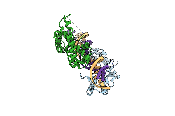
Dna Binding Domain Of Hnf-1A Bound To P2-Hnf4A Promoter Dna
Organism: Homo sapiens
Method: X-RAY DIFFRACTION Resolution:2.30 Å Release Date: 2024-06-19 Classification: DNA BINDING PROTEIN Ligands: GOL |
Organism: Homo sapiens
Method: X-RAY DIFFRACTION
Release Date: 2024-06-19
Ligands: GOL
 |
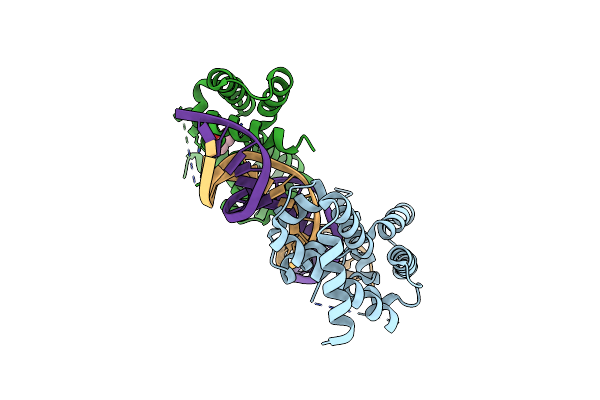
Dna Binding Domain Of Hnf-1A Bound To P2-Hnf4A Promoter Dna Variant (P2 -181G>A)
Organism: Homo sapiens
Method: X-RAY DIFFRACTION Resolution:2.80 Å Release Date: 2024-06-19 Classification: DNA BINDING PROTEIN |
Organism: Homo sapiens
Method: X-RAY DIFFRACTION
Release Date: 2024-06-19
 |
Dna Binding Domain Of Hnf-1A Bound To P2-Hnf4A Promoter Dna Variant (P2 -181G>T)
Organism: Homo sapiens
Method: X-RAY DIFFRACTION Resolution:2.80 Å Release Date: 2024-06-19 Classification: DNA BINDING PROTEIN Ligands: GOL |
Organism: Homo sapiens
Method: X-RAY DIFFRACTION
Release Date: 2024-06-19
Ligands: GOL
 |
Crystal Structure Of The Human Gdap1 Cmt2 Mutant-H123R
Organism: Homo sapiens
Method: X-RAY DIFFRACTION Resolution:2.20 Å Release Date: 2022-06-08 Classification: SIGNALING PROTEIN Ligands: PEG |
Organism: Homo sapiens
Method: X-RAY DIFFRACTION
Release Date: 2022-06-08
Ligands: PEG
 |
Crystal Structure Of The Human Gdap1 Cmt2 Mutant-R120W
Organism: Homo sapiens
Method: X-RAY DIFFRACTION Resolution:3.41 Å Release Date: 2022-06-08 Classification: SIGNALING PROTEIN |
Organism: Homo sapiens
Method: X-RAY DIFFRACTION
Release Date: 2022-06-08
 |
Human Gdap1 Core Domain, Trigonal Crystal Form
Organism: Homo sapiens
Method: X-RAY DIFFRACTION Resolution:3.20 Å Release Date: 2022-06-08 Classification: MEMBRANE PROTEIN |
Organism: Homo sapiens
Method: X-RAY DIFFRACTION
Release Date: 2022-06-08
 |
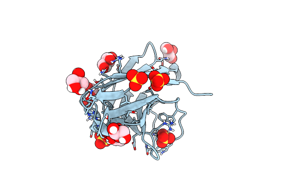
C-Terminal Domain Of Harc In Complex With Nanobodies H11 And C11, Collapsed Crystal Form
Organism: Vicugna pacos, Rattus norvegicus
Method: X-RAY DIFFRACTION Resolution:1.94 Å Release Date: 2022-06-01 Classification: SIGNALING PROTEIN |
Organism: Vicugna pacos, Rattus norvegicus
Method: X-RAY DIFFRACTION
Release Date: 2022-06-01
 |
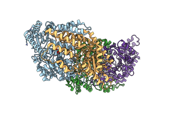
Anti-Arc Nanobody E5
Organism: Vicugna pacos
Method: X-RAY DIFFRACTION Resolution:1.42 Å Release Date: 2022-06-01 Classification: IMMUNE SYSTEM Ligands: GOL, SO4 |
Organism: Vicugna pacos
Method: X-RAY DIFFRACTION
Release Date: 2022-06-01
Ligands: GOL, SO4
 |
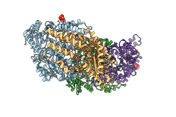
Crystal Structure Of Mouse Csad In Apo Form
Organism: Mus musculus
Method: X-RAY DIFFRACTION Resolution:2.80 Å Release Date: 2021-08-18 Classification: LYASE Ligands: SO4, NA |
Organism: Mus musculus
Method: X-RAY DIFFRACTION
Release Date: 2021-08-18
Ligands: SO4, NA
 |
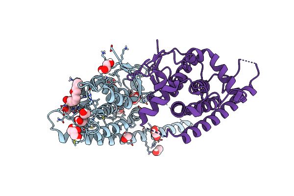
Crystal Structure Of Mouse Csad
Organism: Mus musculus
Method: X-RAY DIFFRACTION Resolution:2.10 Å Release Date: 2021-04-28 Classification: LYASE Ligands: SO4, CO, CL, GOL |
Organism: Mus musculus
Method: X-RAY DIFFRACTION
Release Date: 2021-04-28
Ligands: SO4, CO, CL, GOL
 |
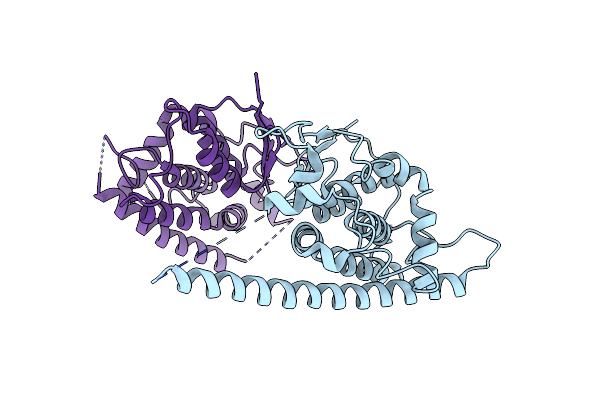
Complex Of Human Gdap1 With Hexadecanedioic Acid
Organism: Homo sapiens
Method: X-RAY DIFFRACTION Resolution:2.20 Å Release Date: 2021-02-24 Classification: SIGNALING PROTEIN Ligands: GOL, EOH, RF8 |
Organism: Homo sapiens
Method: X-RAY DIFFRACTION
Release Date: 2021-02-24
Ligands: GOL, EOH, RF8
 |
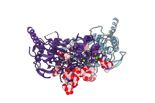
Crystal Structure Of Human Gdap1 At 2.8 Angstrom Resolution.
Organism: Homo sapiens
Method: X-RAY DIFFRACTION Resolution:2.80 Å Release Date: 2021-02-24 Classification: SIGNALING PROTEIN |
Organism: Homo sapiens
Method: X-RAY DIFFRACTION
Release Date: 2021-02-24
 |
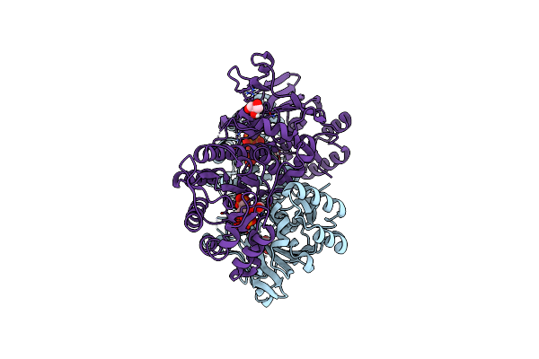
Crystal Structure Of Human Transmembrane Prolyl 4-Hydroxylase
Organism: Homo sapiens
Method: X-RAY DIFFRACTION Resolution:2.25 Å Release Date: 2020-12-23 Classification: OXIDOREDUCTASE Ligands: OGA, FE2, TBU, GLY, CA, CL |
Organism: Homo sapiens
Method: X-RAY DIFFRACTION
Release Date: 2020-12-23
Ligands: OGA, FE2, TBU, GLY, CA, CL
 |
Human Nampt In Complex With Nicotinic Acid And Phosphoribosyl Pyrophosphate
Organism: Homo sapiens
Method: X-RAY DIFFRACTION Resolution:1.58 Å Release Date: 2020-11-18 Classification: TRANSFERASE Ligands: PRP, GOL, NIO |
Organism: Homo sapiens
Method: X-RAY DIFFRACTION
Release Date: 2020-11-18
Ligands: PRP, GOL, NIO