

Search Count: 19
 |
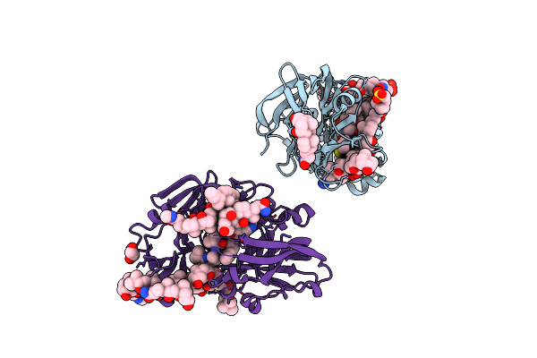
Cyp3A4 Bound To Ritonavir
Organism: Homo sapiens
Method: ELECTRON MICROSCOPY Release Date: 2026-09-30 Classification: OXIDOREDUCTASE Ligands: HEM, RIT |
Organism: Homo sapiens
Method: ELECTRON MICROSCOPY
Release Date: 2026-09-30
Ligands: HEM, RIT
 |
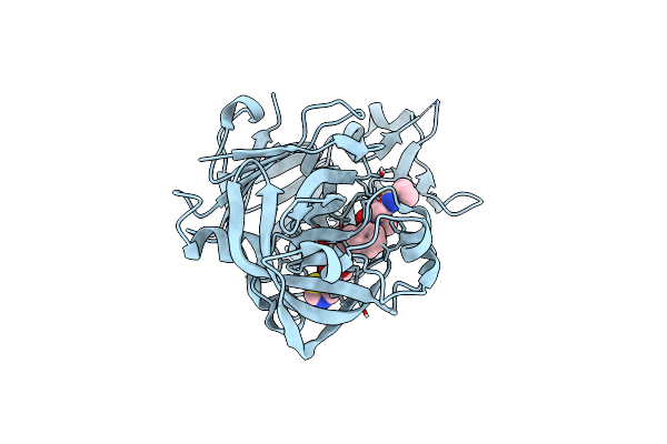
Crystal Structure Of Ritonavir Bound Plasmepsin Ii (Pmii) From Plasmodium Falciparum
Organism: Plasmodium falciparum (isolate 3d7)
Method: X-RAY DIFFRACTION Resolution:1.90 Å Release Date: 2023-02-01 Classification: HYDROLASE Ligands: RIT, CPS, EDO |
Organism: Plasmodium falciparum (isolate 3d7)
Method: X-RAY DIFFRACTION
Release Date: 2023-02-01
Ligands: RIT, CPS, EDO
 |
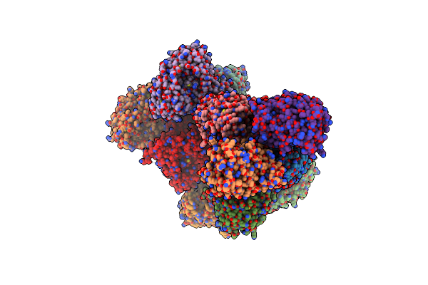
Porcine Pepsin In Complex With Ritonavir
Organism: Sus scrofa
Method: X-RAY DIFFRACTION Resolution:2.05 Å Release Date: 2021-06-16 Classification: HYDROLASE/INHIBITOR Ligands: RIT |
Organism: Sus scrofa
Method: X-RAY DIFFRACTION
Release Date: 2021-06-16
Ligands: RIT
 |
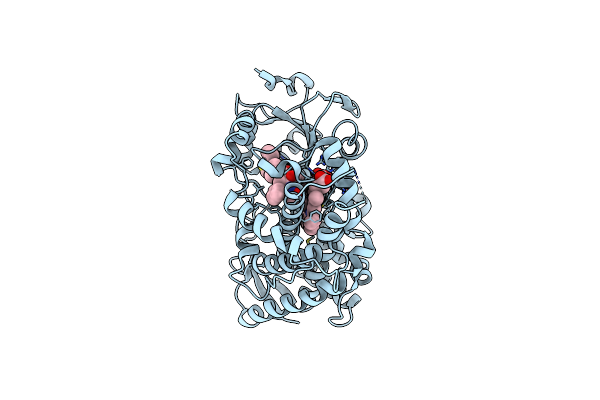
Human Cytochrome P450 3A5 (Cyp3A5)
Organism: Homo sapiens
Method: X-RAY DIFFRACTION Resolution:2.91 Å Release Date: 2017-11-15 Classification: OXIDOREDUCTASE/OXIDOREDUCTASE inhibitor Ligands: HEM, RIT |
Organism: Homo sapiens
Method: X-RAY DIFFRACTION
Release Date: 2017-11-15
Ligands: HEM, RIT
 |
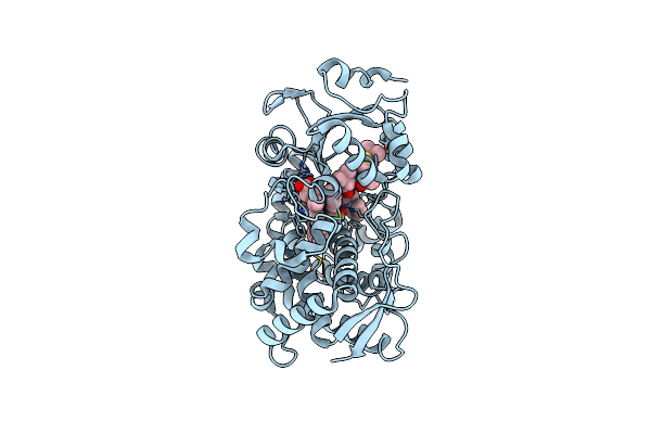
Crystal Structure Of Human Cyp3A4 Bound To Ritonavir
Organism: Homo sapiens
Method: X-RAY DIFFRACTION Resolution:2.70 Å Release Date: 2017-05-31 Classification: OXIDOREDUCTASE/INHIBITOR Ligands: HEM, RIT |
Organism: Homo sapiens
Method: X-RAY DIFFRACTION
Release Date: 2017-05-31
Ligands: HEM, RIT
 |
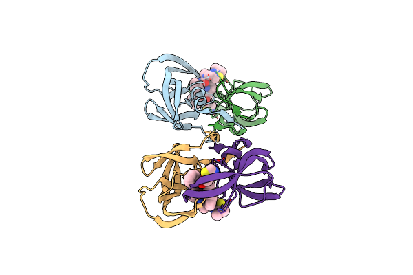
Crystal Structure Of The Cysteine Depleted Cyp3A4 Bound To Ritonavir
Organism: Homo sapiens
Method: X-RAY DIFFRACTION Resolution:2.20 Å Release Date: 2017-05-31 Classification: OXIDOREDUCTASE/INHIBITOR Ligands: HEM, RIT |
Organism: Homo sapiens
Method: X-RAY DIFFRACTION
Release Date: 2017-05-31
Ligands: HEM, RIT
 |
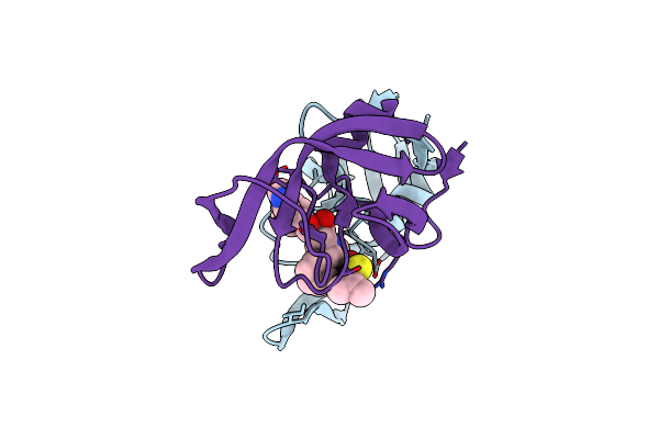
Crystal Structure Of Multidrug-Resistant Clinical Isolate A02 Hiv-1 Protease In Complex With Ritonavir
Organism: Human immunodeficiency virus 1
Method: X-RAY DIFFRACTION Resolution:1.80 Å Release Date: 2014-04-02 Classification: Hydrolase/Hydrolase inhibitor Ligands: RIT |
Organism: Human immunodeficiency virus 1
Method: X-RAY DIFFRACTION
Release Date: 2014-04-02
Ligands: RIT
 |
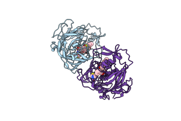
Crystal Structure Of Multidrug-Resistant Clinical Isolate 769 Hiv-1 Protease In Complex With Ritonavir
Organism: Human immunodeficiency virus 1
Method: X-RAY DIFFRACTION Resolution:1.80 Å Release Date: 2013-01-30 Classification: HYDROLASE/HYDROLASE INHIBITOR Ligands: RIT |
Organism: Human immunodeficiency virus 1
Method: X-RAY DIFFRACTION
Release Date: 2013-01-30
Ligands: RIT
 |
The Crystal Structure Of Protease Sapp1P From Candida Parapsilosis In Complex With The Hiv Protease Inhibitor Ritonavir
Organism: Candida parapsilosis
Method: X-RAY DIFFRACTION Resolution:2.40 Å Release Date: 2012-03-07 Classification: HYDROLASE/HYDROLASE INHIBITOR Ligands: RIT |
Organism: Candida parapsilosis
Method: X-RAY DIFFRACTION
Release Date: 2012-03-07
Ligands: RIT
 |
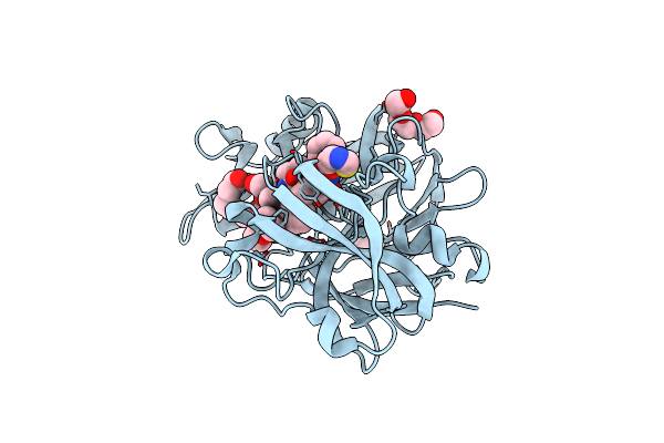
Secreted Aspartic Protease In Complex With Ritonavir
Organism: Candida albicans
Method: X-RAY DIFFRACTION Resolution:1.40 Å Release Date: 2012-01-04 Classification: HYDROLASE/HYDROLASE INHIBITOR Ligands: RIT, ZN, MRD |
Organism: Candida albicans
Method: X-RAY DIFFRACTION
Release Date: 2012-01-04
Ligands: RIT, ZN, MRD
 |
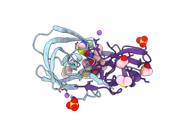
Endothiapepsin In Complex With Ritonavir
Organism: Cryphonectria parasitica
Method: X-RAY DIFFRACTION Resolution:1.38 Å Release Date: 2011-10-26 Classification: HYDROLASE/HYDROLASE INHIBITOR Ligands: RIT, DMS, 1PE, PGE, GOL |
Organism: Cryphonectria parasitica
Method: X-RAY DIFFRACTION
Release Date: 2011-10-26
Ligands: RIT, DMS, 1PE, PGE, GOL
 |
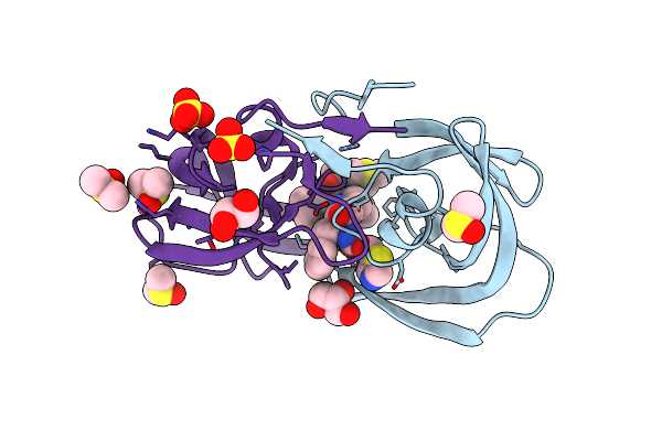
Hiv-1 Protease Saquinavir:Ritonavir 1:15 Complex Structure
Organism: Human immunodeficiency virus 1
Method: X-RAY DIFFRACTION Resolution:1.14 Å Release Date: 2011-07-20 Classification: hydrolase/hydrolase inhibitor Ligands: RIT, DMS, SO4, NA, ACT |
Organism: Human immunodeficiency virus 1
Method: X-RAY DIFFRACTION
Release Date: 2011-07-20
Ligands: RIT, DMS, SO4, NA, ACT
 |
Hiv-1 Protease Saquinavir:Ritonavir 1:50 Complex Structure
Organism: Human immunodeficiency virus 1
Method: X-RAY DIFFRACTION Resolution:1.03 Å Release Date: 2011-07-20 Classification: hydrolase/hydrolase inhibitor Ligands: RIT, DMS, GOL, CL, SO4 |
Organism: Human immunodeficiency virus 1
Method: X-RAY DIFFRACTION
Release Date: 2011-07-20
Ligands: RIT, DMS, GOL, CL, SO4
 |
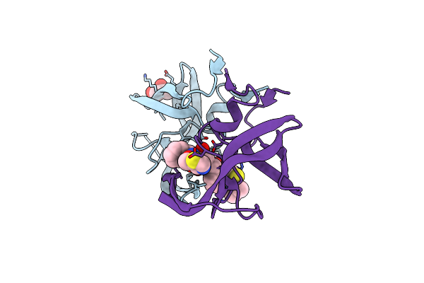
Crystal Structure Of Human Cytochrome P4503A4 Bound To An Inhibitor Ritonavir
Organism: Homo sapiens
Method: X-RAY DIFFRACTION Resolution:2.00 Å Release Date: 2010-10-20 Classification: OXIDOREDUCTASE/OXIDOREDUCTASE INHIBITOR Ligands: DMS, HEM, RIT |
Organism: Homo sapiens
Method: X-RAY DIFFRACTION
Release Date: 2010-10-20
Ligands: DMS, HEM, RIT
 |
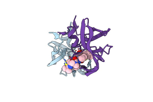
Structure Of Hiv-1 Protease Mutant Bound To Ritonavir
Organism: Human immunodeficiency virus 1
Method: X-RAY DIFFRACTION Resolution:2.20 Å Release Date: 2006-11-14 Classification: HYDROLASE/HYDROLASE INHIBITOR Ligands: GOL, RIT |
Organism: Human immunodeficiency virus 1
Method: X-RAY DIFFRACTION
Release Date: 2006-11-14
Ligands: GOL, RIT
 |
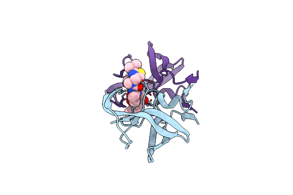
Crystal Structure Of The Complex Of Resistant Strain Of Hiv-1 Protease(V82A Mutant) With Ritonavir
Organism: Human immunodeficiency virus 1
Method: X-RAY DIFFRACTION Resolution:2.00 Å Release Date: 2005-04-19 Classification: HYDROLASE/HYDROLASE INHIBITOR Ligands: RIT |
Organism: Human immunodeficiency virus 1
Method: X-RAY DIFFRACTION
Release Date: 2005-04-19
Ligands: RIT
 |
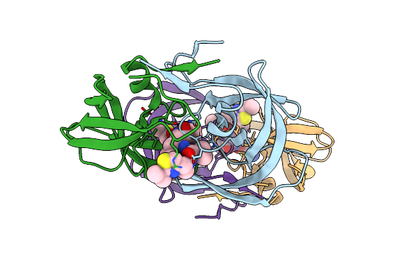
Comparing The Accumulation Of Active Site And Non-Active Site Mutations In The Hiv-1 Protease
Organism: Human immunodeficiency virus 1
Method: X-RAY DIFFRACTION Resolution:2.50 Å Release Date: 2004-10-05 Classification: HYDROLASE/HYDROLASE INHIBITOR Ligands: RIT |
Organism: Human immunodeficiency virus 1
Method: X-RAY DIFFRACTION
Release Date: 2004-10-05
Ligands: RIT
 |
Viability Of A Drug-Resistant Hiv-1 Protease Variant: Structural Insights For Better Anti-Viral Therapy
Organism: Human immunodeficiency virus 1
Method: X-RAY DIFFRACTION Resolution:2.20 Å Release Date: 2003-01-07 Classification: HYDROLASE/HYDROLASE INHIBITOR Ligands: RIT |
Organism: Human immunodeficiency virus 1
Method: X-RAY DIFFRACTION
Release Date: 2003-01-07
Ligands: RIT
 |
Hiv-1 Protease Dimer Complexed With A-84538
Organism: Human immunodeficiency virus 1
Method: X-RAY DIFFRACTION Resolution:1.80 Å Release Date: 1998-02-04 Classification: HYDROLASE/HYDROLASE INHIBITOR Ligands: RIT |
Organism: Human immunodeficiency virus 1
Method: X-RAY DIFFRACTION
Release Date: 1998-02-04
Ligands: RIT