
Search Count: 133
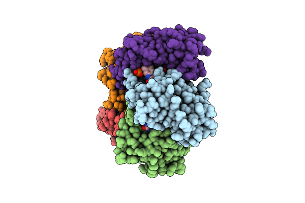 |
Structure Of 6,7-Dimethyl-8-Ribityllumazine Synthase From Schizosaccharomyces Pombe Mutant W27Y With Bound Riboflavin
Organism: Schizosaccharomyces pombe
Method: X-RAY DIFFRACTION Resolution:2.80 Å Release Date: 2005-07-19 Classification: TRANSFERASE Ligands: PO4, RBF |
 |
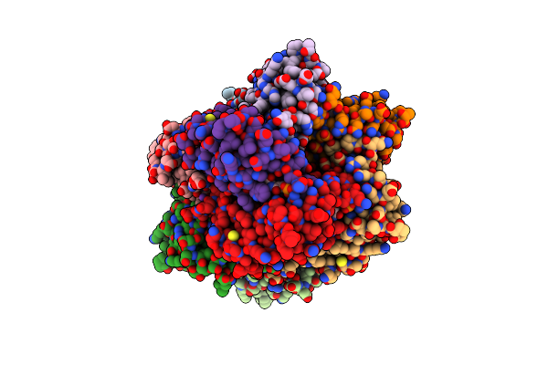
Organism: Bacillus subtilis
Method: X-RAY DIFFRACTION Resolution:2.40 Å Release Date: 1996-12-07 Classification: RIBOFLAVIN SYNTHASE Ligands: PO4, INI |
 |
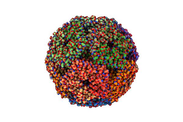
Organism: Schizosaccharomyces pombe
Method: X-RAY DIFFRACTION Resolution:2.10 Å Release Date: 2002-11-06 Classification: TRANSFERASE Ligands: HG, CRM |
 |
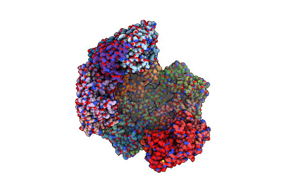
Structure Of 6,7-Dimthyl-8-Ribityllumazine Synthase From Schizosaccharomyces Pombe Mutant W27Y With Bound Ligand 6-Carboxyethyl-7-Oxo-8-Ribityllumazine
Organism: Schizosaccharomyces pombe
Method: X-RAY DIFFRACTION Resolution:2.75 Å Release Date: 2005-07-19 Classification: TRANSFERASE Ligands: CRM, PO4 |
 |
Lumazine Synthase From Mycobacterium Tuberculosis Bound To N-6-(Ribitylamino)Pyrimidine-2,4(1H,3H)-Dione-5-Yl-Propionamide
Organism: Mycobacterium tuberculosis
Method: X-RAY DIFFRACTION Resolution:2.30 Å Release Date: 2008-04-08 Classification: TRANSFERASE Ligands: Y19, K, PO4 |
 |
Organism: Salmonella typhimurium
Method: X-RAY DIFFRACTION Resolution:3.57 Å Release Date: 2011-02-02 Classification: TRANSFERASE Ligands: SO4 |
 |
Organism: Salmonella typhimurium
Method: X-RAY DIFFRACTION Resolution:3.50 Å Release Date: 2011-06-08 Classification: TRANSFERASE Ligands: SO4 |
 |
Lumazine Synthase From Mycobacterium Tuberculosis Bound To 3-(1,3,7- Trihydro-9-D-Ribityl-2,6,8-Purinetrione-7-Yl) Pentane 1 Phosphate
Organism: Mycobacterium tuberculosis
Method: X-RAY DIFFRACTION Resolution:1.60 Å Release Date: 2006-12-13 Classification: TRANSFERASE Ligands: TP6, K, DTU, MPD |
 |
Lumazine Synthase From Mycobacterium Tuberculosis Bound To 3-(1,3,7- Trihydro-9-D-Ribityl-2,6,8-Purinetrione-7-Yl)Hexane 1-Phosphate
Organism: Mycobacterium tuberculosis
Method: X-RAY DIFFRACTION Resolution:2.80 Å Release Date: 2006-12-13 Classification: TRANSFERASE Ligands: PHR, K, MPD |
 |
Lumazine Synthase From Mycobacterium Tuberculosis Bound To 3-(1,3,7- Trihydro-9-D-Ribityl-2,6,8-Purinetrione-7-Yl) 1,1 Difluoropentane-1- Phosphate
Organism: Mycobacterium tuberculosis
Method: X-RAY DIFFRACTION Resolution:1.90 Å Release Date: 2006-12-13 Classification: TRANSFERASE Ligands: TSF, K |
 |
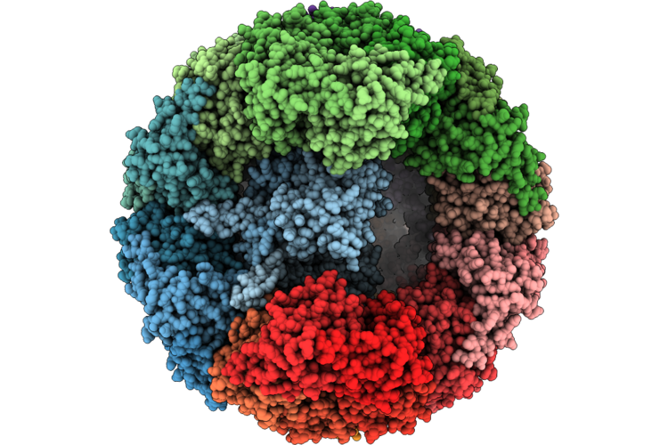
Aquifex Aeolicus Lumazine Synthase 11-Pentamer Cage In Complex With Riboflavin Synthase Trimer
Organism: Aquifex aeolicus vf5
Method: ELECTRON MICROSCOPY Release Date: 2026-05-27 Classification: BIOSYNTHETIC PROTEIN |
 |
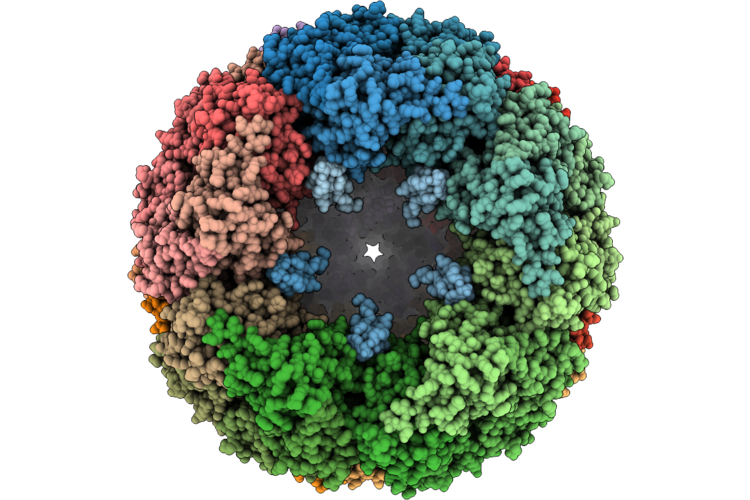
Aquifex Aeolicus Lumazine Synthase 10-Pentamer Cage In Complex With Riboflavin Synthase Trimer
Organism: Aquifex aeolicus vf5
Method: ELECTRON MICROSCOPY Release Date: 2026-05-27 Classification: BIOSYNTHETIC PROTEIN |
 |
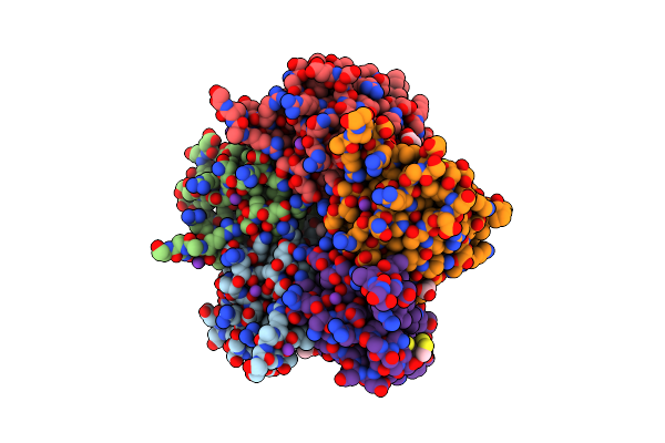
Aquifex Aeolicus Lumazine Synthase 11-Pentamer Cage In Complex With C5-Symmetrized Riboflavin Synthase C-Termini
Organism: Aquifex aeolicus vf5
Method: ELECTRON MICROSCOPY Release Date: 2026-05-27 Classification: BIOSYNTHETIC PROTEIN |
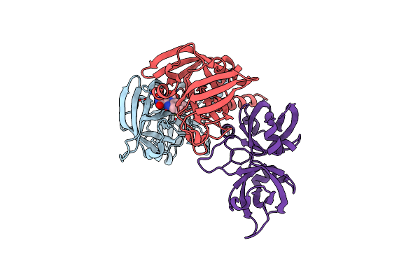 |
Crystallographic Structure Of Trimeric Riboflavin Synthase From Brucella Abortus In Complex With Riboflavin
Organism: Brucella abortus
Method: X-RAY DIFFRACTION Resolution:2.85 Å Release Date: 2013-10-16 Classification: TRANSFERASE Ligands: RBF |
 |
Crystal Structure Of A Probable Riboflavin Synthase, Beta Chain Ribh (6,7-Dimethyl-8-Ribityllumazine Synthase, Dmrl Synthase, Lumazine Synthase) From Mycobacterium Leprae
Organism: Mycobacterium leprae
Method: X-RAY DIFFRACTION Resolution:1.95 Å Release Date: 2013-03-06 Classification: TRANSFERASE |
 |
Lumazine Synthase From Mycobacterium Tuberculosis Bound To 4-(6- Chloro-2,4-Dioxo-1,2,3,4-Tetrahydropyrimidin-5-Yl)Butyl Phosphate
Organism: Mycobacterium tuberculosis
Method: X-RAY DIFFRACTION Resolution:2.00 Å Release Date: 2006-12-13 Classification: TRANSFERASE Ligands: JCL, K, ACT, MPD, DTD |
 |
Crystal Structures Of A Pentameric Fungal And An Icosahedral Plant Lumazine Synthase Reveals The Structural Basis For Differences In Assembly
Organism: Magnaporthe grisea
Method: X-RAY DIFFRACTION Resolution:3.10 Å Release Date: 2000-08-06 Classification: TRANSFERASE Ligands: SO4, LMZ |
 |
Crystal Structure Of Lumazine Synthase From Aquifex Aeolicus In Complex With Inhibitor: 6,7-Dioxo-5H-8-Ribitylaminolumazine
Organism: Aquifex aeolicus
Method: X-RAY DIFFRACTION Resolution:1.75 Å Release Date: 2004-01-23 Classification: TRANSFERASE Ligands: PO4, RDL |
 |
Crystal Structure Of Lumazine Synthase From Aquifex Aeolicus In Complex With Inhibitor: 3-(7-Hydroxy-8-Ribityllumazine-6-Yl)Propionic Acid
Organism: Aquifex aeolicus
Method: X-RAY DIFFRACTION Resolution:1.82 Å Release Date: 2004-01-23 Classification: TRANSFERASE Ligands: PO4, RLP |
 |
Crystal Structure Of Lumazine Synthase From Aquifex Aeolicus In Complex With Inhibitor: 5-Nitroso-6-Ribityl-Amino-2,4(1H,3H)Pyrimidinedione
Organism: Aquifex aeolicus
Method: X-RAY DIFFRACTION Resolution:2.05 Å Release Date: 2004-01-23 Classification: TRANSFERASE Ligands: PO4, LMZ |