
Search Count: 52
 |
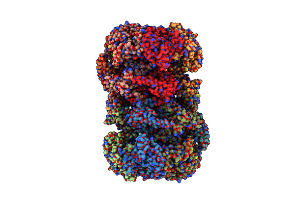
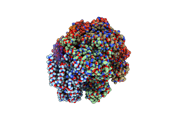
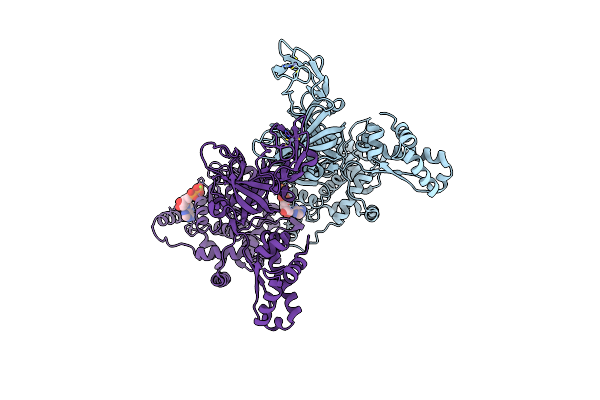
Cryo-Em Structure Of The Mcm2-7 Double Hexamer
Organism: Saccharomyces cerevisiae s288c
Method: ELECTRON MICROSCOPY Release Date: 2015-08-05 Classification: HYDROLASE Ligands: ADP |
Organism: Saccharomyces cerevisiae s288c
Method: ELECTRON MICROSCOPY
Release Date: 2015-08-05
Ligands: ADP
 |
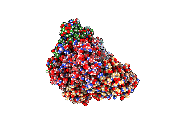
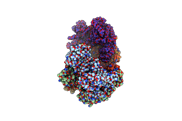
Cryo-Em Structure Of Mcm2-7 Double Hexamer On Dsdna
Organism: Saccharomyces cerevisiae
Method: ELECTRON MICROSCOPY Release Date: 2017-10-25 Classification: HYDROLASE/DNA Ligands: ADP |
Organism: Saccharomyces cerevisiae
Method: ELECTRON MICROSCOPY
Release Date: 2017-10-25
Ligands: ADP
 |
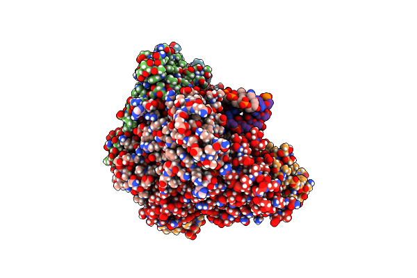
Cryo-Em Structure Of The Cdt1-Mcm2-7 Complex In Amppnp State
Organism: Saccharomyces cerevisiae (strain atcc 204508 / s288c)
Method: ELECTRON MICROSCOPY Release Date: 2017-05-03 Classification: HYDROLASE |
 |
Cryo-Em Structure Of Pcv2 Replicase Bound To Ssdna
Organism: Porcine circovirus 2, Escherichia coli
Method: ELECTRON MICROSCOPY Release Date: 2021-08-25 Classification: REPLICATION, HYDROLASE/DNA Ligands: ADP, MG |
Organism: Porcine circovirus 2, Escherichia coli
Method: ELECTRON MICROSCOPY
Release Date: 2021-08-25
Ligands: ADP, MG
 |
Cryo-Em Structure Of Pcv2 Replicase Bound To Ssdna
Organism: Porcine circovirus 2, Unidentified
Method: ELECTRON MICROSCOPY Release Date: 2021-08-25 Classification: REPLICATION, HYDROLASE/DNA Ligands: ADP, MG |
Organism: Porcine circovirus 2, Unidentified
Method: ELECTRON MICROSCOPY
Release Date: 2021-08-25
Ligands: ADP, MG
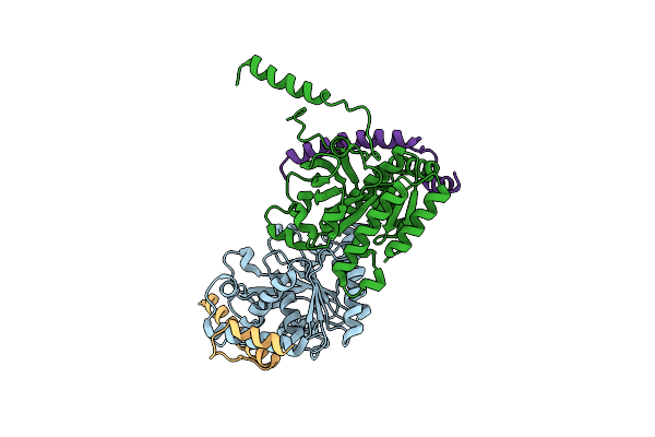 |
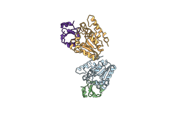
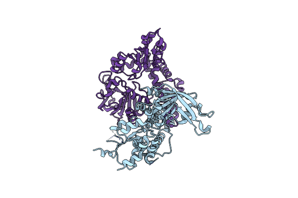
Crystal Structure Of The Complex Of The Interaction Domains Of E. Coli Dnab Helicase And Dnac Helicase Loader
Organism: Escherichia coli
Method: X-RAY DIFFRACTION Resolution:3.10 Å Release Date: 2019-11-20 Classification: HYDROLASE |
Organism: Escherichia coli
Method: X-RAY DIFFRACTION
Release Date: 2019-11-20
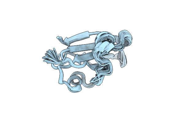 |
Nmr Solution Structure Of The Master-Rep Protein Nuclease Domain (2-95) From The Faba Bean Necrotic Yellows Virus
Organism: Faba bean necrotic yellows virus
Method: SOLUTION NMR Release Date: 2007-06-26 Classification: replication, hydrolase |
Organism: Faba bean necrotic yellows virus
Method: SOLUTION NMR
Release Date: 2007-06-26
 |
Structure Of A Dimer Of The Sulfolobus Solfataricus Mcm N-Terminal Domain Reveals Potential Role In Mcm Ring Opening
Organism: Saccharolobus solfataricus (strain atcc 35092 / dsm 1617 / jcm 11322 / p2)
Method: X-RAY DIFFRACTION Resolution:2.00 Å Release Date: 2021-06-16 Classification: REPLICATION, HYDROLASE Ligands: ZN |
Organism: Saccharolobus solfataricus (strain atcc 35092 / dsm 1617 / jcm 11322 / p2)
Method: X-RAY DIFFRACTION
Release Date: 2021-06-16
Ligands: ZN
 |
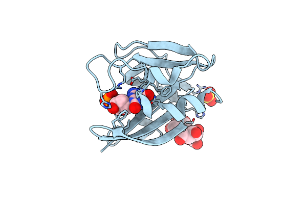
Escherichia Coli Uracil-Dna Glycosylase:Inhibitor Complex With Wild-Type Udg And Wild-Type Ugi
Organism: Escherichia coli k12, Bacillus phage pbs2
Method: X-RAY DIFFRACTION Resolution:2.40 Å Release Date: 1999-03-25 Classification: REPLICATION, HYDROLASE |
Organism: Escherichia coli k12, Bacillus phage pbs2
Method: X-RAY DIFFRACTION
Release Date: 1999-03-25
 |
Escherichia Coli Uracil-Dna Glycosylase:Inhibitor Complex With H187D Mutant Udg And Wild-Type Ugi
Organism: Escherichia coli k12, Bacillus phage pbs2
Method: X-RAY DIFFRACTION Resolution:2.60 Å Release Date: 1999-03-25 Classification: REPLICATION, HYDROLASE |
Organism: Escherichia coli k12, Bacillus phage pbs2
Method: X-RAY DIFFRACTION
Release Date: 1999-03-25
 |
The Crystal Structure Of The Monomeric Reverse Transcriptase From Moloney Murine Leukemia Virus
Organism: Moloney murine leukemia virus
Method: X-RAY DIFFRACTION Resolution:3.00 Å Release Date: 2013-10-16 Classification: TRANSFERASE, HYDROLASE, REPLICATION |
Organism: Moloney murine leukemia virus
Method: X-RAY DIFFRACTION
Release Date: 2013-10-16
 |
Mcm In Complex With Dsdna In Presence Of Atp.
Organism: Thermococcus kodakarensis
Method: ELECTRON MICROSCOPY Release Date: 2024-01-17 Classification: HYDROLASE Ligands: MG, ATP, ADP |
Organism: Thermococcus kodakarensis
Method: ELECTRON MICROSCOPY
Release Date: 2024-01-17
Ligands: MG, ATP, ADP
 |
Mcm In The Apo State.
Organism: Thermococcus kodakarensis
Method: ELECTRON MICROSCOPY Release Date: 2024-01-17 Classification: HYDROLASE |
Organism: Thermococcus kodakarensis
Method: ELECTRON MICROSCOPY
Release Date: 2024-01-17
 |
Vaccinia Virus His-D4/A20(1-50) In Complex With Uracil
Organism: Vaccinia virus (strain copenhagen)
Method: X-RAY DIFFRACTION Resolution:1.85 Å Release Date: 2015-06-10 Classification: HYDROLASE Ligands: SO4, URA |
Organism: Vaccinia virus (strain copenhagen)
Method: X-RAY DIFFRACTION
Release Date: 2015-06-10
Ligands: SO4, URA
 |
Crystal Structure Of Restriction Endonuclease Ecorii Mutant R88A
Organism: Escherichia coli
Method: X-RAY DIFFRACTION Resolution:2.10 Å Release Date: 2003-12-16 Classification: HYDROLASE |
Organism: Escherichia coli
Method: X-RAY DIFFRACTION
Release Date: 2003-12-16
 |
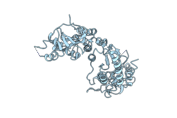
Pfmcm-Aaa Double-Octamer
Organism: Pyrococcus furiosus
Method: X-RAY DIFFRACTION Resolution:3.80 Å Release Date: 2014-10-08 Classification: HYDROLASE Ligands: ADP, MG |
Organism: Pyrococcus furiosus
Method: X-RAY DIFFRACTION
Release Date: 2014-10-08
Ligands: ADP, MG
 |
Vaccinia Virus D4/A20(1-50) In Complex With Dsdna Containing An Abasic Site And Free Uracyl
Organism: Vaccinia virus (strain copenhagen), Vaccinia virus, Synthetic construct
Method: X-RAY DIFFRACTION Resolution:2.70 Å Release Date: 2015-06-10 Classification: HYDROLASE Ligands: URA |
Organism: Vaccinia virus (strain copenhagen), Vaccinia virus, Synthetic construct
Method: X-RAY DIFFRACTION
Release Date: 2015-06-10
Ligands: URA
 |
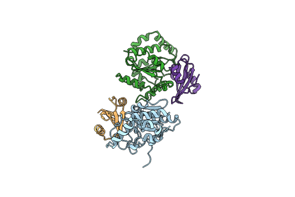
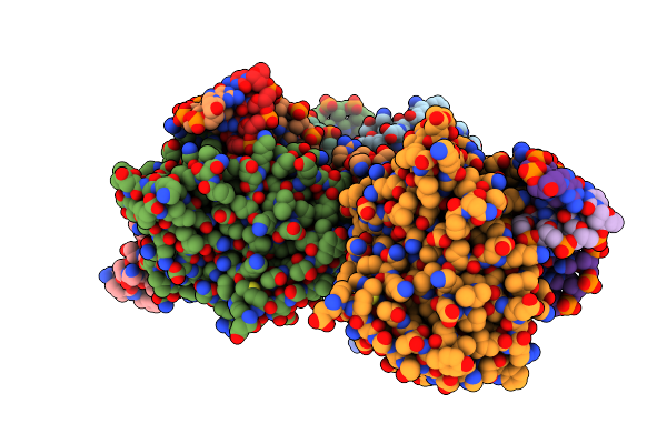
Crystal Structure Of An Active Mcm Hexamer
Organism: Sulfolobus solfataricus, Pyrococcus furiosus
Method: X-RAY DIFFRACTION Resolution:2.70 Å Release Date: 2014-10-08 Classification: HYDROLASE Ligands: ADP, MG, ZN, CL |
Organism: Sulfolobus solfataricus, Pyrococcus furiosus
Method: X-RAY DIFFRACTION
Release Date: 2014-10-08
Ligands: ADP, MG, ZN, CL
 |
Crystal Structure Of The Mutt Protein In Mn(Ii) Bound Holo Form
Organism: Escherichia coli k-12
Method: X-RAY DIFFRACTION Resolution:2.00 Å Release Date: 2009-10-27 Classification: HYDROLASE Ligands: MN, NA, TLA |
Organism: Escherichia coli k-12
Method: X-RAY DIFFRACTION
Release Date: 2009-10-27
Ligands: MN, NA, TLA
 |
Crystal Structure Of Mutt-8-Oxo-Dgmp Complex
Organism: Escherichia coli k-12
Method: X-RAY DIFFRACTION Resolution:1.96 Å Release Date: 2009-10-27 Classification: HYDROLASE |
Organism: Escherichia coli k-12
Method: X-RAY DIFFRACTION
Release Date: 2009-10-27