

Search Count: 35
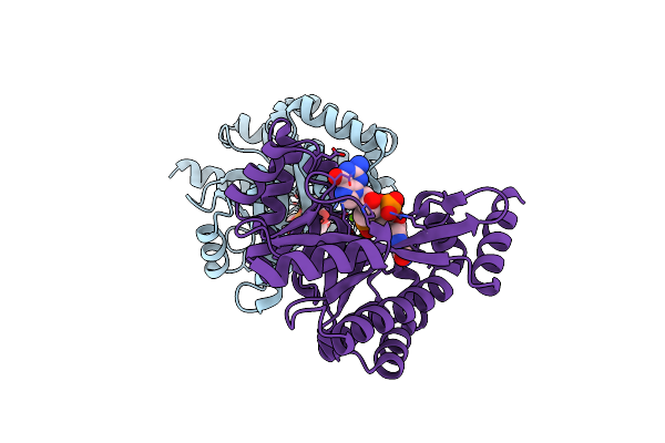 |
Crystal Structure Of Putative Diguanylate Cyclase/Phosphodiesterase Complex With Cyclic Di-Gmp
Organism: Thiobacillus denitrificans
Method: X-RAY DIFFRACTION Resolution:2.35 Å Release Date: 2010-06-16 Classification: Structural Genomics, Unknown function Ligands: C2E, MG, CL |
Organism: Thiobacillus denitrificans
Method: X-RAY DIFFRACTION
Release Date: 2010-06-16
Ligands: C2E, MG, CL
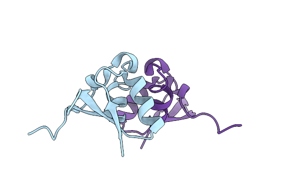 |
Crystal Structure Of A Cystathionine Beta-Synthase Domain Protein Fused To A Zn-Ribbon-Like Domain
Organism: Pyrococcus furiosus
Method: X-RAY DIFFRACTION Resolution:1.81 Å Release Date: 2009-03-31 Classification: Nucleotide binding protein, Metal binding protein |
Organism: Pyrococcus furiosus
Method: X-RAY DIFFRACTION
Release Date: 2009-03-31
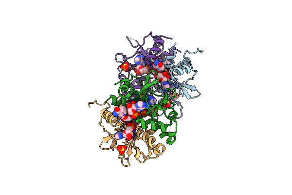 |
Crystal Structure Of The Cbs-Domain Containing Protein Atu1752 From Agrobacterium Tumefaciens
Organism: Agrobacterium tumefaciens str.
Method: X-RAY DIFFRACTION Resolution:2.70 Å Release Date: 2009-01-13 Classification: structural genomics, unknown function, NUCLEOTIDE-BINDING PROTEIN Ligands: AMP, NAI, SO4 |
Organism: Agrobacterium tumefaciens str.
Method: X-RAY DIFFRACTION
Release Date: 2009-01-13
Ligands: AMP, NAI, SO4
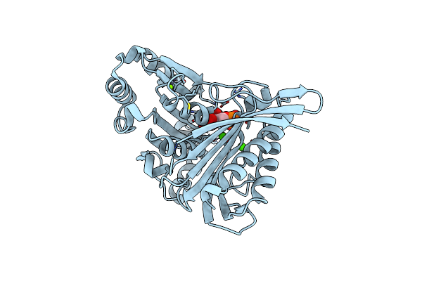 |
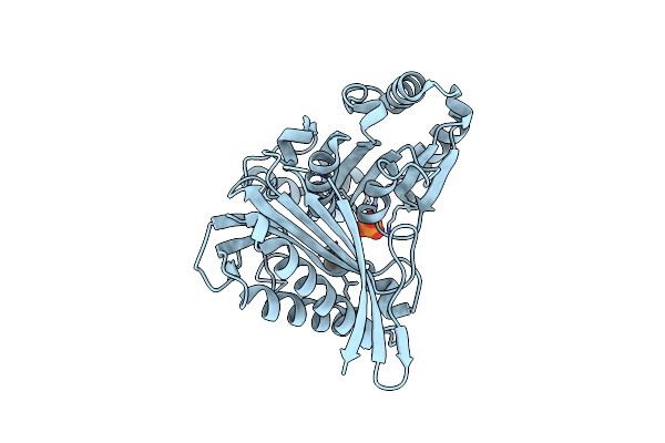
Structure Of E. Coli Glpx With Its Substrate Fructose 1,6-Bisphosphate
Organism: Escherichia coli
Method: X-RAY DIFFRACTION Resolution:1.85 Å Release Date: 2008-12-23 Classification: HYDROLASE Ligands: FBP, CA, MG, CL |
Organism: Escherichia coli
Method: X-RAY DIFFRACTION
Release Date: 2008-12-23
Ligands: FBP, CA, MG, CL
 |
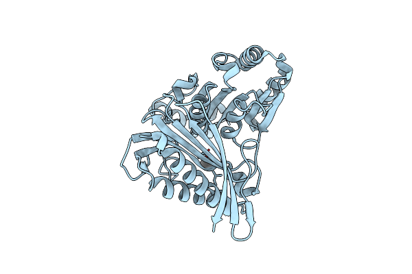
Crystal Structure Of The Fructose-1,6-Bisphosphatase Glpx From E.Coli In Complex With Inorganic Phosphate
Organism: Escherichia coli
Method: X-RAY DIFFRACTION Resolution:1.85 Å Release Date: 2008-12-09 Classification: HYDROLASE Ligands: PO4, UNX |
Organism: Escherichia coli
Method: X-RAY DIFFRACTION
Release Date: 2008-12-09
Ligands: PO4, UNX
 |
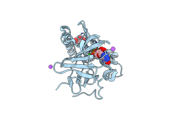
Crystal Structure Of Fructose-1,6-Bisphosphatase From E.Coli Glpx
Organism: Escherichia coli
Method: X-RAY DIFFRACTION Resolution:2.10 Å Release Date: 2008-12-09 Classification: HYDROLASE Ligands: UNX |
Organism: Escherichia coli
Method: X-RAY DIFFRACTION
Release Date: 2008-12-09
Ligands: UNX
 |
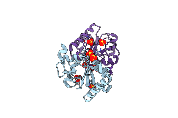
Crystal Structure Of The Phosphatase Sc4828 With The Non-Hydrolyzable Nucleotide Gpcp
Organism: Streptomyces coelicolor a3(2)
Method: X-RAY DIFFRACTION Resolution:1.65 Å Release Date: 2008-12-09 Classification: HYDROLASE Ligands: CA, NA, GP2, GOL |
Organism: Streptomyces coelicolor a3(2)
Method: X-RAY DIFFRACTION
Release Date: 2008-12-09
Ligands: CA, NA, GP2, GOL
 |
Structure Of Ygr203W, A Yeast Protein Tyrosine Phosphatase Of The Rhodanese Family
Organism: Saccharomyces cerevisiae
Method: X-RAY DIFFRACTION Resolution:1.80 Å Release Date: 2008-11-25 Classification: HYDROLASE Ligands: SO4, CL, NH4 |
Organism: Saccharomyces cerevisiae
Method: X-RAY DIFFRACTION
Release Date: 2008-11-25
Ligands: SO4, CL, NH4
 |
Structure Of The Rna'Se Sso8090 From Sulfolobus Solfataricus
Organism: Sulfolobus solfataricus
Method: X-RAY DIFFRACTION Resolution:2.25 Å Release Date: 2008-11-11 Classification: HYDROLASE Ligands: CL, NA |
Organism: Sulfolobus solfataricus
Method: X-RAY DIFFRACTION
Release Date: 2008-11-11
Ligands: CL, NA
 |
Crystal Structure Of Putative Polyphosphate Kinase 2 From Pseudomonas Aeruginosa Pa01
Organism: Pseudomonas aeruginosa pao1
Method: X-RAY DIFFRACTION Resolution:2.00 Å Release Date: 2008-07-01 Classification: TRANSFERASE Ligands: ACT, EDO, GOL, MLI |
Organism: Pseudomonas aeruginosa pao1
Method: X-RAY DIFFRACTION
Release Date: 2008-07-01
Ligands: ACT, EDO, GOL, MLI
 |
Crystal Structure Of Putative Polyphosphate Kinase 2 From Sinorhizobium Meliloti
Organism: Sinorhizobium meliloti
Method: X-RAY DIFFRACTION Resolution:2.23 Å Release Date: 2008-07-01 Classification: TRANSFERASE Ligands: GOL, FMT |
Organism: Sinorhizobium meliloti
Method: X-RAY DIFFRACTION
Release Date: 2008-07-01
Ligands: GOL, FMT
 |
Crystal Structure Of The Covalent Complex Between The Bacillus Subtilis Glutaminase Ybgj And 5-Oxo-L-Norleucine Formed By Reaction Of The Protein With 6-Diazo-5-Oxo-L-Norleucine
Organism: Bacillus subtilis
Method: X-RAY DIFFRACTION Resolution:2.29 Å Release Date: 2008-05-20 Classification: HYDROLASE Ligands: ONL |
Organism: Bacillus subtilis
Method: X-RAY DIFFRACTION
Release Date: 2008-05-20
Ligands: ONL
 |
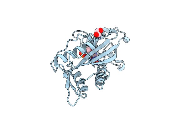
Crystal Structure Of Sc4828, A Unique Phosphatase From Streptomyces Coelicolor
Organism: Streptomyces coelicolor a3(2)
Method: X-RAY DIFFRACTION Resolution:1.70 Å Release Date: 2008-03-25 Classification: HYDROLASE Ligands: MG, NA |
Organism: Streptomyces coelicolor a3(2)
Method: X-RAY DIFFRACTION
Release Date: 2008-03-25
Ligands: MG, NA
 |
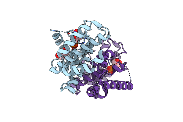
Structure Of The E. Coli Inosine Triphosphate Pyrophosphatase Rgdb In Complex With Imp
Organism: Escherichia coli
Method: X-RAY DIFFRACTION Resolution:2.02 Å Release Date: 2008-03-18 Classification: HYDROLASE Ligands: IMP, EDO |
Organism: Escherichia coli
Method: X-RAY DIFFRACTION
Release Date: 2008-03-18
Ligands: IMP, EDO
 |
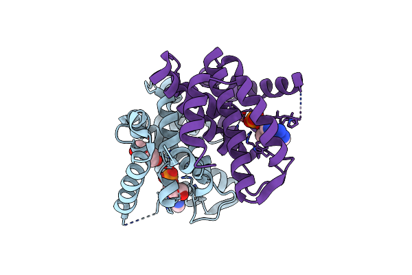
Crystal Structure Of The 5'-Deoxynucleotidase Yfbr Mutant E72A Complexed With Co(2+) And Tmp
Organism: Escherichia coli
Method: X-RAY DIFFRACTION Resolution:2.10 Å Release Date: 2008-03-04 Classification: HYDROLASE Ligands: CO, TMP, PEG |
Organism: Escherichia coli
Method: X-RAY DIFFRACTION
Release Date: 2008-03-04
Ligands: CO, TMP, PEG
 |
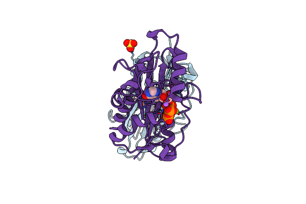
Crystal Structure Of The 5'-Deoxynucleotidase Yfbr Mutant E72A Complexed With Co(2+) And Damp
Organism: Escherichia coli
Method: X-RAY DIFFRACTION Resolution:2.10 Å Release Date: 2008-03-04 Classification: HYDROLASE Ligands: CO, D5M, PEG |
Organism: Escherichia coli
Method: X-RAY DIFFRACTION
Release Date: 2008-03-04
Ligands: CO, D5M, PEG
 |
Structure Of The E. Coli Inosine Triphosphate Pyrophosphatase Rgdb In Complex With Itp
Organism: Escherichia coli
Method: X-RAY DIFFRACTION Resolution:1.95 Å Release Date: 2008-02-19 Classification: HYDROLASE Ligands: CA, NA, SO4, ITT |
Organism: Escherichia coli
Method: X-RAY DIFFRACTION
Release Date: 2008-02-19
Ligands: CA, NA, SO4, ITT
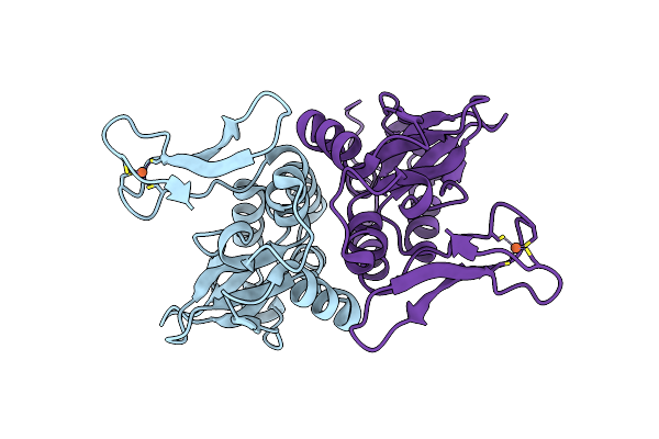 |
Structure Of Ta289, A Cbs-Rubredoxin-Like Protein, In Its Fe+2-Bound State
Organism: Thermoplasma acidophilum
Method: X-RAY DIFFRACTION Resolution:2.00 Å Release Date: 2008-02-19 Classification: UNKNOWN FUNCTION Ligands: FE2 |
Organism: Thermoplasma acidophilum
Method: X-RAY DIFFRACTION
Release Date: 2008-02-19
Ligands: FE2
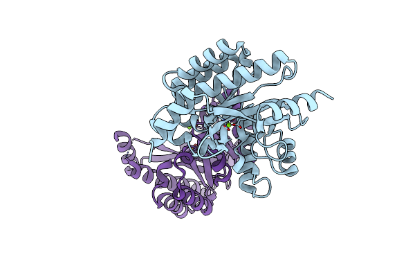 |
Crystal Structure Of Putative Diguanylate Cyclase/Phosphodiesterase From Thiobacillus Denitrificans
Organism: Thiobacillus denitrificans
Method: X-RAY DIFFRACTION Resolution:1.80 Å Release Date: 2007-09-18 Classification: STRUCTURAL GENOMICS, UNKNOWN FUNCTION Ligands: MG, CL |
Organism: Thiobacillus denitrificans
Method: X-RAY DIFFRACTION
Release Date: 2007-09-18
Ligands: MG, CL
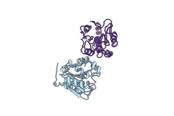 |
Crystal Structure Of The Human Retinoblastoma-Binding Protein 9 (Rbbp-9). Nesg Target Hr2978
Organism: Homo sapiens
Method: X-RAY DIFFRACTION Resolution:1.72 Å Release Date: 2007-08-14 Classification: STRUCTURAL PROTEIN |
Organism: Homo sapiens
Method: X-RAY DIFFRACTION
Release Date: 2007-08-14