

Search Count: 29
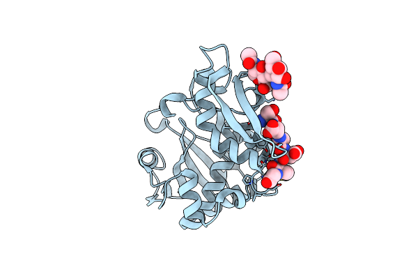 |
Crystal Structure Of The Complex Of Type I Ribosome Inactivating Protein With Hexapeptide Ser-Asp-Asp-Asp-Met-Gly At 2.2 A Resolution
Organism: Saccharomyces cerevisiae, Momordica balsamina
Method: X-RAY DIFFRACTION Resolution:2.22 Å Release Date: 2010-07-28 Classification: HYDROLASE |
Organism: Saccharomyces cerevisiae, Momordica balsamina
Method: X-RAY DIFFRACTION
Release Date: 2010-07-28
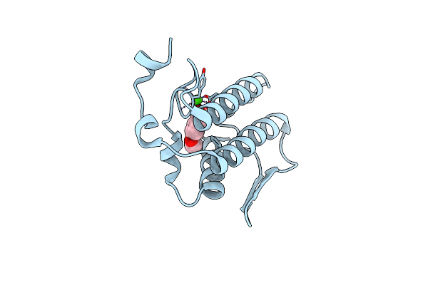 |
Crystal Structure Of The Complex Of Group I Phospholipase A2 With 4-Methoxy-Benzoicacid At 1.4A Resolution
Organism: Naja sagittifera
Method: X-RAY DIFFRACTION Resolution:1.40 Å Release Date: 2010-07-14 Classification: HYDROLASE Ligands: ANN, CA |
Organism: Naja sagittifera
Method: X-RAY DIFFRACTION
Release Date: 2010-07-14
Ligands: ANN, CA
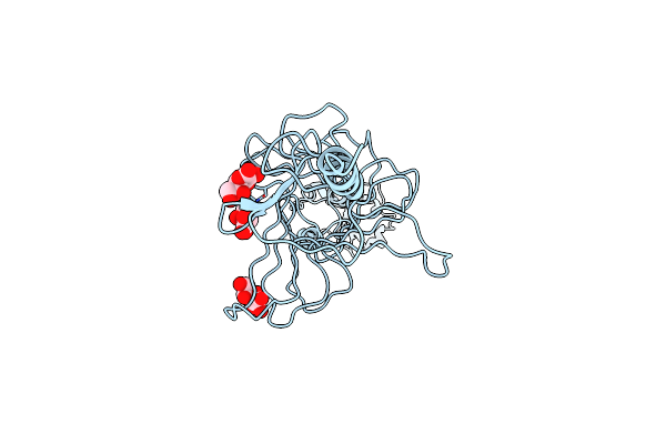 |
Crystal Structure Of The Complex Of Hyaluranidase Trimer With Ascorbic Acid At 3.1 A Resolution Reveals The Locations Of Three Binding Sites
Organism: Streptococcus pyogenes
Method: X-RAY DIFFRACTION Resolution:3.10 Å Release Date: 2009-09-29 Classification: LYASE Ligands: ASC |
Organism: Streptococcus pyogenes
Method: X-RAY DIFFRACTION
Release Date: 2009-09-29
Ligands: ASC
 |
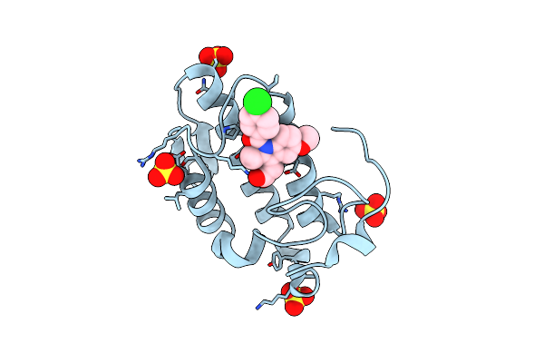
Crystal Structure Of The Complex Formed Between A Group Ii Phospholipase A2 And Designed Peptide Inhibitor Carbobenzoxy-Dehydro-Val-Ala-Arg-Ser At 1.2 A Resolution
Organism: Daboia russellii russellii
Method: X-RAY DIFFRACTION Resolution:1.25 Å Release Date: 2009-03-10 Classification: HYDROLASE/HYDROLASE INHIBITOR Ligands: SO4 |
Organism: Daboia russellii russellii
Method: X-RAY DIFFRACTION
Release Date: 2009-03-10
Ligands: SO4
 |
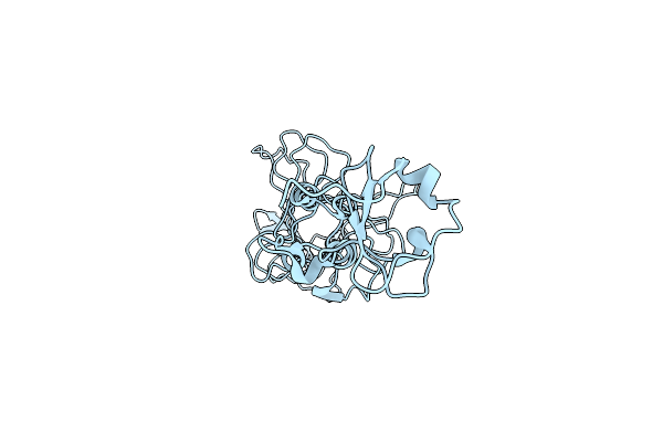
Simultaneous Inhibition Of Anti-Coagulation And Inflammation: Crystal Structure Of Phospholipase A2 Complexed With Indomethacin At 1.4 A Resolution Reveals The Presence Of The New Common Ligand Binding Site
Organism: Daboia russelli pulchella
Method: X-RAY DIFFRACTION Resolution:1.40 Å Release Date: 2009-01-20 Classification: HYDROLASE Ligands: SO4, IMN |
Organism: Daboia russelli pulchella
Method: X-RAY DIFFRACTION
Release Date: 2009-01-20
Ligands: SO4, IMN
 |
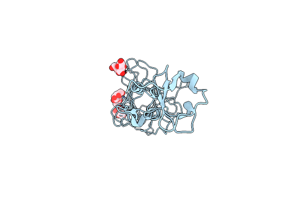
Crystal Structure Of Cloned Trimeric Hyluranidase From Streptococcus Pyogenes At 2.8 A Resolution
Organism: Streptococcus pyogenes
Method: X-RAY DIFFRACTION Resolution:2.80 Å Release Date: 2007-05-08 Classification: LYASE |
Organism: Streptococcus pyogenes
Method: X-RAY DIFFRACTION
Release Date: 2007-05-08
 |
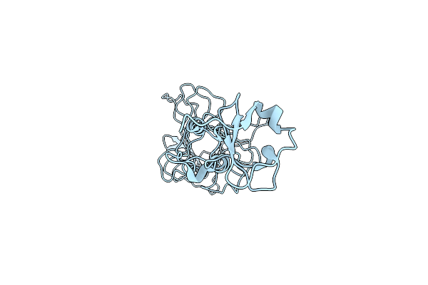
Crystal Structure Of Hyluranidase Complexed With Lactose At 2.6 A Resolution Reveals Three Specific Sugar Recognition Sites
Organism: Streptococcus pyogenes
Method: X-RAY DIFFRACTION Resolution:2.60 Å Release Date: 2007-05-01 Classification: LYASE |
Organism: Streptococcus pyogenes
Method: X-RAY DIFFRACTION
Release Date: 2007-05-01
 |
Crystal Structure Of Hyluranidase Trimer At 2.6 A Resolution
Organism: Streptococcus pyogenes serotype m1
Method: X-RAY DIFFRACTION Resolution:2.60 Å Release Date: 2007-05-01 Classification: LYASE |
Organism: Streptococcus pyogenes serotype m1
Method: X-RAY DIFFRACTION
Release Date: 2007-05-01
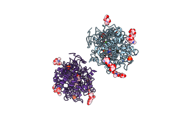 |
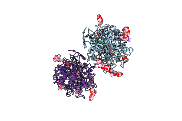
Crystal Structure Of The Complex Of Goat Lactoperoxidase With Phosphate At 2.94 A Resolution
Organism: Capra hircus
Method: X-RAY DIFFRACTION Resolution:2.94 Å Release Date: 2007-03-06 Classification: OXIDOREDUCTASE Ligands: PO4, CO3, CA, HEM, CYN, SCN, OSM |
Organism: Capra hircus
Method: X-RAY DIFFRACTION
Release Date: 2007-03-06
Ligands: PO4, CO3, CA, HEM, CYN, SCN, OSM
 |
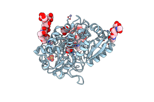
Crystal Structure Of The Complex Of Goat Lactoperoxidase With Nitrate At 3.25 A Resolution
Organism: Capra hircus
Method: X-RAY DIFFRACTION Resolution:3.25 Å Release Date: 2007-02-06 Classification: METAL BINDING PROTEIN, OXIDOREDUCTASE Ligands: NO3, CO3, CA, CYN, SCN, HEM, OSM |
Organism: Capra hircus
Method: X-RAY DIFFRACTION
Release Date: 2007-02-06
Ligands: NO3, CO3, CA, CYN, SCN, HEM, OSM
 |
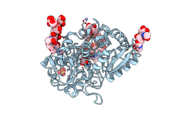
Crystal Structure Of The Complex Of C-Terminal Half Of Bovine Lactoferrin And Arabinose At 2.7 A Resolution
Organism: Bos taurus
Method: X-RAY DIFFRACTION Resolution:2.70 Å Release Date: 2006-11-14 Classification: METAL BINDING PROTEIN Ligands: ARA, FE, CO3, ZN, SO4 |
Organism: Bos taurus
Method: X-RAY DIFFRACTION
Release Date: 2006-11-14
Ligands: ARA, FE, CO3, ZN, SO4
 |
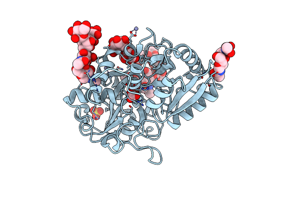
Carbohydrate Recognition Of C-Terminal Half Of Lactoferrin: Crystal Structure Of The Complex Of C-Lobe With Rhamnose At 2.15 A Resolution
Organism: Bos taurus
Method: X-RAY DIFFRACTION Resolution:2.15 Å Release Date: 2006-10-24 Classification: METAL BINDING PROTEIN Ligands: RAM, FE, CO3, ZN, SO4 |
Organism: Bos taurus
Method: X-RAY DIFFRACTION
Release Date: 2006-10-24
Ligands: RAM, FE, CO3, ZN, SO4
 |
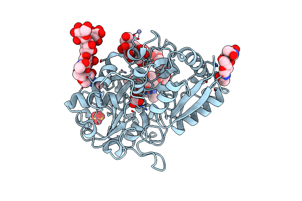
Structure Of The Complex Of Lactoferrin C-Lobe With Melibiose At 2.0 A Resolution
Organism: Bos taurus
Method: X-RAY DIFFRACTION Resolution:2.00 Å Release Date: 2006-10-03 Classification: METAL BINDING PROTEIN Ligands: FE, CO3, ZN, SO4 |
Organism: Bos taurus
Method: X-RAY DIFFRACTION
Release Date: 2006-10-03
Ligands: FE, CO3, ZN, SO4
 |
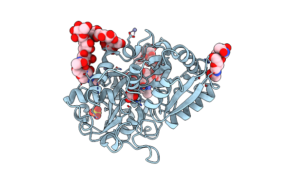
Crystal Structure Of The Complex Formed Between C-Terminal Half Of Bovine Lactoferrin And Sorbitol At 2.85 A Resolution
Organism: Bos taurus
Method: X-RAY DIFFRACTION Resolution:2.85 Å Release Date: 2006-09-12 Classification: METAL BINDING PROTEIN Ligands: SOR, FE, CO3, ZN, SO4 |
Organism: Bos taurus
Method: X-RAY DIFFRACTION
Release Date: 2006-09-12
Ligands: SOR, FE, CO3, ZN, SO4
 |
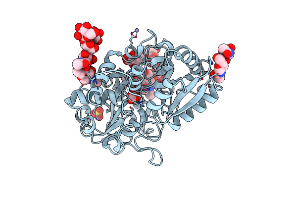
Crystal Structure Of The Complex Formed Between C-Terminal Half Of Bovine Lactoferrin And Cellobiose At 2.2 A Resolution
Organism: Bos taurus
Method: X-RAY DIFFRACTION Resolution:2.20 Å Release Date: 2006-09-05 Classification: METAL BINDING PROTEIN Ligands: FE, CO3, ZN, SO4 |
Organism: Bos taurus
Method: X-RAY DIFFRACTION
Release Date: 2006-09-05
Ligands: FE, CO3, ZN, SO4
 |
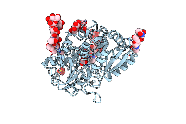
Structure Of The Complex Of Lactoferrin C-Terminal Half With Fucose At 2.07 A Resolution
Organism: Bos taurus
Method: X-RAY DIFFRACTION Resolution:2.07 Å Release Date: 2006-08-29 Classification: METAL BINDING PROTEIN Ligands: FUC, FE, CO3, ZN, SO4 |
Organism: Bos taurus
Method: X-RAY DIFFRACTION
Release Date: 2006-08-29
Ligands: FUC, FE, CO3, ZN, SO4
 |
Structure Of The Bovine Lactoferrin C-Lobe Complex With Sucrose At 3.0 A Resolution
Organism: Bos taurus
Method: X-RAY DIFFRACTION Resolution:3.00 Å Release Date: 2006-08-15 Classification: METAL BINDING PROTEIN Ligands: FE, CO3, ZN, SO4 |
Organism: Bos taurus
Method: X-RAY DIFFRACTION
Release Date: 2006-08-15
Ligands: FE, CO3, ZN, SO4
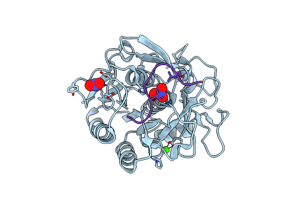 |
Crystal Structure Of Proteinase K Complex With A Synthetic Peptide Klkllvvirlk At 1.69 A Resolution
Organism: Engyodontium album
Method: X-RAY DIFFRACTION Resolution:1.69 Å Release Date: 2006-08-01 Classification: HYDROLASE Ligands: CA, NO3 |
Organism: Engyodontium album
Method: X-RAY DIFFRACTION
Release Date: 2006-08-01
Ligands: CA, NO3
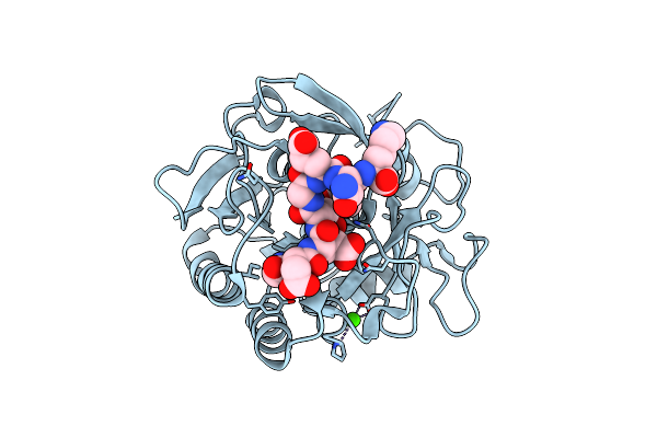 |
Crystal Structure Of Proteinase K Inhibited By A Lactoferrin Octapeptide Gly-Asp-Glu-Gln-Gly-Glu-Asn-Lys At 2.15 A Resolution
Organism: Engyodontium album
Method: X-RAY DIFFRACTION Resolution:2.15 Å Release Date: 2006-07-04 Classification: HYDROLASE Ligands: CA, ACY |
Organism: Engyodontium album
Method: X-RAY DIFFRACTION
Release Date: 2006-07-04
Ligands: CA, ACY
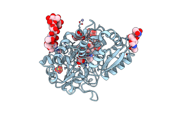 |
Crystal Structure Of Bovine Lactoferrin C-Lobe Liganded With Glucose At 2.8 A Resolution
Organism: Bos taurus
Method: X-RAY DIFFRACTION Resolution:2.80 Å Release Date: 2006-06-27 Classification: HYDROLASE Ligands: GLC, FE, CO3, ZN, SO4 |
Organism: Bos taurus
Method: X-RAY DIFFRACTION
Release Date: 2006-06-27
Ligands: GLC, FE, CO3, ZN, SO4