

Search Count: 21
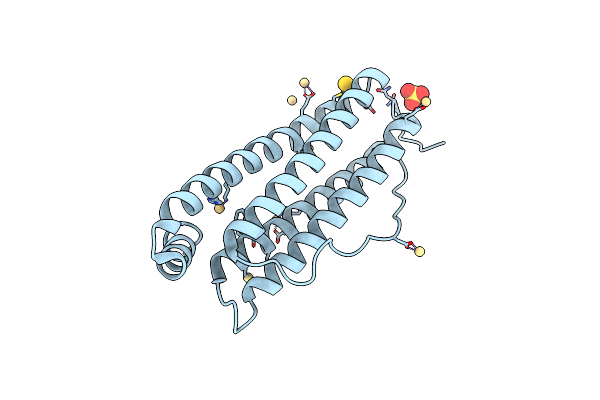 |
X-Ray Structure Of Auranofin-Horse Spleen Ferritin
Organism: Equus caballus
Method: X-RAY DIFFRACTION Resolution:1.24 Å Release Date: 2022-12-28 Classification: TRANSPORT PROTEIN Ligands: CD, SO4, AU, CL |
Organism: Equus caballus
Method: X-RAY DIFFRACTION
Release Date: 2022-12-28
Ligands: CD, SO4, AU, CL
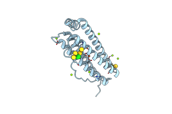 |
X-Ray Structure Of Auranofin-Human H-Chain Ferritin
Organism: Homo sapiens
Method: X-RAY DIFFRACTION Resolution:1.17 Å Release Date: 2022-12-28 Classification: TRANSPORT PROTEIN Ligands: CL, MG, FE, AU |
Organism: Homo sapiens
Method: X-RAY DIFFRACTION
Release Date: 2022-12-28
Ligands: CL, MG, FE, AU
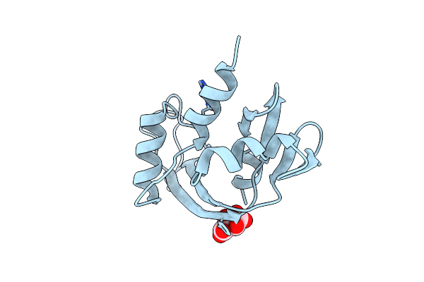 |
X-Ray Structure Of The Adduct Formed Upon Reaction Of Oxaliplatin With Human Angiogenin
Organism: Homo sapiens
Method: X-RAY DIFFRACTION Resolution:1.86 Å Release Date: 2022-03-23 Classification: HYDROLASE Ligands: PT, TAR |
Organism: Homo sapiens
Method: X-RAY DIFFRACTION
Release Date: 2022-03-23
Ligands: PT, TAR
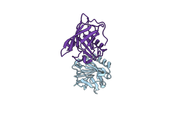 |
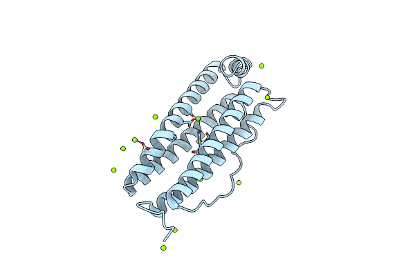
X.Ray Structure Of The Oxaliplatin/Beta-Lactoglobulin Adduct
Organism: Bos taurus
Method: X-RAY DIFFRACTION Resolution:2.01 Å Release Date: 2022-03-23 Classification: TRANSPORT PROTEIN Ligands: PT |
Organism: Bos taurus
Method: X-RAY DIFFRACTION
Release Date: 2022-03-23
Ligands: PT
 |
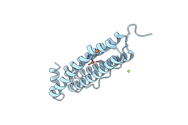
High Resolution Crystal Structure Of Human Mitochondrial Ferritin (Hmtf)
Organism: Homo sapiens
Method: X-RAY DIFFRACTION Resolution:1.16 Å Release Date: 2021-10-13 Classification: OXIDOREDUCTASE Ligands: MG, CL |
Organism: Homo sapiens
Method: X-RAY DIFFRACTION
Release Date: 2021-10-13
Ligands: MG, CL
 |
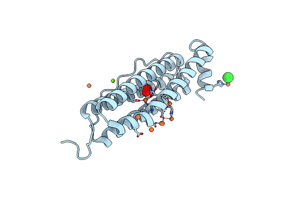
Crystal Structure Of Human Mitochondrial Ferritin (Hmtf) Fe(Ii)-Loaded For 1 Minute.
Organism: Homo sapiens
Method: X-RAY DIFFRACTION Resolution:1.96 Å Release Date: 2021-10-13 Classification: OXIDOREDUCTASE Ligands: FE2, CL, MG |
Organism: Homo sapiens
Method: X-RAY DIFFRACTION
Release Date: 2021-10-13
Ligands: FE2, CL, MG
 |
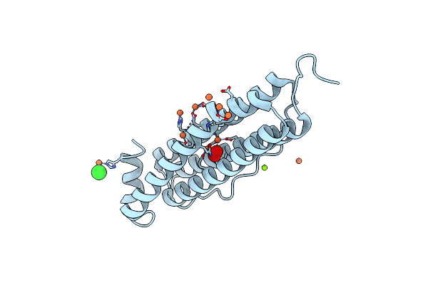
Crystal Structure Of Human Mitochondrial Ferritin (Hmtf) Fe(Ii)-Loaded For 90 Minutes Showing Either A Dioxygen Or A Superoxide Anion Coordinated To Iron Ions In The Ferroxidase Site
Organism: Homo sapiens
Method: X-RAY DIFFRACTION Resolution:1.70 Å Release Date: 2021-10-13 Classification: OXIDOREDUCTASE Ligands: FE2, OXY, CL, MG |
Organism: Homo sapiens
Method: X-RAY DIFFRACTION
Release Date: 2021-10-13
Ligands: FE2, OXY, CL, MG
 |
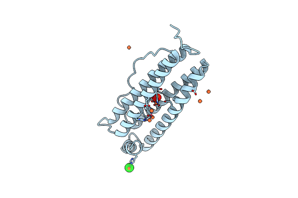
Crystal Structure Of Human Mitochondrial Ferritin (Hmtf) Fe(Ii)-Loaded For 60 Minutes Showing Either A Dioxygen Or A Superoxide Anion Coordinated To Iron Ions In The Ferroxidase Site
Organism: Homo sapiens
Method: X-RAY DIFFRACTION Resolution:1.60 Å Release Date: 2021-10-13 Classification: OXIDOREDUCTASE Ligands: OXY, FE2, MG, CL |
Organism: Homo sapiens
Method: X-RAY DIFFRACTION
Release Date: 2021-10-13
Ligands: OXY, FE2, MG, CL
 |
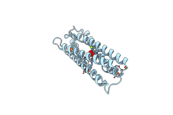
Crystal Structure Of Human Mitochondrial Ferritin (Hmtf) Fe(Ii)-Loaded For 15 Minutes Showing Either A Dioxygen Or A Superoxide Anion Coordinated To Iron Ions In The Ferroxidase Site
Organism: Homo sapiens
Method: X-RAY DIFFRACTION Resolution:1.86 Å Release Date: 2021-10-13 Classification: OXIDOREDUCTASE Ligands: OXY, FE2, CL |
Organism: Homo sapiens
Method: X-RAY DIFFRACTION
Release Date: 2021-10-13
Ligands: OXY, FE2, CL
 |
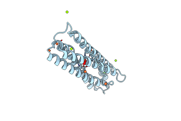
Crystal Structure Of Human Mitochondrial Ferritin (Hmtf) Fe(Ii)-Loaded For 120 Minutes Showing Either A Dioxygen Or A Superoxide Anion Coordinated To Iron Ions In The Ferroxidase Site.
Organism: Homo sapiens
Method: X-RAY DIFFRACTION Resolution:1.68 Å Release Date: 2021-10-13 Classification: OXIDOREDUCTASE Ligands: FE2, OXY, CL, MG |
Organism: Homo sapiens
Method: X-RAY DIFFRACTION
Release Date: 2021-10-13
Ligands: FE2, OXY, CL, MG
 |
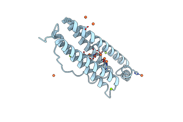
Crystal Structure Of Human Mitochondrial Ferritin (Hmtf) Fe(Ii)-Loaded For 5 Minutes Showing A Peroxide Anion As Bridging Species Of Iron Ions In The Ferroxidase Site
Organism: Homo sapiens
Method: X-RAY DIFFRACTION Resolution:1.35 Å Release Date: 2021-10-13 Classification: OXIDOREDUCTASE Ligands: FE2, PEO, MG, CL |
Organism: Homo sapiens
Method: X-RAY DIFFRACTION
Release Date: 2021-10-13
Ligands: FE2, PEO, MG, CL
 |
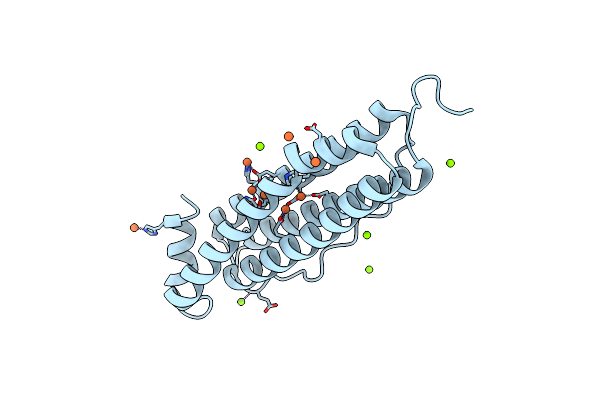
Crystal Structure Of Human Mitochondrial Ferritin (Hmtf) Fe(Ii)-Loaded For 5 Minutes Under Anaerobic Environment
Organism: Homo sapiens
Method: X-RAY DIFFRACTION Resolution:1.40 Å Release Date: 2021-10-13 Classification: OXIDOREDUCTASE Ligands: FE2, MG, CL |
Organism: Homo sapiens
Method: X-RAY DIFFRACTION
Release Date: 2021-10-13
Ligands: FE2, MG, CL
 |
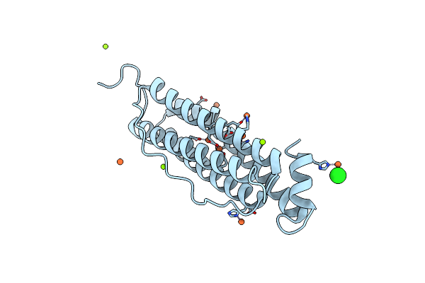
Crystal Structure Of Human Mitochondrial Ferritin (Hmtf) Fe(Ii)-Loaded For 15 Minutes Under Anaerobic Environment
Organism: Homo sapiens
Method: X-RAY DIFFRACTION Resolution:1.20 Å Release Date: 2021-10-13 Classification: OXIDOREDUCTASE Ligands: FE2, MG, CL |
Organism: Homo sapiens
Method: X-RAY DIFFRACTION
Release Date: 2021-10-13
Ligands: FE2, MG, CL
 |
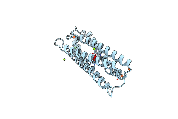
Crystal Structure Of Human Mitochondrial Ferritin (Hmtf) Fe(Ii)-Loaded For 3 Minutes Under Anaerobic Environment
Organism: Homo sapiens
Method: X-RAY DIFFRACTION Resolution:1.47 Å Release Date: 2021-10-13 Classification: OXIDOREDUCTASE Ligands: FE2, MG, CL |
Organism: Homo sapiens
Method: X-RAY DIFFRACTION
Release Date: 2021-10-13
Ligands: FE2, MG, CL
 |
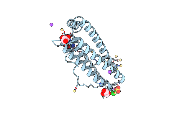
Crystal Structure Of Human Mitochondrial Ferritin (Hmtf) Fe(Ii)-Loaded For 3 Minutes Showing A Peroxide Anion As Bridging Species Of Iron Ions In The Ferroxidase Site
Organism: Homo sapiens
Method: X-RAY DIFFRACTION Resolution:1.55 Å Release Date: 2021-10-13 Classification: OXIDOREDUCTASE Ligands: FE2, PEO, MG, CL |
Organism: Homo sapiens
Method: X-RAY DIFFRACTION
Release Date: 2021-10-13
Ligands: FE2, PEO, MG, CL
 |
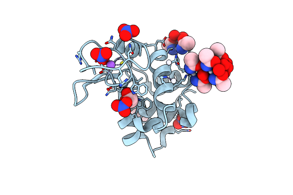
X-Ray Structure Of Arsenoplatin-1-Encapsulated Horse Spleen Ferritin
Organism: Equus caballus
Method: X-RAY DIFFRACTION Resolution:1.50 Å Release Date: 2021-03-17 Classification: TRANSPORT PROTEIN |
Organism: Equus caballus
Method: X-RAY DIFFRACTION
Release Date: 2021-03-17
 |
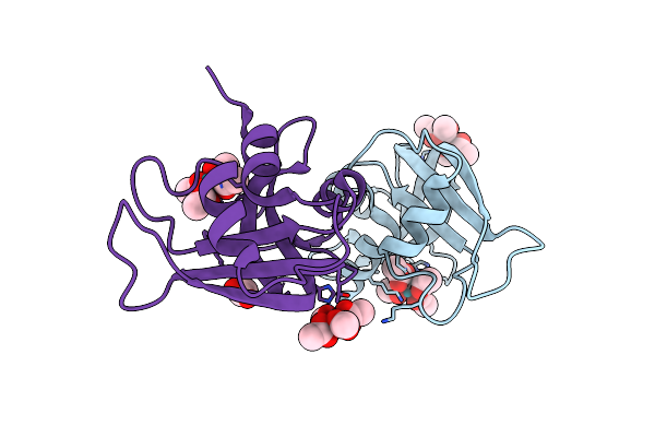
X-Ray Structure Of The Adduct Formed Upon Treating Lysozyme With An Aged Solution Of Arsenoplatin-1
Organism: Gallus gallus
Method: X-RAY DIFFRACTION Resolution:2.15 Å Release Date: 2020-12-23 Classification: HYDROLASE Ligands: NO3, EDO, A6R, DMS, NA |
Organism: Gallus gallus
Method: X-RAY DIFFRACTION
Release Date: 2020-12-23
Ligands: NO3, EDO, A6R, DMS, NA
 |
X-Ray Structure Obtained Upon Reaction Of Dirhodium Tetraacetate With Rnase A (High Resolution)
Organism: Bos taurus
Method: X-RAY DIFFRACTION Resolution:1.40 Å Release Date: 2020-02-19 Classification: UNKNOWN FUNCTION Ligands: ACT, RH3 |
Organism: Bos taurus
Method: X-RAY DIFFRACTION
Release Date: 2020-02-19
Ligands: ACT, RH3
 |
X-Ray Structure Obtained Upon Reaction Of Dirhodium Tetraacetate With Rnase A (Low Resolution)
Organism: Bos taurus
Method: X-RAY DIFFRACTION Resolution:1.80 Å Release Date: 2020-02-19 Classification: UNKNOWN FUNCTION Ligands: RH3, ACT |
Organism: Bos taurus
Method: X-RAY DIFFRACTION
Release Date: 2020-02-19
Ligands: RH3, ACT
 |
The X-Ray Structure Of The Gold/Serum Albumin Adduct Obtained Upon Reaction Of The Protein With Aul12, A Gold(Iii) Dithiocarbamate Complex
Organism: Bos taurus
Method: X-RAY DIFFRACTION Resolution:3.21 Å Release Date: 2019-08-14 Classification: TRANSPORT PROTEIN Ligands: MG, AU |
Organism: Bos taurus
Method: X-RAY DIFFRACTION
Release Date: 2019-08-14
Ligands: MG, AU