

Search Count: 28
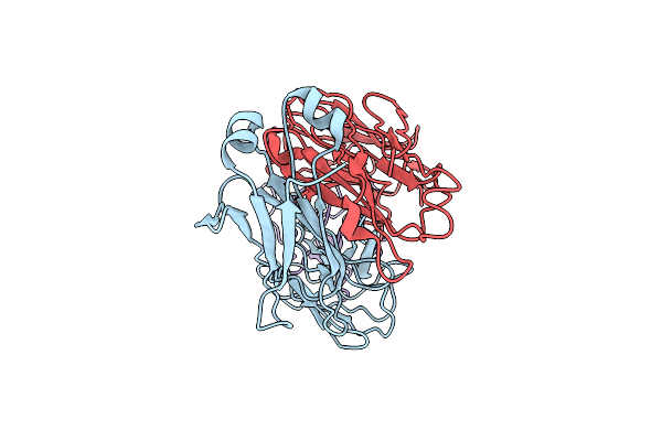 |
Cryo-Em Structure Of Pomab, A Type-I Anti-Prothrombin Antiphospholipid Antibody, Bound To Kringle-1 Of Human Prothrombin
Organism: Mus musculus, Homo sapiens
Method: ELECTRON MICROSCOPY Release Date: 2024-02-14 Classification: BLOOD CLOTTING |
Organism: Mus musculus, Homo sapiens
Method: ELECTRON MICROSCOPY
Release Date: 2024-02-14
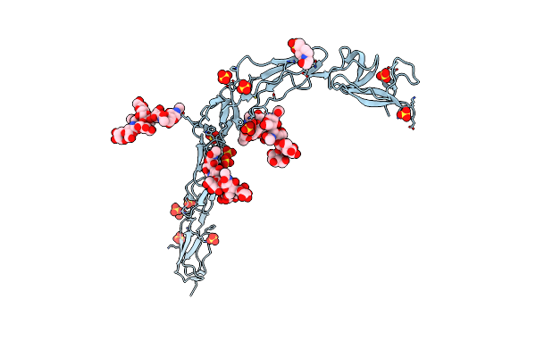 |
Crystal Structure Of Beta-2 Glycoprotein I Purified From Plasma (Pb2Gpi)
Organism: Homo sapiens
Method: X-RAY DIFFRACTION Resolution:2.40 Å Release Date: 2020-06-17 Classification: BLOOD CLOTTING Ligands: SO4, EPE |
Organism: Homo sapiens
Method: X-RAY DIFFRACTION
Release Date: 2020-06-17
Ligands: SO4, EPE
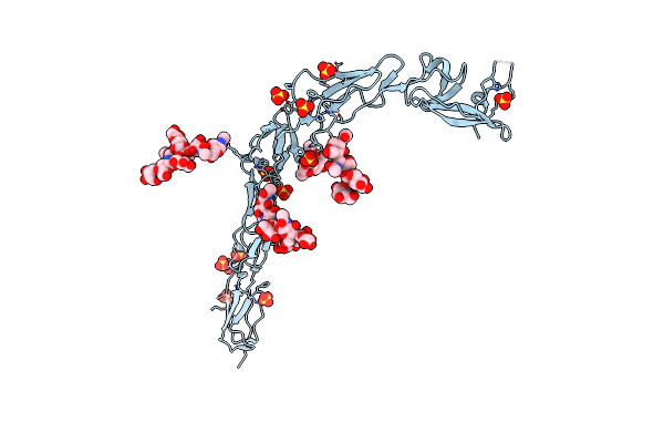 |
Crystal Structure Of Human Recombinant Beta-2 Glycoprotein I (Hrb2Gpi)
Organism: Homo sapiens
Method: X-RAY DIFFRACTION Resolution:2.58 Å Release Date: 2020-06-17 Classification: BLOOD CLOTTING Ligands: SO4 |
Organism: Homo sapiens
Method: X-RAY DIFFRACTION
Release Date: 2020-06-17
Ligands: SO4
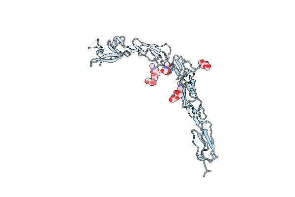 |
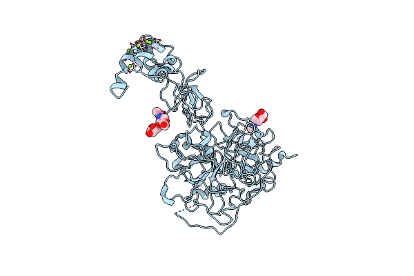
Crystal Structure Of Human Recombinant Beta-2 Glycoprotein I Short Tag (St-B2Gpi)
Organism: Homo sapiens
Method: X-RAY DIFFRACTION Resolution:2.99 Å Release Date: 2020-06-17 Classification: BLOOD CLOTTING Ligands: NAG, SO4 |
Organism: Homo sapiens
Method: X-RAY DIFFRACTION
Release Date: 2020-06-17
Ligands: NAG, SO4
 |
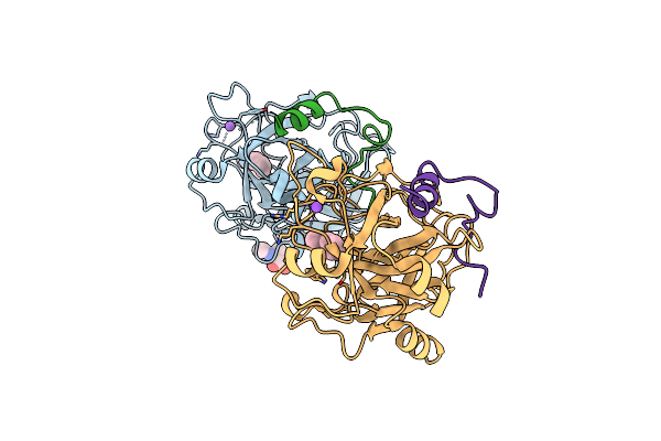
Crystal Structure Of Prothrombin Mutant S101C/A470C
Organism: Homo sapiens
Method: X-RAY DIFFRACTION Resolution:6.00 Å Release Date: 2018-06-27 Classification: HYDROLASE Ligands: MG, NAG |
Organism: Homo sapiens
Method: X-RAY DIFFRACTION
Release Date: 2018-06-27
Ligands: MG, NAG
 |
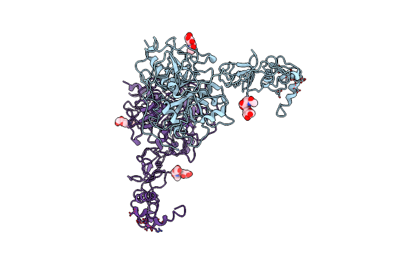
Reversible Covalent Direct Thrombin Inhibitors
Organism: Homo sapiens
Method: X-RAY DIFFRACTION Resolution:2.90 Å Release Date: 2018-05-09 Classification: Hydrolase/Hydrolase Inhibitor Ligands: 71F, NAG, NA |
Organism: Homo sapiens
Method: X-RAY DIFFRACTION
Release Date: 2018-05-09
Ligands: 71F, NAG, NA
 |
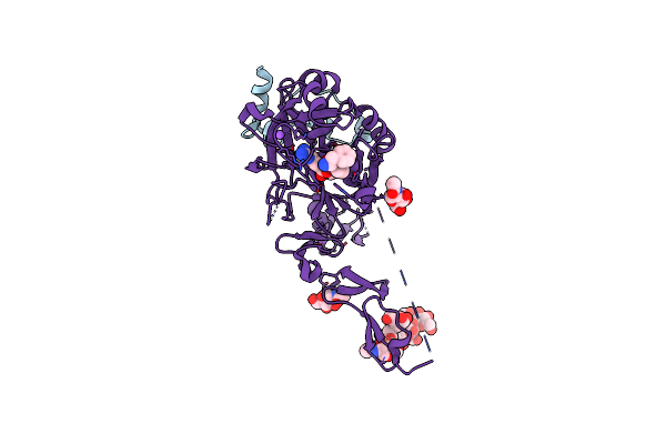
Crystal Structure Of Human Prothrombin Mutant S101C/A470C
Organism: Homo sapiens
Method: X-RAY DIFFRACTION Resolution:4.12 Å Release Date: 2018-02-28 Classification: BLOOD CLOTTING Ligands: MG, NAG |
Organism: Homo sapiens
Method: X-RAY DIFFRACTION
Release Date: 2018-02-28
Ligands: MG, NAG
 |
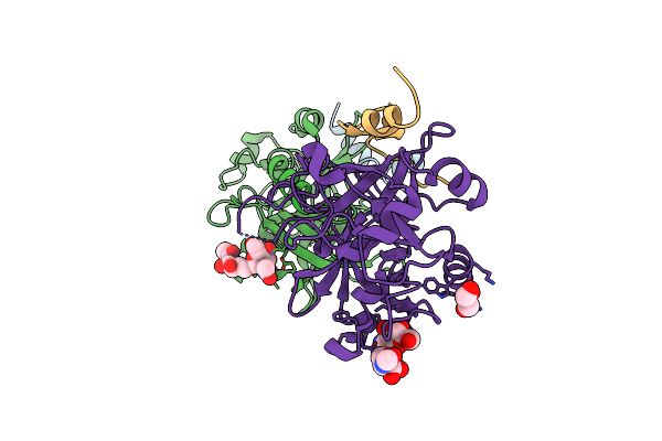
Crystal Structure Of Thrombin Mutant W215A/E217A Fused To Egf456 Of Thrombomodulin Via A 31-Residue Linker And Bound To Ppack
Organism: Homo sapiens
Method: X-RAY DIFFRACTION Resolution:2.34 Å Release Date: 2017-03-29 Classification: HYDROLASE Ligands: 0G6, K, NA, NAG |
Organism: Homo sapiens
Method: X-RAY DIFFRACTION
Release Date: 2017-03-29
Ligands: 0G6, K, NA, NAG
 |
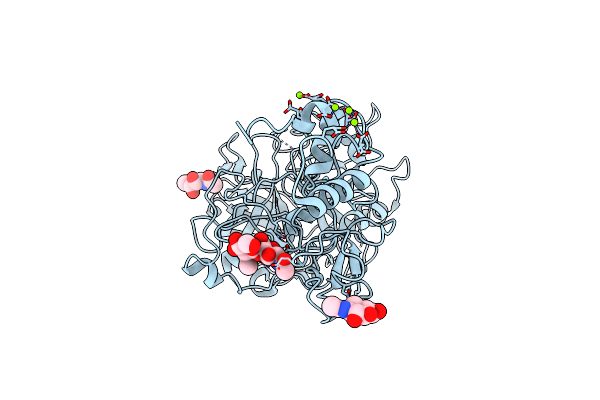
Crystal Structure For Human Thrombin Mutant D189A
Organism: Homo sapiens
Method: X-RAY DIFFRACTION Resolution:1.70 Å Release Date: 2016-07-13 Classification: HYDROLASE Ligands: CL, GOL |
Organism: Homo sapiens
Method: X-RAY DIFFRACTION
Release Date: 2016-07-13
Ligands: CL, GOL
 |
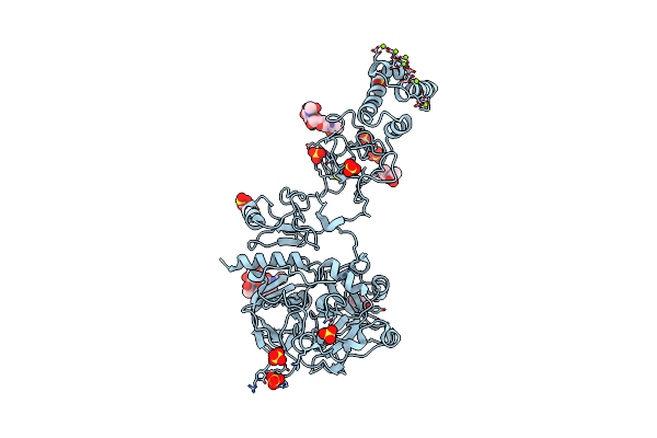
Crystal Structure Of Prothrombin Deletion Mutant Residues 146-167 ( Form Ii ).
Organism: Homo sapiens
Method: X-RAY DIFFRACTION Resolution:3.21 Å Release Date: 2016-01-20 Classification: HYDROLASE Ligands: NAG, MG |
Organism: Homo sapiens
Method: X-RAY DIFFRACTION
Release Date: 2016-01-20
Ligands: NAG, MG
 |
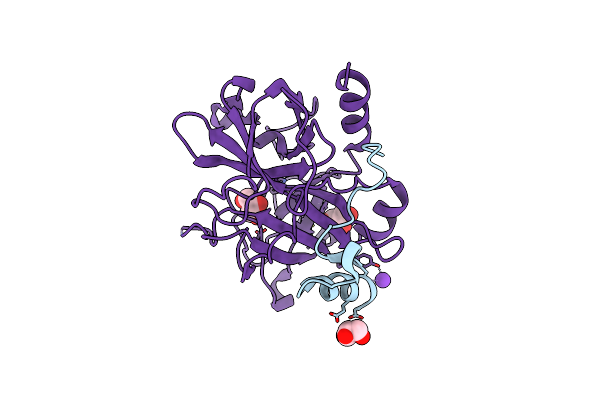
Crystal Structure Of Prothrombin Deletion Mutant Residues 154-167 ( Form I )
Organism: Homo sapiens
Method: X-RAY DIFFRACTION Resolution:2.20 Å Release Date: 2016-01-20 Classification: HYDROLASE Ligands: MG, NAG, SO4, GOL |
Organism: Homo sapiens
Method: X-RAY DIFFRACTION
Release Date: 2016-01-20
Ligands: MG, NAG, SO4, GOL
 |
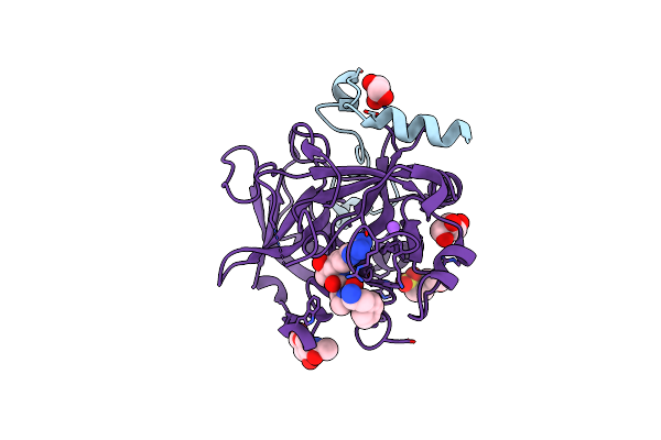
Crystal Structure Of Thrombin Mutant S195T (Free Form)
Organism: Homo sapiens
Method: X-RAY DIFFRACTION Resolution:1.70 Å Release Date: 2015-03-11 Classification: HYDROLASE Ligands: K, GOL |
Organism: Homo sapiens
Method: X-RAY DIFFRACTION
Release Date: 2015-03-11
Ligands: K, GOL
 |
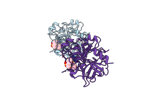
Crystal Structure Of Thrombin Mutant S195T Bound With Ppack
Organism: Homo sapiens
Method: X-RAY DIFFRACTION Resolution:1.84 Å Release Date: 2015-03-11 Classification: HYDROLASE/HYDROLASE INHIBITOR Ligands: 0G6, MES, GOL, NA, NAG |
Organism: Homo sapiens
Method: X-RAY DIFFRACTION
Release Date: 2015-03-11
Ligands: 0G6, MES, GOL, NA, NAG
 |
Structure Of Prethrombin-2 Mutant S195A Bound To The Active Site Inhibitor Argatroban
Organism: Homo sapiens
Method: X-RAY DIFFRACTION Resolution:3.00 Å Release Date: 2014-11-05 Classification: HYDROLASE/HYDROLASE INHIBITOR Ligands: 15U |
Organism: Homo sapiens
Method: X-RAY DIFFRACTION
Release Date: 2014-11-05
Ligands: 15U
 |
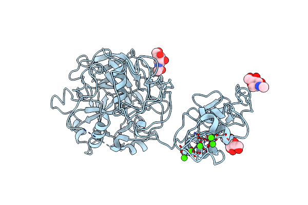
Crystal Structure Of Ca2+-Free Prothrombin Deletion Mutant Residues 146-167
Organism: Homo sapiens
Method: X-RAY DIFFRACTION Resolution:2.81 Å Release Date: 2014-05-21 Classification: HYDROLASE Ligands: NAG |
Organism: Homo sapiens
Method: X-RAY DIFFRACTION
Release Date: 2014-05-21
Ligands: NAG
 |
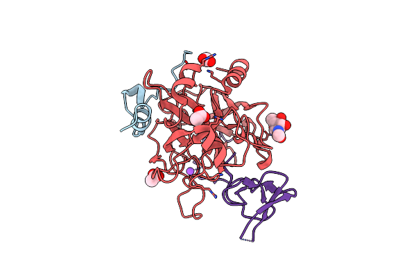
Crystal Structure Of Ca2+ Bound Prothrombin Deletion Mutant Residues 146-167
Organism: Homo sapiens
Method: X-RAY DIFFRACTION Resolution:3.38 Å Release Date: 2014-05-21 Classification: HYDROLASE Ligands: CA, NAG |
Organism: Homo sapiens
Method: X-RAY DIFFRACTION
Release Date: 2014-05-21
Ligands: CA, NAG
 |
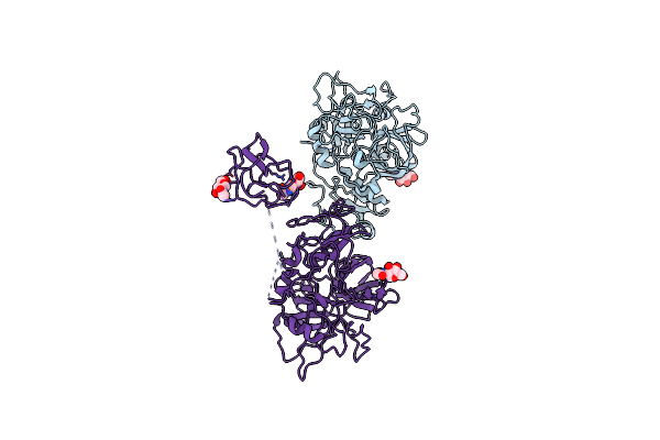
Crystal Structure For The Complex Of Thrombin Mutant D102N And Hirudin
Organism: Homo sapiens, Hirudo medicinalis
Method: X-RAY DIFFRACTION Resolution:2.20 Å Release Date: 2013-09-25 Classification: HYDROLASE/HYDROLASE INHIBITOR |
Organism: Homo sapiens, Hirudo medicinalis
Method: X-RAY DIFFRACTION
Release Date: 2013-09-25
 |
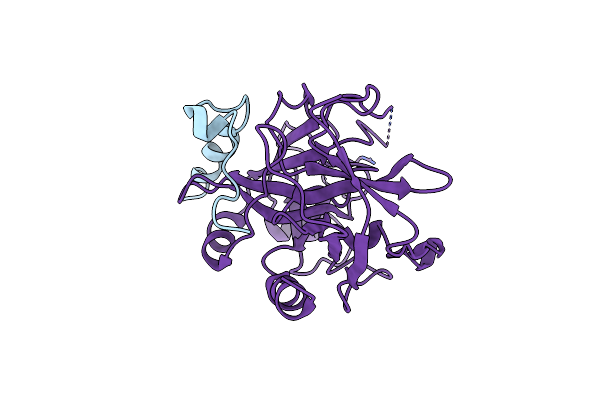
Structure Of Recombinant Gla-Domainless Prothrombin Mutant S525A
Organism: Homo sapiens
Method: X-RAY DIFFRACTION Resolution:3.30 Å Release Date: 2013-06-26 Classification: HYDROLASE Ligands: NAG |
Organism: Homo sapiens
Method: X-RAY DIFFRACTION
Release Date: 2013-06-26
Ligands: NAG
 |
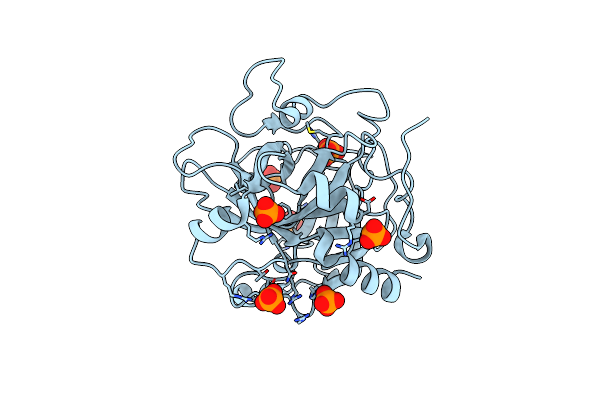
Crystal Structure Of Thrombin Mutant E14Ea/D14La/E18A/S195A
Organism: Homo sapiens
Method: X-RAY DIFFRACTION Resolution:2.19 Å Release Date: 2013-03-13 Classification: HYDROLASE/PEPTIDE |
Organism: Homo sapiens
Method: X-RAY DIFFRACTION
Release Date: 2013-03-13
 |
Crystal Structure Of Prethrombin-2 Mutant E14Ea/D14La/E18A/S195A
Organism: Homo sapiens
Method: X-RAY DIFFRACTION Resolution:2.40 Å Release Date: 2013-03-13 Classification: HYDROLASE Ligands: PO4 |
Organism: Homo sapiens
Method: X-RAY DIFFRACTION
Release Date: 2013-03-13
Ligands: PO4