

Search Count: 13
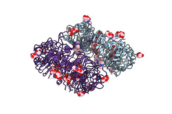 |
Crystal Structure Of Human Tlr8 Ectodomain Bound To Small Molecule Antagonist 21
Organism: Homo sapiens
Method: X-RAY DIFFRACTION Resolution:2.76 Å Release Date: 2022-05-04 Classification: RNA BINDING PROTEIN Ligands: NAG, RQY |
Organism: Homo sapiens
Method: X-RAY DIFFRACTION
Release Date: 2022-05-04
Ligands: NAG, RQY
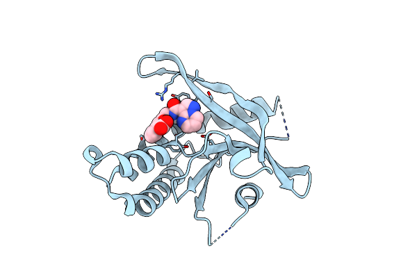 |
Crystal Structure Of Human Sting R232 In Complex With Compound 11
Organism: Homo sapiens
Method: X-RAY DIFFRACTION Resolution:1.96 Å Release Date: 2022-02-09 Classification: IMMUNE SYSTEM Ligands: B7L |
Organism: Homo sapiens
Method: X-RAY DIFFRACTION
Release Date: 2022-02-09
Ligands: B7L
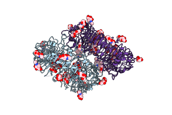 |
Crystal Structure Of Human Tlr8 Ectodomain Bound To Small Molecule Antagonist 14C
Organism: Homo sapiens
Method: X-RAY DIFFRACTION Resolution:2.65 Å Release Date: 2020-08-12 Classification: RNA BINDING PROTEIN Ligands: NAG, QSM |
Organism: Homo sapiens
Method: X-RAY DIFFRACTION
Release Date: 2020-08-12
Ligands: NAG, QSM
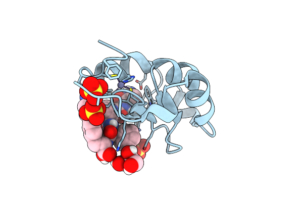 |
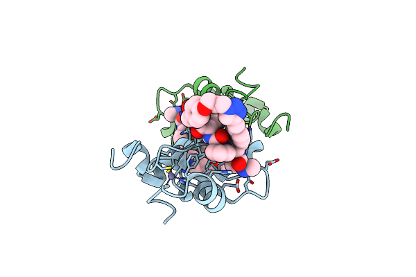
Crystal Structure Of Xiap-Bir2 Domain Complexed With (S)-3-(4-Methoxyphenyl)-2-((S)-2-((S)-1-((S)-2-((S)-2-(Methylamino)Propanamido)Pent-4-Ynoyl)Pyrrolidine-2-Carboxamido)-3-Phenylpropanamido)Propanoic Acid
Organism: Homo sapiens
Method: X-RAY DIFFRACTION Resolution:2.09 Å Release Date: 2015-05-06 Classification: APOPTOSIS Ligands: ZN, SO4, GOL |
Organism: Homo sapiens
Method: X-RAY DIFFRACTION
Release Date: 2015-05-06
Ligands: ZN, SO4, GOL
 |
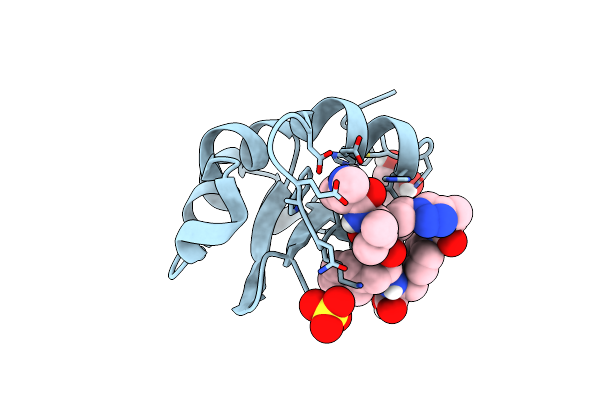
Crystal Structure Of Xiap-Bir2 Domain Complexed With Ligand Bound
Organism: Homo sapiens
Method: X-RAY DIFFRACTION Resolution:1.96 Å Release Date: 2015-03-04 Classification: APOPTOSIS Ligands: ZN |
Organism: Homo sapiens
Method: X-RAY DIFFRACTION
Release Date: 2015-03-04
Ligands: ZN
 |
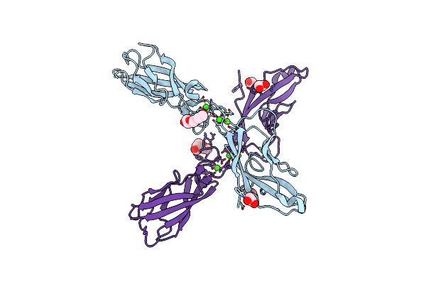
Crystal Structure Of Xiap-Bir2 Domain Complexed With Ligand Bound
Organism: Homo sapiens
Method: X-RAY DIFFRACTION Resolution:2.02 Å Release Date: 2015-03-04 Classification: APOPTOSIS Ligands: ZN, SO4, GOL |
Organism: Homo sapiens
Method: X-RAY DIFFRACTION
Release Date: 2015-03-04
Ligands: ZN, SO4, GOL
 |
Crystal Structure Of E-Cadherin Ec1-2 P5A P6A
Organism: Mus musculus
Method: X-RAY DIFFRACTION Resolution:1.80 Å Release Date: 2011-05-18 Classification: CELL ADHESION Ligands: CA, EDO, PGE |
Organism: Mus musculus
Method: X-RAY DIFFRACTION
Release Date: 2011-05-18
Ligands: CA, EDO, PGE
 |
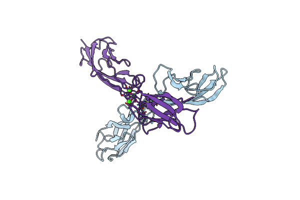
Crystal Structure Of Mouse T-Cadherin Ec1 Ec2
Organism: Mus musculus
Method: X-RAY DIFFRACTION Resolution:2.00 Å Release Date: 2010-03-02 Classification: STRUCTURAL PROTEIN |
Organism: Mus musculus
Method: X-RAY DIFFRACTION
Release Date: 2010-03-02
 |
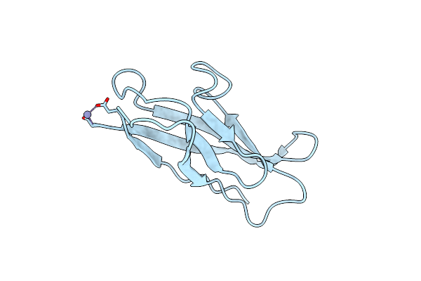
Crystal Structure Of Chicken T-Cadherin Ec1 Ec2
Organism: Gallus gallus
Method: X-RAY DIFFRACTION Resolution:2.90 Å Release Date: 2010-03-02 Classification: STRUCTURAL PROTEIN Ligands: CA |
Organism: Gallus gallus
Method: X-RAY DIFFRACTION
Release Date: 2010-03-02
Ligands: CA
 |
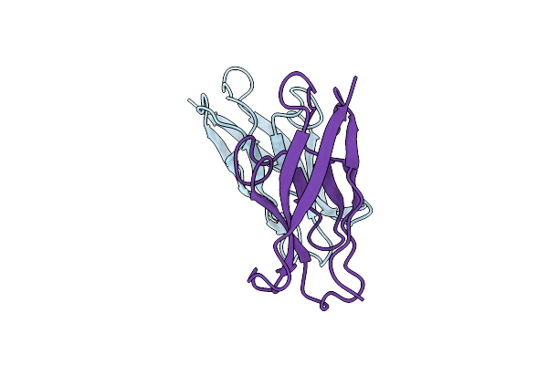
Crystal Structure Of Xenopus Laevis T-Cadherin Ec1
Organism: Xenopus laevis
Method: X-RAY DIFFRACTION Resolution:1.80 Å Release Date: 2010-03-02 Classification: CELL ADHESION Ligands: ZN |
Organism: Xenopus laevis
Method: X-RAY DIFFRACTION
Release Date: 2010-03-02
Ligands: ZN
 |
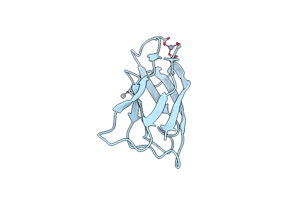
Crystal Structure Of Mouse T-Cadherin Ec1
Organism: Mus musculus
Method: X-RAY DIFFRACTION Resolution:1.81 Å Release Date: 2010-03-02 Classification: CELL ADHESION |
Organism: Mus musculus
Method: X-RAY DIFFRACTION
Release Date: 2010-03-02
 |
Crystal Structure Of Chicken T-Cadherin Ec1
Organism: Gallus gallus
Method: X-RAY DIFFRACTION Resolution:1.13 Å Release Date: 2010-03-02 Classification: CELL ADHESION Ligands: ZN |
Organism: Gallus gallus
Method: X-RAY DIFFRACTION
Release Date: 2010-03-02
Ligands: ZN
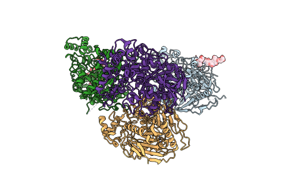 |
Crystal Structure Of The Extracellular Domain Of Neuroligin 2A From Mouse
Organism: Mus musculus
Method: X-RAY DIFFRACTION Resolution:3.30 Å Release Date: 2008-02-19 Classification: CELL ADHESION Ligands: NAG |
Organism: Mus musculus
Method: X-RAY DIFFRACTION
Release Date: 2008-02-19
Ligands: NAG