

Search Count: 24
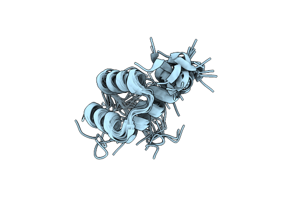 |
Structural Characterization Of Phox2B And Its Dna Interactions Shed Lights Into The Molecular Basis Of The + 7Ala Variant Pathogenicity In Cchs
Organism: Homo sapiens
Method: SOLUTION NMR Release Date: 2024-05-15 Classification: PROTEIN BINDING |
Organism: Homo sapiens
Method: SOLUTION NMR
Release Date: 2024-05-15
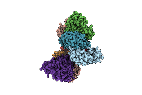 |
Crystal Structure Of The Human Phosposerine Aminotransferase (Psat)
Organism: Homo sapiens
Method: X-RAY DIFFRACTION Resolution:2.46 Å Release Date: 2023-03-29 Classification: TRANSFERASE Ligands: SO4, PMP |
Organism: Homo sapiens
Method: X-RAY DIFFRACTION
Release Date: 2023-03-29
Ligands: SO4, PMP
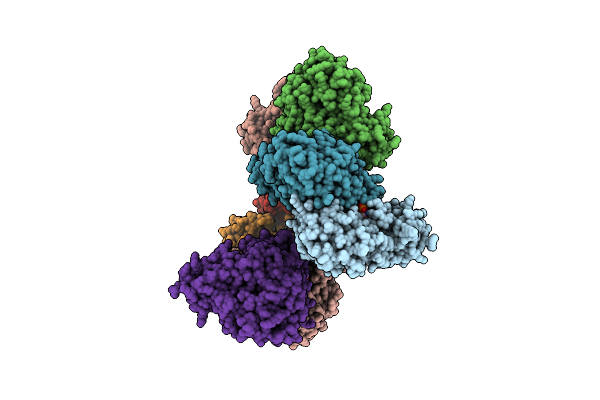 |
Crystal Structure Of The Human Phosphoserine Aminotransferase (Psat) In Complex With O-Phosphoserine
Organism: Homo sapiens
Method: X-RAY DIFFRACTION Resolution:2.78 Å Release Date: 2023-03-29 Classification: TRANSFERASE Ligands: E1U, SEP, PMP, SO4 |
Organism: Homo sapiens
Method: X-RAY DIFFRACTION
Release Date: 2023-03-29
Ligands: E1U, SEP, PMP, SO4
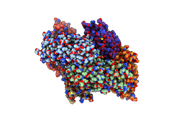 |
Structure Of Human Daspo
Organism: Homo sapiens
Method: X-RAY DIFFRACTION Resolution:3.22 Å Release Date: 2020-03-11 Classification: FLAVOPROTEIN Ligands: FAD, GOL, TRS, PO4 |
Organism: Homo sapiens
Method: X-RAY DIFFRACTION
Release Date: 2020-03-11
Ligands: FAD, GOL, TRS, PO4
 |
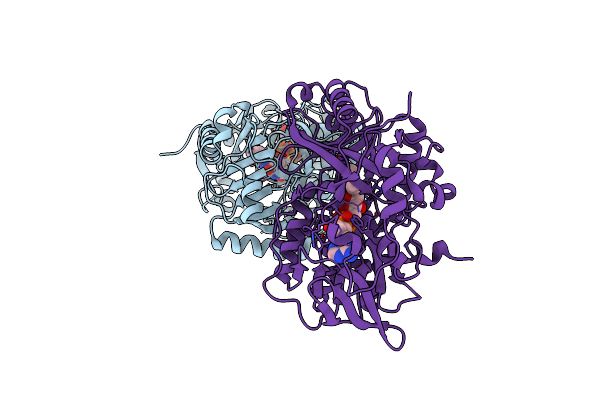
Crystal Structure Of Citrobacter Freundii Methionine Gamma-Lyase With V358Y Replacement
Organism: Citrobacter freundii
Method: X-RAY DIFFRACTION Resolution:1.45 Å Release Date: 2018-10-10 Classification: LYASE Ligands: PLP, PGE, PEG |
Organism: Citrobacter freundii
Method: X-RAY DIFFRACTION
Release Date: 2018-10-10
Ligands: PLP, PGE, PEG
 |
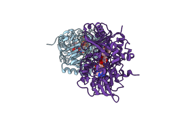
Structure Of L-Amino Acid Deaminase From Proteus Myxofaciens
Organism: Proteus myxofaciens
Method: X-RAY DIFFRACTION Resolution:2.00 Å Release Date: 2016-04-06 Classification: HYDROLASE Ligands: FAD |
Organism: Proteus myxofaciens
Method: X-RAY DIFFRACTION
Release Date: 2016-04-06
Ligands: FAD
 |
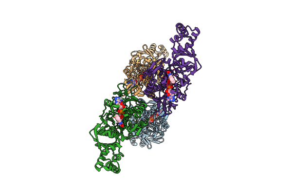
Structure Of L-Amino Acid Deaminase From Proteus Myxofaciens In Complex With Anthranilate
Organism: Proteus myxofaciens
Method: X-RAY DIFFRACTION Resolution:1.75 Å Release Date: 2016-04-06 Classification: HYDROLASE Ligands: FAD, BE2 |
Organism: Proteus myxofaciens
Method: X-RAY DIFFRACTION
Release Date: 2016-04-06
Ligands: FAD, BE2
 |
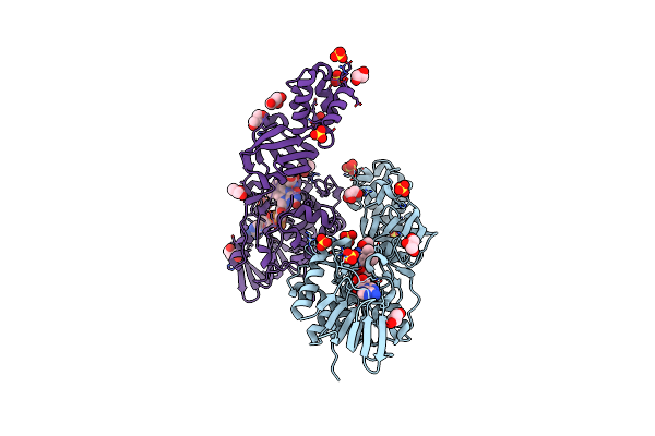
L-Aminoacetone Oxidase From Streptococcus Oligofermentans Belongs To A New 3-Domain Family Of Bacterial Flavoproteins
Organism: Streptococcus oligofermentans
Method: X-RAY DIFFRACTION Resolution:2.70 Å Release Date: 2014-10-15 Classification: OXIDOREDUCTASE Ligands: FAD |
Organism: Streptococcus oligofermentans
Method: X-RAY DIFFRACTION
Release Date: 2014-10-15
Ligands: FAD
 |
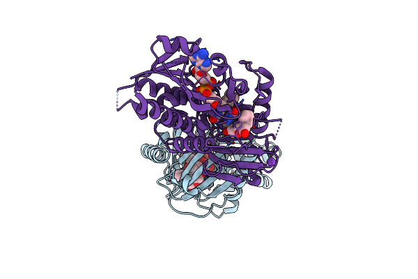
L-Aminoacetone Oxidase From Streptococcus Oligofermentans Belongs To A New 3-Domain Family Of Bacterial Flavoproteins
Organism: Streptococcus oligofermentans
Method: X-RAY DIFFRACTION Resolution:2.00 Å Release Date: 2014-10-15 Classification: OXIDOREDUCTASE Ligands: FAD, MEU, SO4, GOL |
Organism: Streptococcus oligofermentans
Method: X-RAY DIFFRACTION
Release Date: 2014-10-15
Ligands: FAD, MEU, SO4, GOL
 |
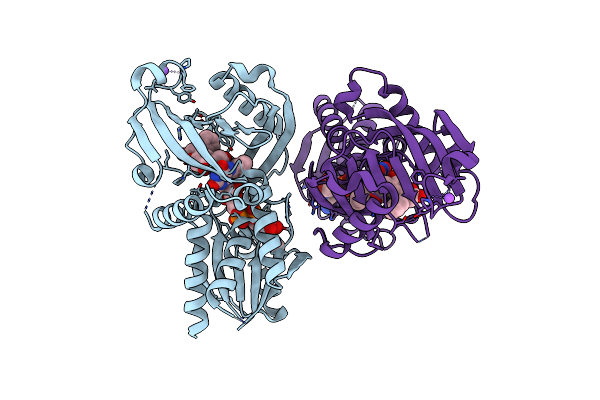
Co-Crystal Structure Of Compound 3 (4-Hydroxy-6-[2-(7-Hydroxy-2-Oxo-4-Phenyl-2H-Chromen-6-Yl)Ethyl]Pyridazin-3(2H)-One) And Fad Bound To Human Daao At 2.4A
Organism: Homo sapiens
Method: X-RAY DIFFRACTION Resolution:2.40 Å Release Date: 2014-07-16 Classification: oxidoreductase/oxidoreductase inhibitor Ligands: FAD, 31T, K |
Organism: Homo sapiens
Method: X-RAY DIFFRACTION
Release Date: 2014-07-16
Ligands: FAD, 31T, K
 |
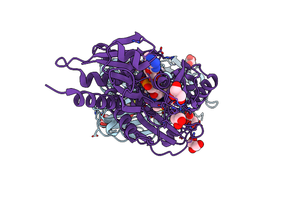
Co-Crystal Structure Of Compound 2 (3-(7-Hydroxy-2-Oxo-4-Phenyl-2H-Chromen-6-Yl)Propanoic Acid) And Fad Bound To Human Daao At 2.85A
Organism: Homo sapiens
Method: X-RAY DIFFRACTION Resolution:2.85 Å Release Date: 2014-07-16 Classification: oxidoreductase/oxidoreductase inhibitor |
Organism: Homo sapiens
Method: X-RAY DIFFRACTION
Release Date: 2014-07-16
 |
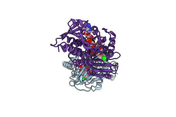
In Vitro And In Vivo Inhibition Of Human D-Amino Acid Oxidase: Regulation Of D-Serine Concentration In The Brain
Organism: Homo sapiens
Method: X-RAY DIFFRACTION Resolution:1.90 Å Release Date: 2013-05-15 Classification: OXIDOREDUCTASE Ligands: FAD, 4WL, GOL |
Organism: Homo sapiens
Method: X-RAY DIFFRACTION
Release Date: 2013-05-15
Ligands: FAD, 4WL, GOL
 |
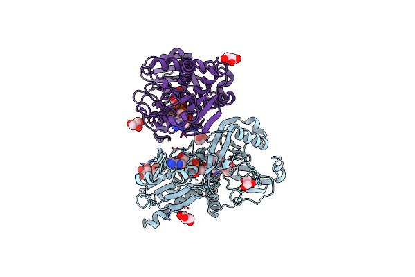
In Vitro And In Vivo Inhibition Of Human D-Amino Acid Oxidase: Regulation Of D-Serine Concentration In The Brain
Organism: Homo sapiens
Method: X-RAY DIFFRACTION Resolution:2.30 Å Release Date: 2013-05-15 Classification: OXIDOREDUCTASE Ligands: FAD, SE5, GOL |
Organism: Homo sapiens
Method: X-RAY DIFFRACTION
Release Date: 2013-05-15
Ligands: FAD, SE5, GOL
 |
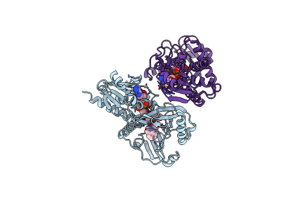
In Vitro And In Vivo Inhibition Of Human D-Amino Acid Oxidase: Regulation Of D-Serine Concentration In The Brain
Organism: Homo sapiens
Method: X-RAY DIFFRACTION Resolution:2.40 Å Release Date: 2013-05-15 Classification: OXIDOREDUCTASE Ligands: FAD, SE2, GOL |
Organism: Homo sapiens
Method: X-RAY DIFFRACTION
Release Date: 2013-05-15
Ligands: FAD, SE2, GOL
 |
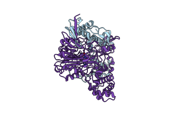
In Vitro And In Vivo Inhibition Of Human D-Amino Acid Oxidase: Regulation Of D-Serine Concentration In The Brain
Organism: Homo sapiens
Method: X-RAY DIFFRACTION Resolution:2.75 Å Release Date: 2013-05-15 Classification: OXIDOREDUCTASE Ligands: FAD, SS8 |
Organism: Homo sapiens
Method: X-RAY DIFFRACTION
Release Date: 2013-05-15
Ligands: FAD, SS8
 |
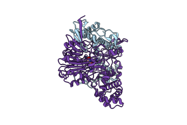
Crystal Structure Of A Class Iii Engineered Cephalosporin Acylase
Organism: Pseudomonas
Method: X-RAY DIFFRACTION Resolution:2.13 Å Release Date: 2013-02-27 Classification: HYDROLASE Ligands: GLJ |
Organism: Pseudomonas
Method: X-RAY DIFFRACTION
Release Date: 2013-02-27
Ligands: GLJ
 |
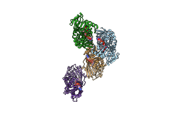
Crystal Structure Of A Double Mutant Of A Class Iii Engineered Cephalosporin Acylase
Organism: Pseudomonas
Method: X-RAY DIFFRACTION Resolution:1.57 Å Release Date: 2013-02-27 Classification: HYDROLASE Ligands: GLJ |
Organism: Pseudomonas
Method: X-RAY DIFFRACTION
Release Date: 2013-02-27
Ligands: GLJ
 |
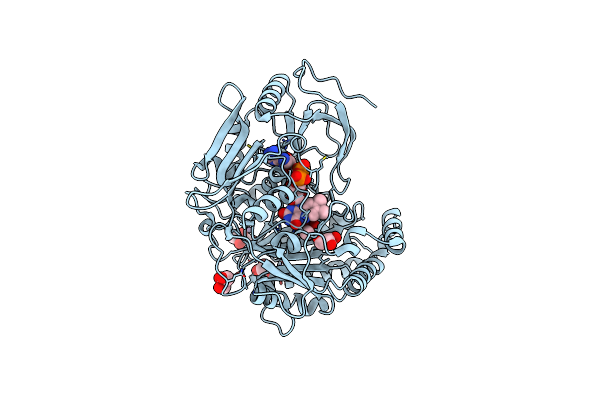
Crystal Structure Of Glycine Oxidase G51S/A54R/H244A Mutant In Complex With Inhibitor Glycolate
Organism: Bacillus subtilis
Method: X-RAY DIFFRACTION Resolution:2.60 Å Release Date: 2009-10-27 Classification: OXIDOREDUCTASE Ligands: GOA, FAD |
Organism: Bacillus subtilis
Method: X-RAY DIFFRACTION
Release Date: 2009-10-27
Ligands: GOA, FAD
 |
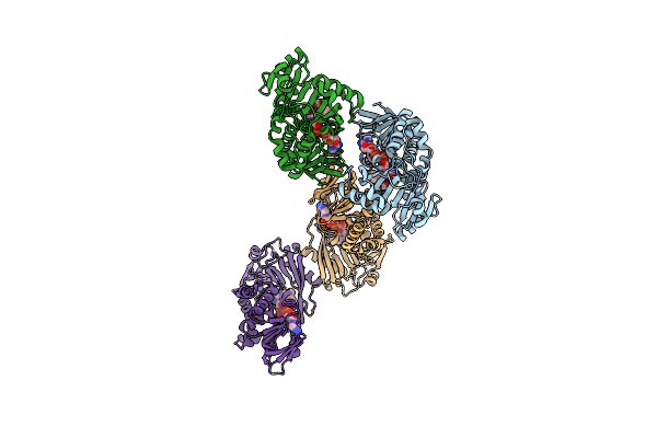
Cholesterol Oxidase From Brevibacterium Sterolicum- His121Ala Mutant
Organism: Brevibacterium sterolicum
Method: X-RAY DIFFRACTION Resolution:1.60 Å Release Date: 2007-12-11 Classification: OXIDOREDUCTASE Ligands: MN, CAC, FAD, GOL |
Organism: Brevibacterium sterolicum
Method: X-RAY DIFFRACTION
Release Date: 2007-12-11
Ligands: MN, CAC, FAD, GOL
 |
Structure Of Glycine Oxidase With Bound Inhibitor Glycolate
Organism: Bacillus subtilis
Method: X-RAY DIFFRACTION Resolution:1.80 Å Release Date: 2005-02-22 Classification: OXIDOREDUCTASE Ligands: FAD, GOA |
Organism: Bacillus subtilis
Method: X-RAY DIFFRACTION
Release Date: 2005-02-22
Ligands: FAD, GOA