

Search Count: 24
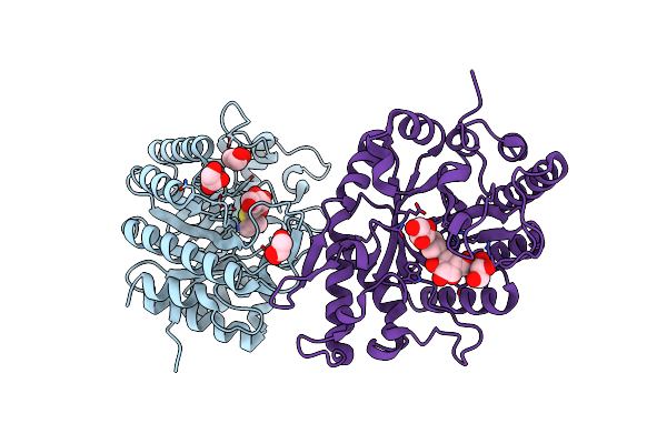 |
Structure Of Xyn11 From Pseudothermotoga Thermarum
Organism: Pseudothermotoga thermarum dsm 5069
Method: X-RAY DIFFRACTION Resolution:1.80 Å Release Date: 2021-06-09 Classification: HYDROLASE Ligands: GOL, IPT |
Organism: Pseudothermotoga thermarum dsm 5069
Method: X-RAY DIFFRACTION
Release Date: 2021-06-09
Ligands: GOL, IPT
 |
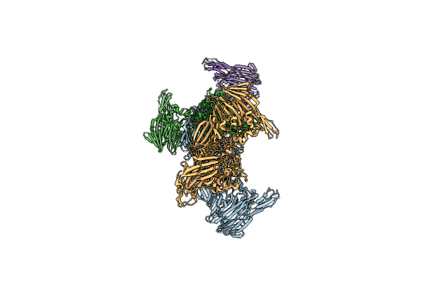
Structure Of Beta-Galactosidase From Thermotoga Maritima
Organism: Thermotoga maritima msb8
Method: ELECTRON MICROSCOPY Resolution:2.00 Å Release Date: 2020-03-18 Classification: HYDROLASE Ligands: MG |
Organism: Thermotoga maritima msb8
Method: ELECTRON MICROSCOPY
Release Date: 2020-03-18
Ligands: MG
 |
Structure Of Beta-Galactosidase From Thermotoga Maritima.
Organism: Thermotoga maritima msb8
Method: X-RAY DIFFRACTION Resolution:3.70 Å Release Date: 2020-01-15 Classification: HYDROLASE Ligands: MG |
Organism: Thermotoga maritima msb8
Method: X-RAY DIFFRACTION
Release Date: 2020-01-15
Ligands: MG
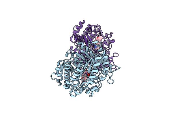 |
Structure Of Beta-Glucosidase A From Paenibacillus Polymyxa Complexed With Multivalent Inhibitors.
Organism: Paenibacillus polymyxa
Method: X-RAY DIFFRACTION Resolution:2.85 Å Release Date: 2019-07-03 Classification: HYDROLASE Ligands: JJW |
Organism: Paenibacillus polymyxa
Method: X-RAY DIFFRACTION
Release Date: 2019-07-03
Ligands: JJW
 |
Structure Of Beta-Glucosidase A From Paenibacillus Polymyxa Complexed With A Monovalent Inhibitor
Organism: Paenibacillus polymyxa
Method: X-RAY DIFFRACTION Resolution:2.13 Å Release Date: 2019-07-03 Classification: HYDROLASE Ligands: JSK |
Organism: Paenibacillus polymyxa
Method: X-RAY DIFFRACTION
Release Date: 2019-07-03
Ligands: JSK
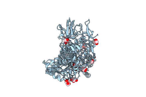 |
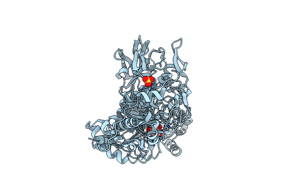
Structure Of A Gh3 B-Glucosidase From Cow Rumen Metagenome
Organism: Metagenome
Method: X-RAY DIFFRACTION Resolution:1.83 Å Release Date: 2016-10-05 Classification: HYDROLASE Ligands: GOL |
Organism: Metagenome
Method: X-RAY DIFFRACTION
Release Date: 2016-10-05
Ligands: GOL
 |
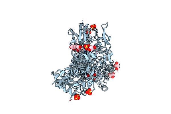
Structure Of A Gh3 B-Glicosidase From Cow Rumen Metagenome In Complex With Glucose
Organism: Metagenome
Method: X-RAY DIFFRACTION Resolution:2.17 Å Release Date: 2016-10-05 Classification: HYDROLASE Ligands: BGC, SO4 |
Organism: Metagenome
Method: X-RAY DIFFRACTION
Release Date: 2016-10-05
Ligands: BGC, SO4
 |
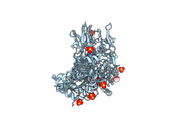
Structure Of A Gh3 B-Glicosidase From Cow Rumen Metagenome In Complexed With Xylose
Organism: Metagenome
Method: X-RAY DIFFRACTION Resolution:2.08 Å Release Date: 2016-10-05 Classification: HYDROLASE Ligands: XYP, SO4, GOL |
Organism: Metagenome
Method: X-RAY DIFFRACTION
Release Date: 2016-10-05
Ligands: XYP, SO4, GOL
 |
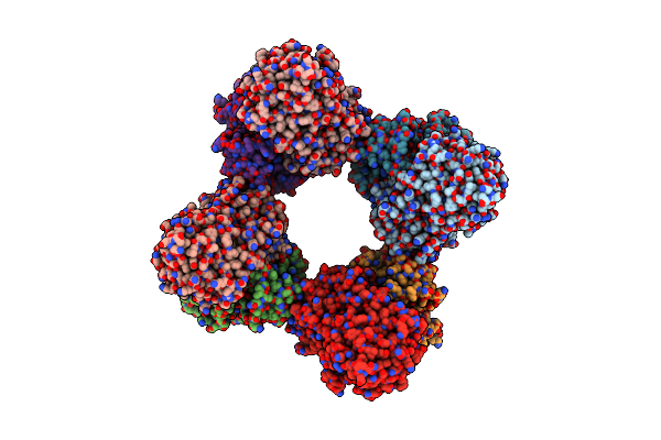
Structure Of A Gh3 B-Glucosidase From Cow Rumen Metagenome In Complex With Galactose
Organism: Metagenome
Method: X-RAY DIFFRACTION Resolution:2.29 Å Release Date: 2016-10-05 Classification: HYDROLASE Ligands: GAL, SO4, EDO |
Organism: Metagenome
Method: X-RAY DIFFRACTION
Release Date: 2016-10-05
Ligands: GAL, SO4, EDO
 |
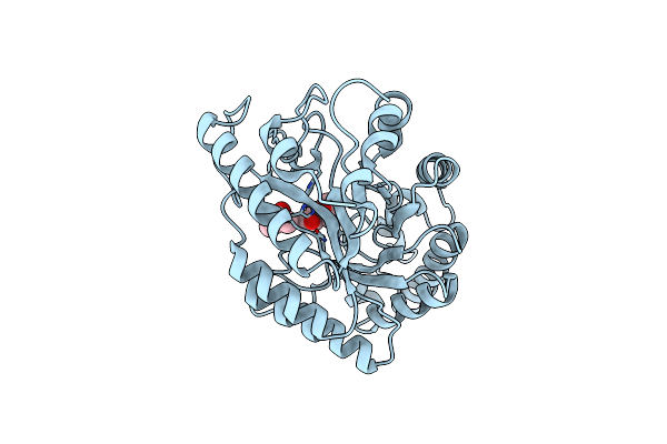
Structure Of Saccharomyces Cerevisiae Invertase
Organism: Saccharomyces cerevisiae
Method: X-RAY DIFFRACTION Resolution:3.40 Å Release Date: 2013-03-06 Classification: HYDROLASE |
Organism: Saccharomyces cerevisiae
Method: X-RAY DIFFRACTION
Release Date: 2013-03-06
 |
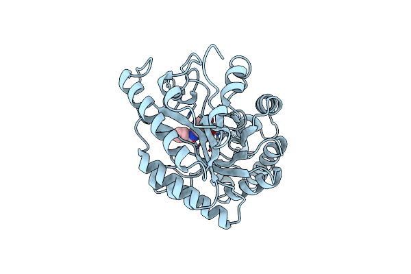
Crystal Structure Of Xynb, An Intracellular Xylanase From Paenibacillus Barcinonensis
Organism: Bacillus sp. bp-23
Method: X-RAY DIFFRACTION Resolution:2.10 Å Release Date: 2009-09-29 Classification: HYDROLASE Ligands: MPD |
Organism: Bacillus sp. bp-23
Method: X-RAY DIFFRACTION
Release Date: 2009-09-29
Ligands: MPD
 |
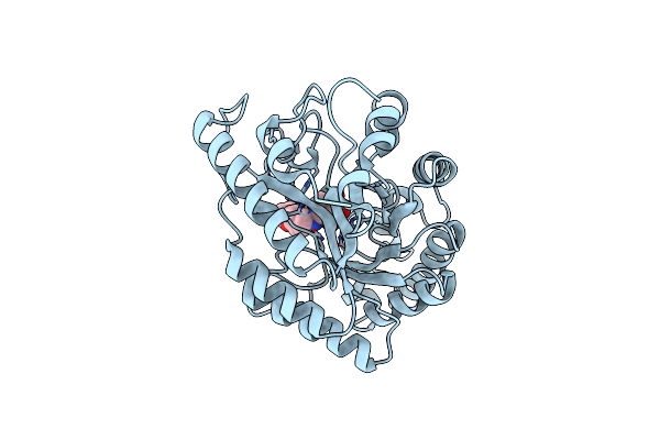
Crystal Structure Of Xilanase Xynb From Paenibacillus Barcelonensis Complexed With An Inhibitor
Organism: Bacillus sp. bp-23
Method: X-RAY DIFFRACTION Resolution:2.73 Å Release Date: 2009-09-29 Classification: HYDROLASE Ligands: HAH |
Organism: Bacillus sp. bp-23
Method: X-RAY DIFFRACTION
Release Date: 2009-09-29
Ligands: HAH
 |
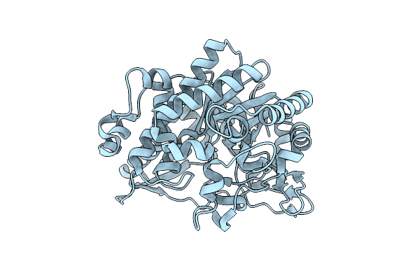
Crystal Structure Of Xylanase Xynb From Paenibacillus Barcinonensis Complexed With A Conduramine Derivative
Organism: Bacillus sp. bp-23
Method: X-RAY DIFFRACTION Resolution:2.08 Å Release Date: 2009-09-29 Classification: HYDROLASE Ligands: HXH |
Organism: Bacillus sp. bp-23
Method: X-RAY DIFFRACTION
Release Date: 2009-09-29
Ligands: HXH
 |
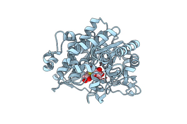
Beta-Glucosidase B From Paenibacillus Polymyxa
Organism: Paenibacillus polymyxa
Method: X-RAY DIFFRACTION Resolution:2.10 Å Release Date: 2007-10-02 Classification: HYDROLASE |
Organism: Paenibacillus polymyxa
Method: X-RAY DIFFRACTION
Release Date: 2007-10-02
 |
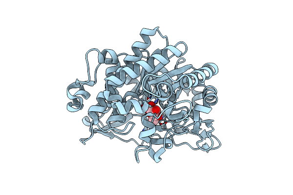
Beta-Glucosidase B Complexed With Thiocellobiose
Organism: Paenibacillus polymyxa
Method: X-RAY DIFFRACTION Resolution:2.30 Å Release Date: 2007-10-02 Classification: HYDROLASE |
Organism: Paenibacillus polymyxa
Method: X-RAY DIFFRACTION
Release Date: 2007-10-02
 |
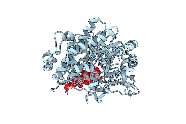
Beta-Glucosidase B From Bacillus Polymyxa Complexed With Glucose
Organism: Paenibacillus polymyxa
Method: X-RAY DIFFRACTION Resolution:2.15 Å Release Date: 2007-10-02 Classification: HYDROLASE Ligands: BGC |
Organism: Paenibacillus polymyxa
Method: X-RAY DIFFRACTION
Release Date: 2007-10-02
Ligands: BGC
 |
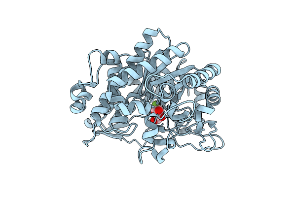
Beta-Glucosidase B From Paenibacillus Polymyxa Complexed With Cellotetraose
Organism: Paenibacillus polymyxa
Method: X-RAY DIFFRACTION Resolution:2.46 Å Release Date: 2007-10-02 Classification: HYDROLASE |
Organism: Paenibacillus polymyxa
Method: X-RAY DIFFRACTION
Release Date: 2007-10-02
 |
Beta-Glucosidase B From Bacillus Polymyxa Complexed With 2-F-Glucose
Organism: Paenibacillus polymyxa
Method: X-RAY DIFFRACTION Resolution:2.30 Å Release Date: 2007-07-03 Classification: HYDROLASE Ligands: G2F |
Organism: Paenibacillus polymyxa
Method: X-RAY DIFFRACTION
Release Date: 2007-07-03
Ligands: G2F
 |
Mutated B-Glucosidase A From Paenibacillus Polymyxa Showing Increased Stability
Organism: Paenibacillus polymyxa
Method: X-RAY DIFFRACTION Resolution:2.20 Å Release Date: 2005-03-03 Classification: HYDROLASE Ligands: G2F, NFG |
Organism: Paenibacillus polymyxa
Method: X-RAY DIFFRACTION
Release Date: 2005-03-03
Ligands: G2F, NFG
 |
Crystal Structure Of Mutated Beta-Glycosidase From Sulfolobus Solfataricus, Working At Moderate Temperature
Organism: Sulfolobus solfataricus
Method: X-RAY DIFFRACTION Resolution:2.55 Å Release Date: 2005-02-09 Classification: HYDROLASE |
Organism: Sulfolobus solfataricus
Method: X-RAY DIFFRACTION
Release Date: 2005-02-09