

Search Count: 22
 |
Cryoem Structure Of The Ring-Shaped Virulence Factor Espb From Mycobacterium Tuberculosis
Organism: Mycobacterium tuberculosis (strain atcc 25618 / h37rv)
Method: ELECTRON MICROSCOPY Release Date: 2020-06-17 Classification: TRANSPORT PROTEIN |
Organism: Mycobacterium tuberculosis (strain atcc 25618 / h37rv)
Method: ELECTRON MICROSCOPY
Release Date: 2020-06-17
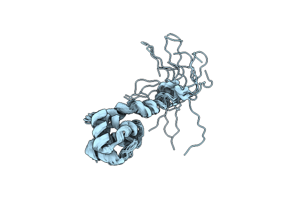 |
Structural And Dna Binding Properties Of Mycobacterial Integration Host Factor Mihf
Organism: Mycobacterium tuberculosis
Method: SOLUTION NMR Release Date: 2019-12-25 Classification: DNA BINDING PROTEIN |
Organism: Mycobacterium tuberculosis
Method: SOLUTION NMR
Release Date: 2019-12-25
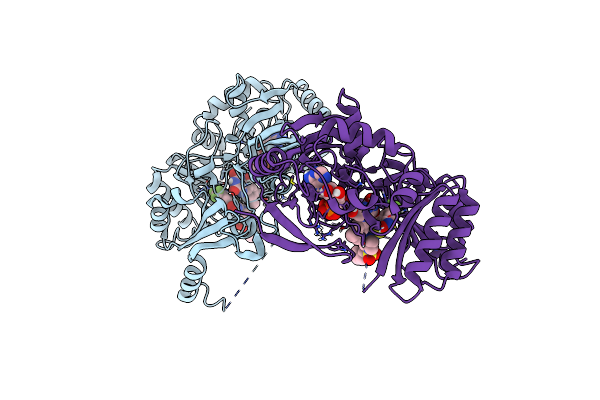 |
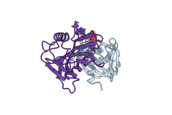
Crystal Structure Of M. Tuberculosis Dpre1 In Complex With Spbtz169 (Sulfonylpbtz)
Organism: Mycobacterium tuberculosis (strain atcc 25618 / h37rv)
Method: X-RAY DIFFRACTION Resolution:2.40 Å Release Date: 2018-08-01 Classification: OXIDOREDUCTASE Ligands: FAD, EQ8 |
Organism: Mycobacterium tuberculosis (strain atcc 25618 / h37rv)
Method: X-RAY DIFFRACTION
Release Date: 2018-08-01
Ligands: FAD, EQ8
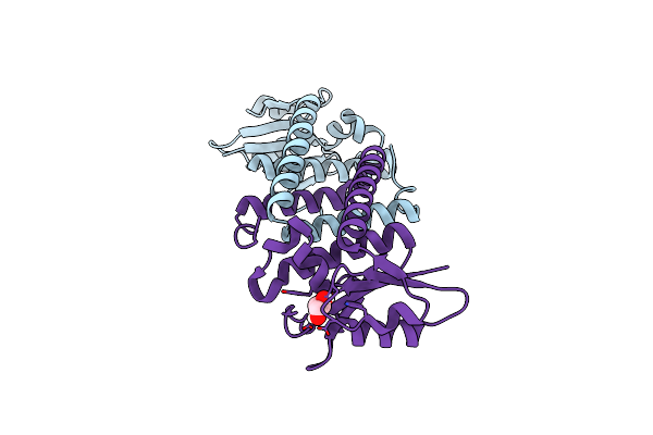 |
Crystal Structure Of Rae1 (Yacp) From Bacillus Subtilis
Organism: Bacillus subtilis (strain 168)
Method: X-RAY DIFFRACTION Resolution:2.25 Å Release Date: 2017-04-19 Classification: TRANSLATION Ligands: GOL |
Organism: Bacillus subtilis (strain 168)
Method: X-RAY DIFFRACTION
Release Date: 2017-04-19
Ligands: GOL
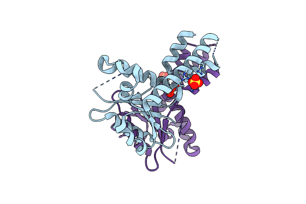 |
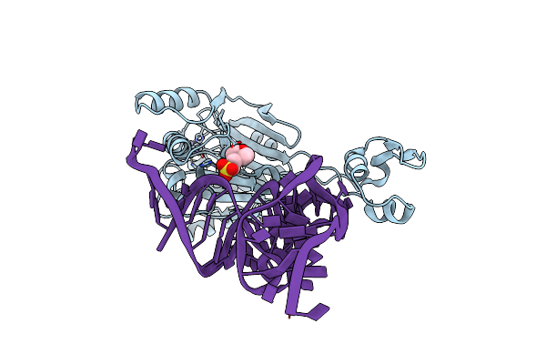
Crystal Structure Of Rae1 (Yacp) From Bacillus Subtilis (W164L Mutant)
Organism: Bacillus subtilis subsp. subtilis str. 168
Method: X-RAY DIFFRACTION Resolution:3.17 Å Release Date: 2017-04-19 Classification: TRANSLATION Ligands: SO4 |
Organism: Bacillus subtilis subsp. subtilis str. 168
Method: X-RAY DIFFRACTION
Release Date: 2017-04-19
Ligands: SO4
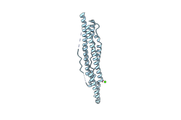 |
Structure Of Pe-Ppe Domains Of Esx-1 Secreted Protein Espb, C2221 In Presence Of Ca
Organism: Mycobacterium tuberculosis
Method: X-RAY DIFFRACTION Resolution:1.82 Å Release Date: 2015-02-18 Classification: PROTEIN TRANSPORT Ligands: CA |
Organism: Mycobacterium tuberculosis
Method: X-RAY DIFFRACTION
Release Date: 2015-02-18
Ligands: CA
 |
Structure Of Pe-Ppe Domains Of Esx-1 Secreted Protein Espb, I222
Organism: Mycobacterium tuberculosis
Method: X-RAY DIFFRACTION Resolution:2.14 Å Release Date: 2015-02-18 Classification: PROTEIN TRANSPORT Ligands: CL, NA |
Organism: Mycobacterium tuberculosis
Method: X-RAY DIFFRACTION
Release Date: 2015-02-18
Ligands: CL, NA
 |
Structure Of Pe-Ppe Domains Of Esx-1 Secreted Protein Espb, C2221
Organism: Mycobacterium tuberculosis
Method: X-RAY DIFFRACTION Resolution:1.50 Å Release Date: 2015-02-18 Classification: PROTEIN TRANSPORT Ligands: CL, GOL |
Organism: Mycobacterium tuberculosis
Method: X-RAY DIFFRACTION
Release Date: 2015-02-18
Ligands: CL, GOL
 |
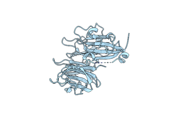
Structure Of Esx-1 Secreted Protein Espb
Organism: Mycobacterium tuberculosis
Method: X-RAY DIFFRACTION Resolution:3.04 Å Release Date: 2015-02-18 Classification: PROTEIN TRANSPORT |
Organism: Mycobacterium tuberculosis
Method: X-RAY DIFFRACTION
Release Date: 2015-02-18
 |
Mycobacterium Tuberculosis Gyrase Type Iia Topoisomerase C-Terminal Domain At 1.4 A Resolution
Organism: Mycobacterium tuberculosis
Method: X-RAY DIFFRACTION Resolution:1.40 Å Release Date: 2013-08-07 Classification: ISOMERASE |
Organism: Mycobacterium tuberculosis
Method: X-RAY DIFFRACTION
Release Date: 2013-08-07
 |
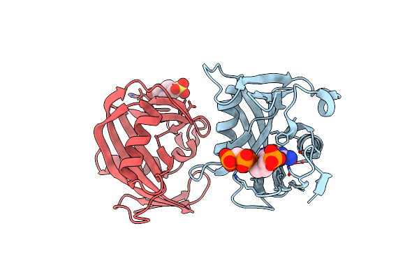
Crystal Structure Of The Bacillus Subtilis Pyrophosphohydrolase Bsrpph (E68A Mutant)
Organism: Bacillus subtilis subsp. subtilis
Method: X-RAY DIFFRACTION Resolution:2.20 Å Release Date: 2013-05-08 Classification: HYDROLASE Ligands: EPE |
Organism: Bacillus subtilis subsp. subtilis
Method: X-RAY DIFFRACTION
Release Date: 2013-05-08
Ligands: EPE
 |
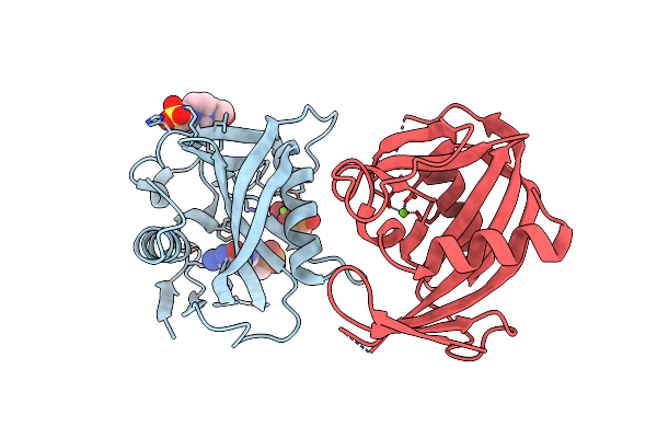
Crystal Structure Of The Bacillus Subtilis Pyrophosphohydrolase Bsrpph Bound To A Non-Hydrolysable Triphosphorylated Dinucleotide Rna (Pcp-Pgpg) - First Guanosine Residue In Guanosine Binding Pocket
Organism: Bacillus subtilis subsp. subtilis
Method: X-RAY DIFFRACTION Resolution:1.70 Å Release Date: 2013-05-08 Classification: HYDROLASE/RNA Ligands: EPE |
Organism: Bacillus subtilis subsp. subtilis
Method: X-RAY DIFFRACTION
Release Date: 2013-05-08
Ligands: EPE
 |
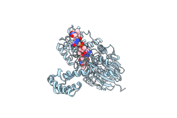
Crystal Structure Of The Bacillus Subtilis Pyrophosphohydrolase Bsrpph Bound To A Non-Hydrolysable Triphosphorylated Dinucleotide Rna (Pcp-Pgpg) - Second Guanosine Residue In Guanosine Binding Pocket
Organism: Bacillus subtilis subsp. subtilis
Method: X-RAY DIFFRACTION Resolution:2.20 Å Release Date: 2013-05-08 Classification: HYDROLASE/RNA Ligands: MG, EPE |
Organism: Bacillus subtilis subsp. subtilis
Method: X-RAY DIFFRACTION
Release Date: 2013-05-08
Ligands: MG, EPE
 |
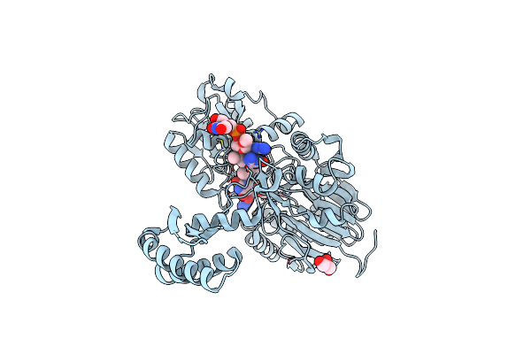
Crystal Structure Of The Bacillus Subtilis Pyrophosphohydrolase Bsrpph (E68A Mutant) Bound To Gtp
Organism: Bacillus subtilis subsp. subtilis
Method: X-RAY DIFFRACTION Resolution:2.90 Å Release Date: 2013-05-01 Classification: HYDROLASE Ligands: GTP |
Organism: Bacillus subtilis subsp. subtilis
Method: X-RAY DIFFRACTION
Release Date: 2013-05-01
Ligands: GTP
 |
Crystal Structure Of Rnase Z In Complex With Precursor Trna(Thr)
Organism: Bacillus subtilis subsp. subtilis
Method: X-RAY DIFFRACTION Resolution:3.00 Å Release Date: 2012-10-31 Classification: HYDROLASE/RNA Ligands: ZN, MES |
Organism: Bacillus subtilis subsp. subtilis
Method: X-RAY DIFFRACTION
Release Date: 2012-10-31
Ligands: ZN, MES
 |
Molecular Basis For The Recognition And Cleavage Of Rna (Uuccgu) By The Bifunctional 5'-3' Exo/Endoribonuclease Rnase J
Organism: Thermus thermophilus hb27
Method: X-RAY DIFFRACTION Resolution:3.09 Å Release Date: 2011-10-19 Classification: HYDROLASE/RNA Ligands: ZN |
Organism: Thermus thermophilus hb27
Method: X-RAY DIFFRACTION
Release Date: 2011-10-19
Ligands: ZN
 |
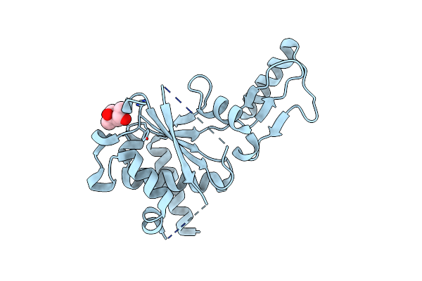
Molecular Basis For The Recognition And Cleavage Of Rna (Cugg) By The Bifunctional 5'-3' Exo/Endoribonuclease Rnase J
Organism: Thermus thermophilus hb27
Method: X-RAY DIFFRACTION Resolution:2.50 Å Release Date: 2011-10-19 Classification: HYDROLASE/RNA Ligands: ZN, GOL |
Organism: Thermus thermophilus hb27
Method: X-RAY DIFFRACTION
Release Date: 2011-10-19
Ligands: ZN, GOL
 |
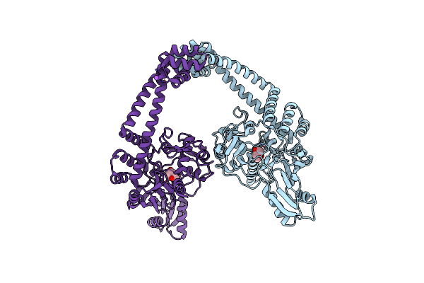
Crystal Structure Of The Second Part Of The Mycobacterium Tuberculosis Dna Gyrase Reaction Core: The Toprim Domain At 1.95 A Resolution
Organism: Mycobacterium tuberculosis
Method: X-RAY DIFFRACTION Resolution:1.95 Å Release Date: 2010-09-29 Classification: ISOMERASE Ligands: MPD |
Organism: Mycobacterium tuberculosis
Method: X-RAY DIFFRACTION
Release Date: 2010-09-29
Ligands: MPD
 |
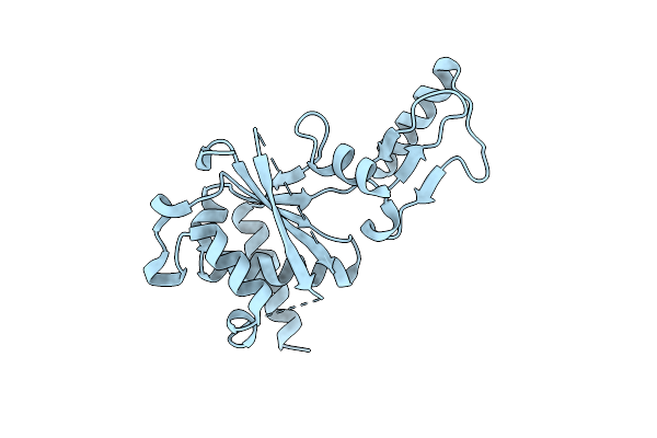
Crystal Structure Of The First Part Of The Mycobacterium Tuberculosis Dna Gyrase Reaction Core: The Breakage And Reunion Domain At 2.7 A Resolution
Organism: Mycobacterium tuberculosis
Method: X-RAY DIFFRACTION Resolution:2.70 Å Release Date: 2010-07-28 Classification: ISOMERASE Ligands: MPD, NA |
Organism: Mycobacterium tuberculosis
Method: X-RAY DIFFRACTION
Release Date: 2010-07-28
Ligands: MPD, NA
 |
Crystal Structure Of The Second Part Of The Mycobacterium Tuberculosis Dna Gyrase Reaction Core: The Toprim Domain At 2.1 A Resolution
Organism: Mycobacterium tuberculosis
Method: X-RAY DIFFRACTION Resolution:2.10 Å Release Date: 2010-07-28 Classification: ISOMERASE |
Organism: Mycobacterium tuberculosis
Method: X-RAY DIFFRACTION
Release Date: 2010-07-28