

Search Count: 17
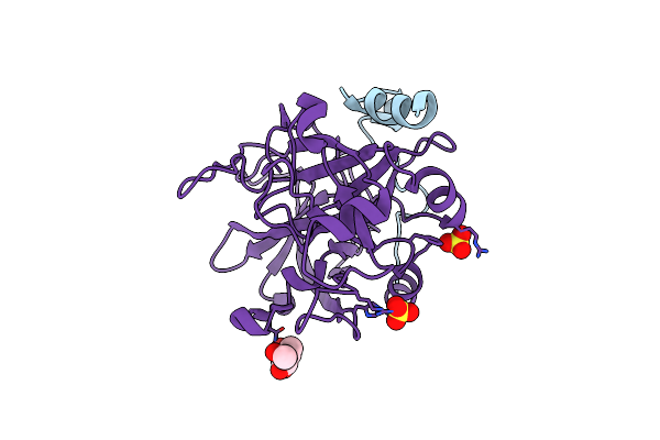 |
Inhibitor-Free Human Thrombin Mutant C191A-C220A
Organism: Homo sapiens
Method: X-RAY DIFFRACTION Resolution:1.54 Å Release Date: 2007-07-17 Classification: HYDROLASE Ligands: NAG, SO4 |
Organism: Homo sapiens
Method: X-RAY DIFFRACTION
Release Date: 2007-07-17
Ligands: NAG, SO4
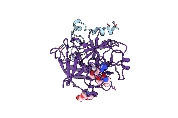 |
Human Thrombin Mutant C191A-C220A In Complex With The Inhibitor Ppack
Organism: Homo sapiens
Method: X-RAY DIFFRACTION Resolution:1.80 Å Release Date: 2007-07-17 Classification: HYDROLASE/HYDROLASE INHIBITOR Ligands: ZN, 0G6, NAG |
Organism: Homo sapiens
Method: X-RAY DIFFRACTION
Release Date: 2007-07-17
Ligands: ZN, 0G6, NAG
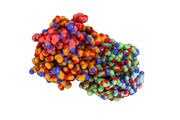 |
Crystal Structure Of Thrombin In Complex With Fibrinogen Gamma' Peptide
Organism: Homo sapiens
Method: X-RAY DIFFRACTION Resolution:2.40 Å Release Date: 2006-09-19 Classification: HYDROLASE |
Organism: Homo sapiens
Method: X-RAY DIFFRACTION
Release Date: 2006-09-19
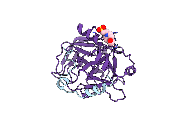 |
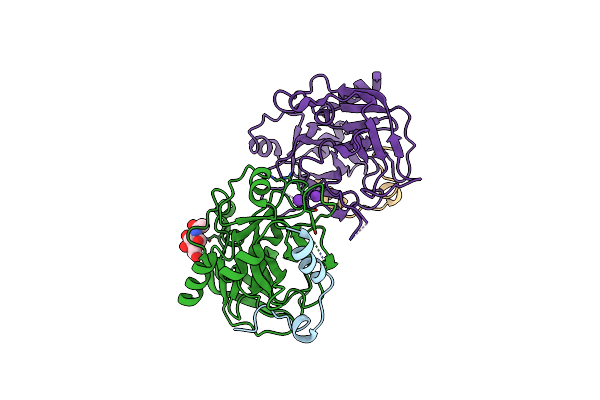
Crystal Structure Of The Slow Form Of Thrombin In A Self-Inhibited Conformation
Organism: Homo sapiens
Method: X-RAY DIFFRACTION Resolution:1.87 Å Release Date: 2006-09-12 Classification: HYDROLASE Ligands: EPE |
Organism: Homo sapiens
Method: X-RAY DIFFRACTION
Release Date: 2006-09-12
Ligands: EPE
 |
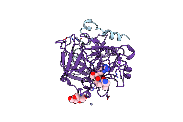
Structure Of Thrombin In 400 Mm Potassium Chloride
Organism: Homo sapiens
Method: X-RAY DIFFRACTION Resolution:1.90 Å Release Date: 2005-07-12 Classification: HYDROLASE Ligands: NDG, K, CL |
Organism: Homo sapiens
Method: X-RAY DIFFRACTION
Release Date: 2005-07-12
Ligands: NDG, K, CL
 |
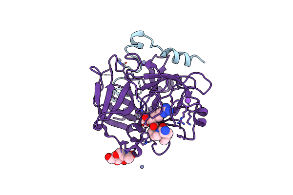
Crystal Structure Of The Thrombin Mutant G193A Bound To Ppack
Organism: Homo sapiens
Method: X-RAY DIFFRACTION Resolution:2.00 Å Release Date: 2005-05-17 Classification: HYDROLASE/HYDROLASE INHIBITOR Ligands: ZN, 0G6, NAG, NA |
Organism: Homo sapiens
Method: X-RAY DIFFRACTION
Release Date: 2005-05-17
Ligands: ZN, 0G6, NAG, NA
 |
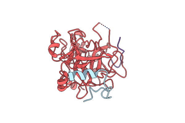
Crystal Structure Of The Thrombin Mutant G193P Bound To Ppack
Organism: Homo sapiens
Method: X-RAY DIFFRACTION Resolution:2.00 Å Release Date: 2005-05-17 Classification: HYDROLASE/HYDROLASE INHIBITOR Ligands: ZN, 0G6, NAG, NA |
Organism: Homo sapiens
Method: X-RAY DIFFRACTION
Release Date: 2005-05-17
Ligands: ZN, 0G6, NAG, NA
 |
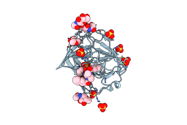
Crystal Structure Of The Thrombin Mutant D221A/D222K
Organism: Homo sapiens
Method: X-RAY DIFFRACTION Resolution:2.40 Å Release Date: 2005-04-19 Classification: HYDROLASE/HYDROLASE INHIBITOR Ligands: NAG |
Organism: Homo sapiens
Method: X-RAY DIFFRACTION
Release Date: 2005-04-19
Ligands: NAG
 |
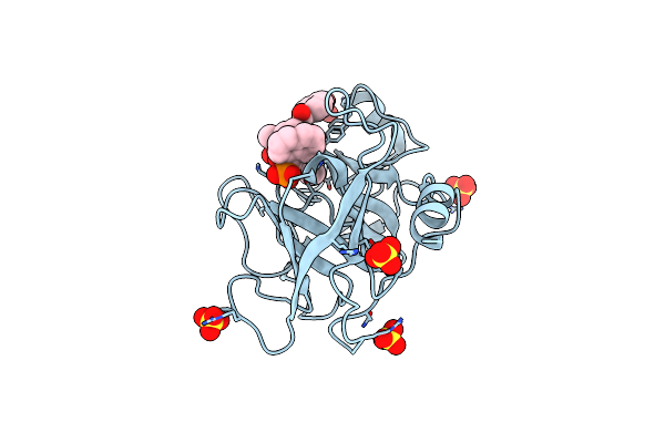
A Dual Inhibitor Of The Leukocyte Proteases Cathepsin G And Chymase With Therapeutic Efficacy In Animals Models Of Inflammation
Organism: Homo sapiens
Method: X-RAY DIFFRACTION Resolution:1.90 Å Release Date: 2005-03-01 Classification: HYDROLASE Ligands: NAG, SO4, CO, OHH, MES |
Organism: Homo sapiens
Method: X-RAY DIFFRACTION
Release Date: 2005-03-01
Ligands: NAG, SO4, CO, OHH, MES
 |
A Dual Inhibitor Of The Leukocyte Proteases Cathepsin G And Chymase With Therapeutic Efficacy In Animals Models Of Inflammation
Organism: Homo sapiens
Method: X-RAY DIFFRACTION Resolution:1.85 Å Release Date: 2005-03-01 Classification: HYDROLASE Ligands: SO4, OHH |
Organism: Homo sapiens
Method: X-RAY DIFFRACTION
Release Date: 2005-03-01
Ligands: SO4, OHH
 |
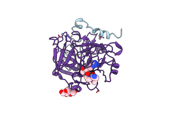
Crystal Structure Of The Potent Anticoagulant Thrombin Mutant W215A/E217A In Free Form
Organism: Homo sapiens
Method: X-RAY DIFFRACTION Resolution:2.80 Å Release Date: 2004-08-03 Classification: BLOOD CLOTTING |
Organism: Homo sapiens
Method: X-RAY DIFFRACTION
Release Date: 2004-08-03
 |
Crystal Structure Of The Anticoagulant Thrombin Mutant W215A/E217A Bound To Ppack
Organism: Homo sapiens
Method: X-RAY DIFFRACTION Resolution:2.40 Å Release Date: 2004-08-03 Classification: HYDROLASE/HYDROLASE INHIBITOR Ligands: ZN, 0G6, NAG |
Organism: Homo sapiens
Method: X-RAY DIFFRACTION
Release Date: 2004-08-03
Ligands: ZN, 0G6, NAG
 |
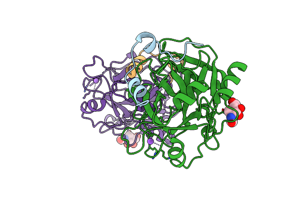
Fast Form Of Thrombin Mutant R(77A)A Bound To Ppack
Organism: Homo sapiens
Method: X-RAY DIFFRACTION Resolution:1.91 Å Release Date: 2004-06-08 Classification: HYDROLASE/hydrolase inhibitor Ligands: 0G6, NAG, NA |
Organism: Homo sapiens
Method: X-RAY DIFFRACTION
Release Date: 2004-06-08
Ligands: 0G6, NAG, NA
 |
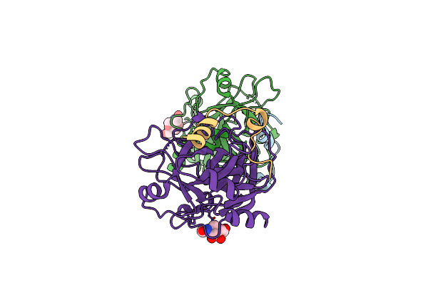
Crystal Structure Of The Procoagulant Fast Form Of Thrombin
Organism: Homo sapiens
Method: X-RAY DIFFRACTION Resolution:2.30 Å Release Date: 2004-06-08 Classification: HYDROLASE Ligands: NAG, NA |
Organism: Homo sapiens
Method: X-RAY DIFFRACTION
Release Date: 2004-06-08
Ligands: NAG, NA
 |
Crystal Structure Of The Anticoagulant Slow Form Of Thrombin
Organism: Homo sapiens
Method: X-RAY DIFFRACTION Resolution:2.30 Å Release Date: 2004-06-08 Classification: HYDROLASE Ligands: NAG |
Organism: Homo sapiens
Method: X-RAY DIFFRACTION
Release Date: 2004-06-08
Ligands: NAG
 |
Slow Form Of Thrombin Bound With Ppack
Organism: Homo sapiens
Method: X-RAY DIFFRACTION Resolution:1.55 Å Release Date: 2004-06-08 Classification: HYDROLASE/hydrolase inhibitor Ligands: 0G6, NAG |
Organism: Homo sapiens
Method: X-RAY DIFFRACTION
Release Date: 2004-06-08
Ligands: 0G6, NAG
 |
Crystal Structure Of The Anticoagulant Slow Form Of Thrombin
Organism: Homo sapiens
Method: X-RAY DIFFRACTION Resolution:2.80 Å Release Date: 2002-11-08 Classification: BLOOD CLOTTING Ligands: NAG |
Organism: Homo sapiens
Method: X-RAY DIFFRACTION
Release Date: 2002-11-08
Ligands: NAG