

Search Count: 266
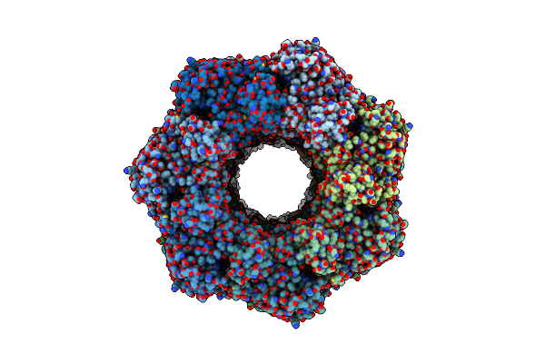 |
Glutamine Synthetase From Pseudomonas Aeruginosa, Filament Double-Unit In Compressed Conformation
Organism: Pseudomonas aeruginosa pao1
Method: ELECTRON MICROSCOPY Release Date: 2024-02-14 Classification: LIGASE |
Organism: Pseudomonas aeruginosa pao1
Method: ELECTRON MICROSCOPY
Release Date: 2024-02-14
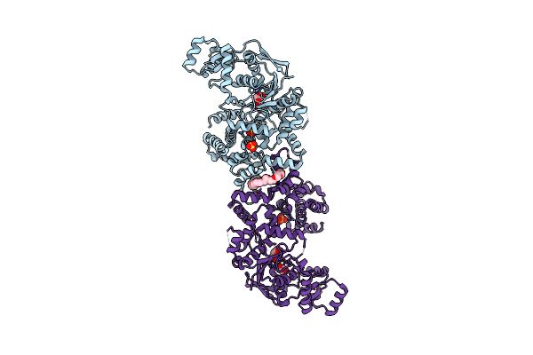 |
Crystal Structure Of Glutamyl-Trna Synthetase Glurs From Pseudomonas Aeruginosa (Zinc Bound)
Organism: Pseudomonas aeruginosa pao1
Method: X-RAY DIFFRACTION Resolution:2.30 Å Release Date: 2023-12-27 Classification: LIGASE Ligands: FLC, SO4, ZN, 2PE, GOL |
Organism: Pseudomonas aeruginosa pao1
Method: X-RAY DIFFRACTION
Release Date: 2023-12-27
Ligands: FLC, SO4, ZN, 2PE, GOL
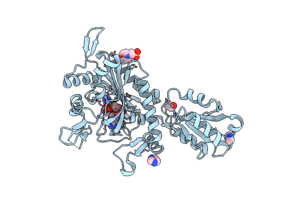 |
Crystal Structure Of Glycine--Trna Ligase From Mycobacterium Tuberculosis (G5A Bound)
Organism: Mycobacterium tuberculosis
Method: X-RAY DIFFRACTION Resolution:2.25 Å Release Date: 2023-09-13 Classification: TRANSFERASE Ligands: G5A, IMD, MES, EDO, MG, CL |
Organism: Mycobacterium tuberculosis
Method: X-RAY DIFFRACTION
Release Date: 2023-09-13
Ligands: G5A, IMD, MES, EDO, MG, CL
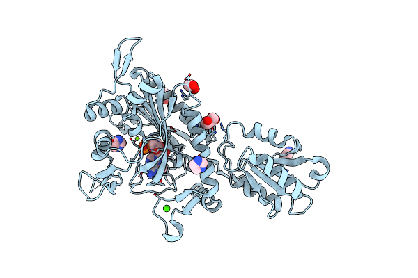 |
Crystal Structure Of Glycine--Trna Ligase From Mycobacterium Tuberculosis (Amp-Mg Bound)
Organism: Mycobacterium tuberculosis
Method: X-RAY DIFFRACTION Resolution:1.65 Å Release Date: 2023-06-28 Classification: LIGASE Ligands: MG, CA, EDO, GOL, IMD, AMP |
Organism: Mycobacterium tuberculosis
Method: X-RAY DIFFRACTION
Release Date: 2023-06-28
Ligands: MG, CA, EDO, GOL, IMD, AMP
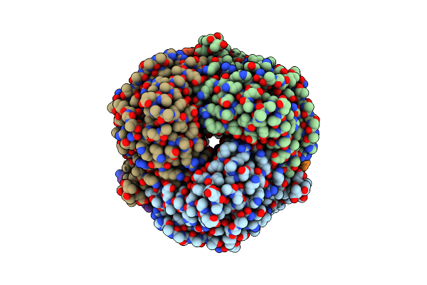 |
Cryo-Em Structure Of Enoyl-Coa Hydratase From Mycobacterium Smegmatis
Organism: Mycolicibacterium smegmatis
Method: ELECTRON MICROSCOPY Release Date: 2023-05-10 Classification: HYDROLASE |
Organism: Mycolicibacterium smegmatis
Method: ELECTRON MICROSCOPY
Release Date: 2023-05-10
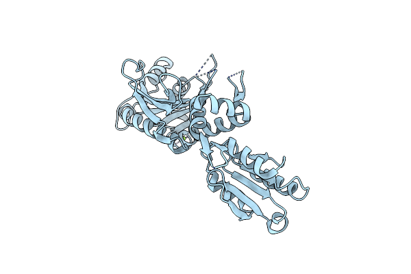 |
Crystal Structure Of Glycine Trna Ligase From Mycobacterium Thermoresistibile (Apo)
Organism: Mycolicibacterium thermoresistibile atcc 19527
Method: X-RAY DIFFRACTION Resolution:2.85 Å Release Date: 2023-05-03 Classification: LIGASE Ligands: MG |
Organism: Mycolicibacterium thermoresistibile atcc 19527
Method: X-RAY DIFFRACTION
Release Date: 2023-05-03
Ligands: MG
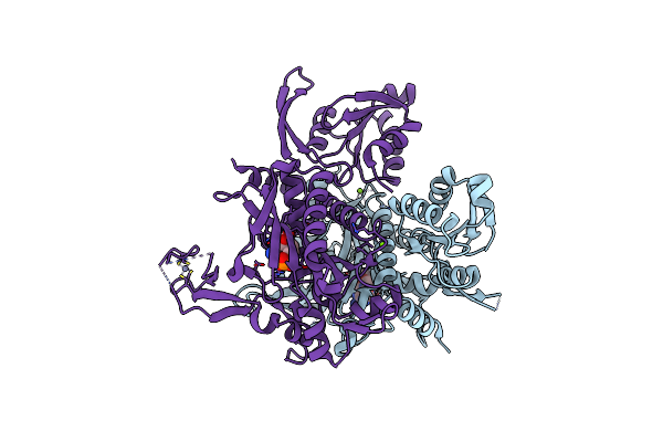 |
Crystal Structure Of Glycine Trna Ligase From Mycobacterium Thermoresistibile (Amp Bound)
Organism: Mycolicibacterium thermoresistibile atcc 19527
Method: X-RAY DIFFRACTION Resolution:2.90 Å Release Date: 2023-05-03 Classification: LIGASE Ligands: AMP, MG, ZN |
Organism: Mycolicibacterium thermoresistibile atcc 19527
Method: X-RAY DIFFRACTION
Release Date: 2023-05-03
Ligands: AMP, MG, ZN
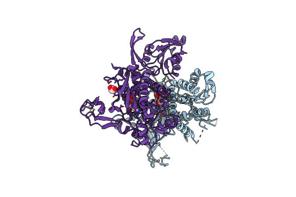 |
Crystal Structure Of Glycine Trna Ligase From Mycobacterium Thermoresistibile (Glycyl Adenylate Bound)
Organism: Mycolicibacterium thermoresistibile atcc 19527
Method: X-RAY DIFFRACTION Resolution:1.95 Å Release Date: 2023-05-03 Classification: LIGASE Ligands: G5A, ZN, MG, GOL |
Organism: Mycolicibacterium thermoresistibile atcc 19527
Method: X-RAY DIFFRACTION
Release Date: 2023-05-03
Ligands: G5A, ZN, MG, GOL
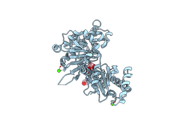 |
Crystal Structure Of Glucokinase From Balamuthia Mandrillaris In Complex With Glucose
Organism: Balamuthia mandrillaris
Method: X-RAY DIFFRACTION Resolution:2.65 Å Release Date: 2020-03-25 Classification: TRANSFERASE Ligands: BGC, CA, EDO |
Organism: Balamuthia mandrillaris
Method: X-RAY DIFFRACTION
Release Date: 2020-03-25
Ligands: BGC, CA, EDO
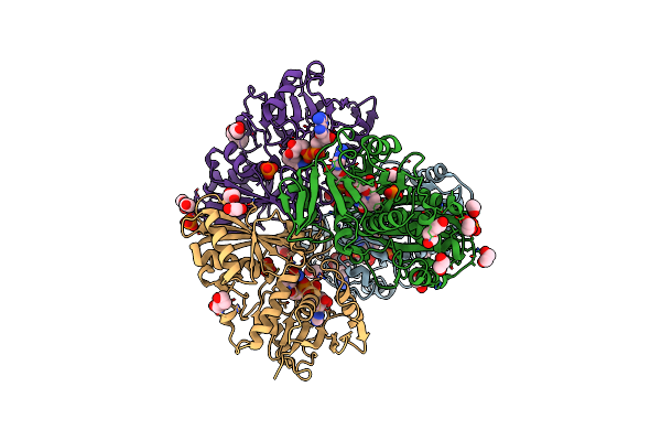 |
Crystal Structure Of Glyceraldehyde-3-Phosphate Dehydrogenase (Gapdh) From Chlamydia Trachomatis With Bound Nad
Organism: Chlamydia trachomatis (strain d/uw-3/cx)
Method: X-RAY DIFFRACTION Resolution:2.40 Å Release Date: 2019-04-24 Classification: OXIDOREDUCTASE Ligands: NAD, EDO, PO4, PEG, CL |
Organism: Chlamydia trachomatis (strain d/uw-3/cx)
Method: X-RAY DIFFRACTION
Release Date: 2019-04-24
Ligands: NAD, EDO, PO4, PEG, CL
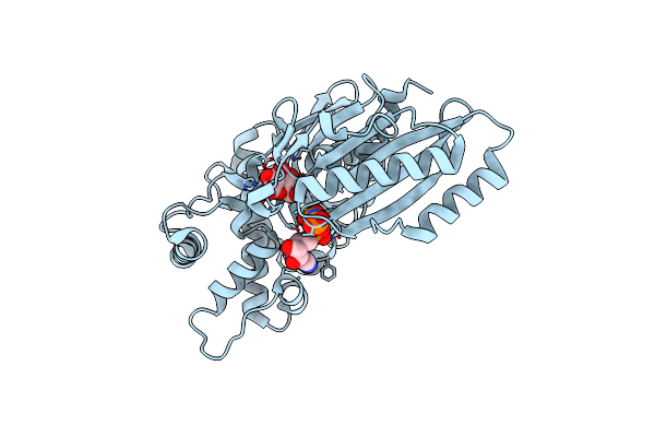 |
Crystal Structure Of Glucokinase (Nfhk) From Naegleria Fowleri
Organism: Naegleria fowleri
Method: X-RAY DIFFRACTION Resolution:2.20 Å Release Date: 2018-05-09 Classification: TRANSFERASE Ligands: BGC, ANP |
Organism: Naegleria fowleri
Method: X-RAY DIFFRACTION
Release Date: 2018-05-09
Ligands: BGC, ANP
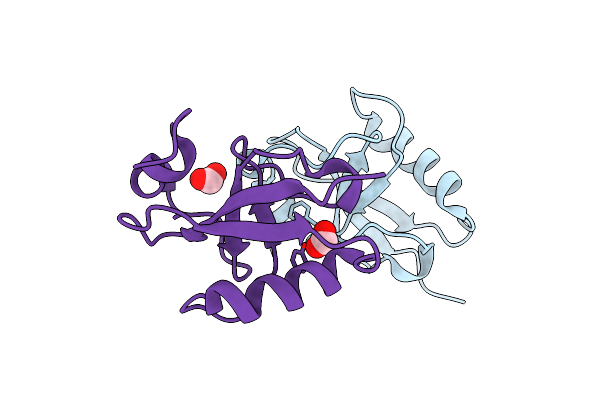 |
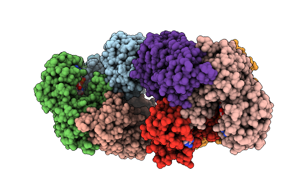
Crystal Structure Of The C-Terminal Domain Of Doublecortin (Tgdcx) From Toxoplasma Gondii Me49
Organism: Toxoplasma gondii
Method: X-RAY DIFFRACTION Resolution:2.00 Å Release Date: 2017-10-18 Classification: STRUCTURAL PROTEIN Ligands: FMT |
Organism: Toxoplasma gondii
Method: X-RAY DIFFRACTION
Release Date: 2017-10-18
Ligands: FMT
 |
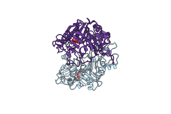
Crystal Structure Of A Glyceraldehyde-3-Phosphate Dehydrogenase From Neisseria Gonorrhoeae Bound To Nad
Organism: Neisseria gonorrhoeae
Method: X-RAY DIFFRACTION Resolution:2.50 Å Release Date: 2017-05-10 Classification: OXIDOREDUCTASE Ligands: NAD, CL |
Organism: Neisseria gonorrhoeae
Method: X-RAY DIFFRACTION
Release Date: 2017-05-10
Ligands: NAD, CL
 |
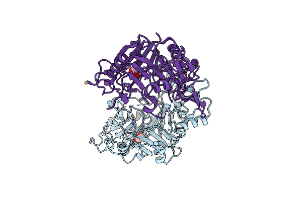
Crystal Structure Of Putative Putative Deoxyribonuclease-2 From Burkholderia Thailandensis, E264
Organism: Burkholderia thailandensis
Method: X-RAY DIFFRACTION Resolution:1.65 Å Release Date: 2017-02-15 Classification: HYDROLASE Ligands: EDO |
Organism: Burkholderia thailandensis
Method: X-RAY DIFFRACTION
Release Date: 2017-02-15
Ligands: EDO
 |
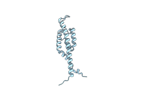
Crystal Structure Of Putative Putative Deoxyribonuclease-2 From Burkholderia Thailandensis In Complex With Copper
Organism: Burkholderia thailandensis (strain atcc 700388 / dsm 13276 / cip 106301 / e264)
Method: X-RAY DIFFRACTION Resolution:1.75 Å Release Date: 2017-02-15 Classification: HYDROLASE Ligands: CU, EDO |
Organism: Burkholderia thailandensis (strain atcc 700388 / dsm 13276 / cip 106301 / e264)
Method: X-RAY DIFFRACTION
Release Date: 2017-02-15
Ligands: CU, EDO
 |
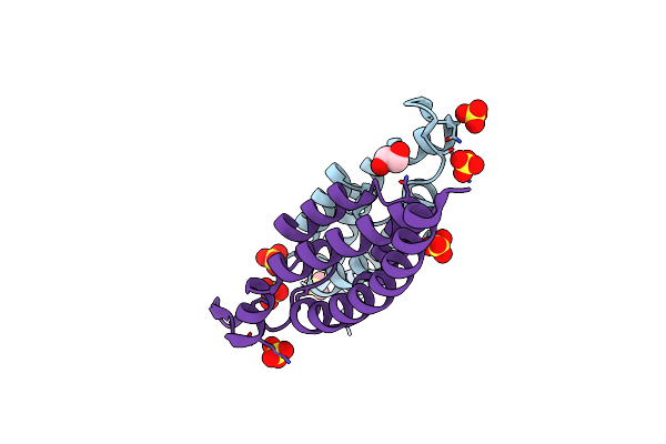
Crystal Structure Of The Bid Domain Of Bep6 From Bartonella Rochalimae
Organism: Bartonella rochalimae atcc baa-1498
Method: X-RAY DIFFRACTION Resolution:2.10 Å Release Date: 2016-03-09 Classification: PROTEIN BINDING |
Organism: Bartonella rochalimae atcc baa-1498
Method: X-RAY DIFFRACTION
Release Date: 2016-03-09
 |
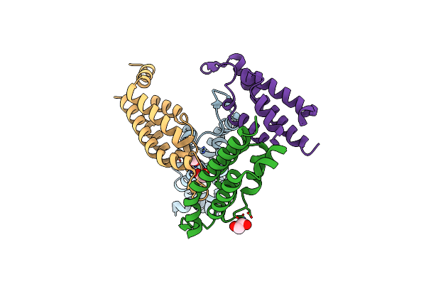
Crystal Structure Of The Bid Domain Of Bep9 From Bartonella Clarridgeiae
Organism: Bartonella clarridgeiae
Method: X-RAY DIFFRACTION Resolution:2.05 Å Release Date: 2016-03-09 Classification: PROTEIN BINDING Ligands: SO4, EDO |
Organism: Bartonella clarridgeiae
Method: X-RAY DIFFRACTION
Release Date: 2016-03-09
Ligands: SO4, EDO
 |
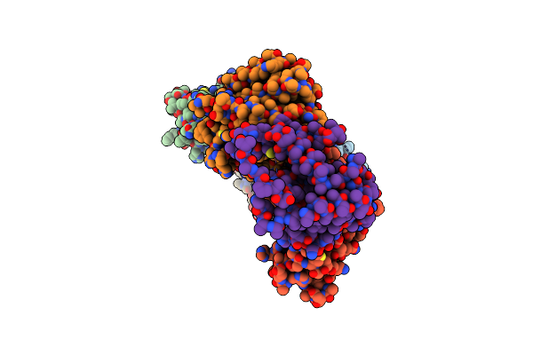
Crystal Structure Of The Bid Domain Of Bepe From Bartonella Henselae
Organism: Bartonella henselae str. houston-1
Method: X-RAY DIFFRACTION Resolution:2.20 Å Release Date: 2016-03-09 Classification: PROTEIN BINDING Ligands: EDO |
Organism: Bartonella henselae str. houston-1
Method: X-RAY DIFFRACTION
Release Date: 2016-03-09
Ligands: EDO
 |
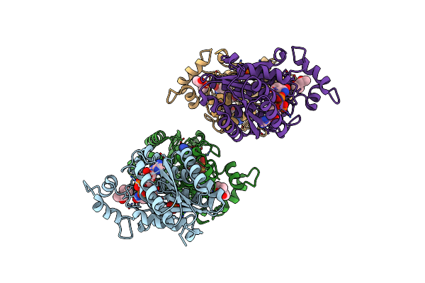
Crystal Structure Of The Fic Domain Of Bep5 Protein (Virb-Translocated Bartonella Effector Protein) From Bartonella Clarridgeiae
Organism: Bartonella clarridgeiae (strain cip 104772 / 73)
Method: X-RAY DIFFRACTION Resolution:2.95 Å Release Date: 2016-01-13 Classification: PROTEIN BINDING Ligands: EDO |
Organism: Bartonella clarridgeiae (strain cip 104772 / 73)
Method: X-RAY DIFFRACTION
Release Date: 2016-01-13
Ligands: EDO
 |
Crystal Structure Of Carveol Dehydrogenase From Mycobacterium Avium In Complex With Nad
Organism: Mycobacterium avium
Method: X-RAY DIFFRACTION Resolution:2.15 Å Release Date: 2015-12-02 Classification: OXIDOREDUCTASE Ligands: NAD, PO4, MPD |
Organism: Mycobacterium avium
Method: X-RAY DIFFRACTION
Release Date: 2015-12-02
Ligands: NAD, PO4, MPD