

Search Count: 34
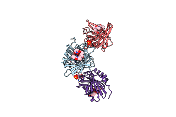 |
Fundamental Characterization Of Chelated And Crystallized Actinium In A Macromolecular Host
Organism: Homo sapiens
Method: X-RAY DIFFRACTION Resolution:2.00 Å Release Date: 2024-09-25 Classification: METAL BINDING PROTEIN Ligands: LA, 4OL, SO4, PIN |
Organism: Homo sapiens
Method: X-RAY DIFFRACTION
Release Date: 2024-09-25
Ligands: LA, 4OL, SO4, PIN
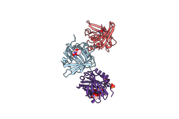 |
Fundamental Characterization Of Chelated And Crystallized Actinium In A Macromolecular Host
Organism: Homo sapiens
Method: X-RAY DIFFRACTION Resolution:2.08 Å Release Date: 2024-09-25 Classification: METAL BINDING PROTEIN Ligands: ZTM, 4OL, SO4, CL |
Organism: Homo sapiens
Method: X-RAY DIFFRACTION
Release Date: 2024-09-25
Ligands: ZTM, 4OL, SO4, CL
 |
Bio-2895 (Brd0705) Bound Gsk3Alpha-Axin Complex
Organism: Homo sapiens
Method: X-RAY DIFFRACTION Resolution:1.94 Å Release Date: 2023-06-14 Classification: HYDROLASE Ligands: 6VL, CA |
Organism: Homo sapiens
Method: X-RAY DIFFRACTION
Release Date: 2023-06-14
Ligands: 6VL, CA
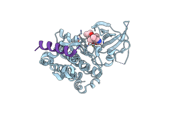 |
Bio-8546 Bound Gsk3Alpha-Axin Complex
Organism: Homo sapiens
Method: X-RAY DIFFRACTION Resolution:2.40 Å Release Date: 2023-06-14 Classification: HYDROLASE Ligands: D1E |
Organism: Homo sapiens
Method: X-RAY DIFFRACTION
Release Date: 2023-06-14
Ligands: D1E
 |
Bio-8546 Bound Gsk3Beta-Axin Complex
Organism: Homo sapiens
Method: X-RAY DIFFRACTION Resolution:2.09 Å Release Date: 2023-06-14 Classification: HYDROLASE Ligands: D1E |
Organism: Homo sapiens
Method: X-RAY DIFFRACTION
Release Date: 2023-06-14
Ligands: D1E
 |
Bio-2895 (Brd0705) Bound Gsk3Beta-Axin Complex
Organism: Homo sapiens
Method: X-RAY DIFFRACTION Resolution:1.85 Å Release Date: 2023-06-14 Classification: HYDROLASE Ligands: 6VL |
Organism: Homo sapiens
Method: X-RAY DIFFRACTION
Release Date: 2023-06-14
Ligands: 6VL
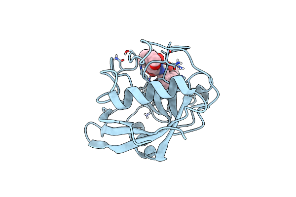 |
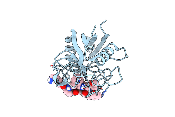
Structure Of Cyclophilin D Peptidyl-Prolyl Isomerase Domain Bound To Macrocyclic Inhibitor Jombt
Organism: Homo sapiens
Method: X-RAY DIFFRACTION Resolution:1.75 Å Release Date: 2022-08-24 Classification: ISOMERASE Ligands: I3Q |
Organism: Homo sapiens
Method: X-RAY DIFFRACTION
Release Date: 2022-08-24
Ligands: I3Q
 |
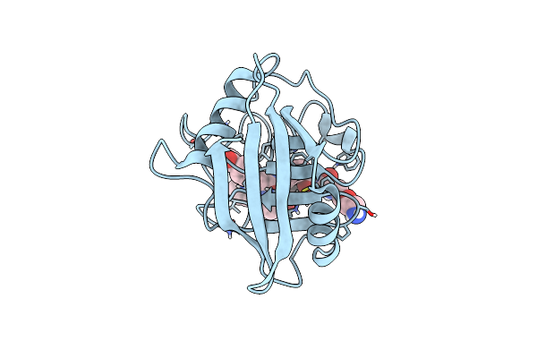
Structure Of Cyclophilin D Peptidyl-Prolyl Isomerase Domain Bound To Macrocyclic Inhibitor A26
Organism: Homo sapiens
Method: X-RAY DIFFRACTION Resolution:1.06 Å Release Date: 2022-08-24 Classification: ISOMERASE Ligands: I4F |
Organism: Homo sapiens
Method: X-RAY DIFFRACTION
Release Date: 2022-08-24
Ligands: I4F
 |
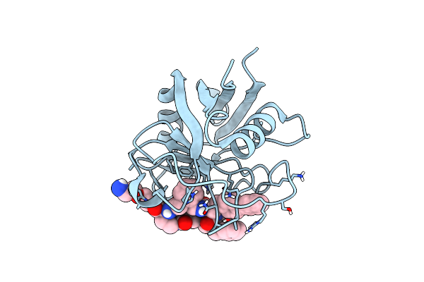
Structure Of Cyclophilin D Peptidyl-Prolyl Isomerase Domain Bound To Macrocyclic Inhibitor B1
Organism: Homo sapiens
Method: X-RAY DIFFRACTION Resolution:1.21 Å Release Date: 2022-08-24 Classification: ISOMERASE/ISOMERASE inhibitor Ligands: I44 |
Organism: Homo sapiens
Method: X-RAY DIFFRACTION
Release Date: 2022-08-24
Ligands: I44
 |
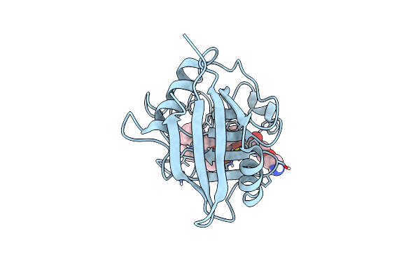
Structure Of Cyclophilin D Peptidyl-Prolyl Isomerase Domain Bound To Macrocyclic Inhibitor B2
Organism: Homo sapiens
Method: X-RAY DIFFRACTION Resolution:1.46 Å Release Date: 2022-08-24 Classification: ISOMERASE/ISOMERASE inhibitor Ligands: I3X |
Organism: Homo sapiens
Method: X-RAY DIFFRACTION
Release Date: 2022-08-24
Ligands: I3X
 |
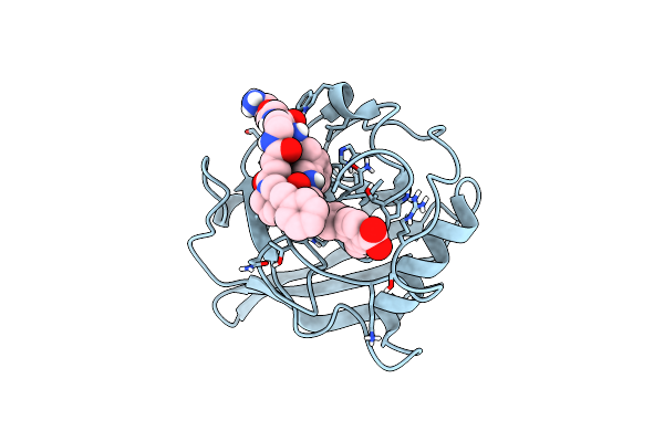
Structure Of Cyclophilin D Peptidyl-Prolyl Isomerase Domain Bound To Macrocyclic Inhibitor B3
Organism: Homo sapiens
Method: X-RAY DIFFRACTION Resolution:1.52 Å Release Date: 2022-08-24 Classification: ISOMERASE Ligands: I5J |
Organism: Homo sapiens
Method: X-RAY DIFFRACTION
Release Date: 2022-08-24
Ligands: I5J
 |
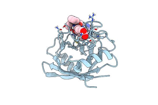
Structure Of Cyclophilin D Peptidyl-Prolyl Isomerase Domain Bound To Macrocyclic Inhibitor B21
Organism: Homo sapiens
Method: X-RAY DIFFRACTION Resolution:0.97 Å Release Date: 2022-08-24 Classification: ISOMERASE Ligands: I56 |
Organism: Homo sapiens
Method: X-RAY DIFFRACTION
Release Date: 2022-08-24
Ligands: I56
 |
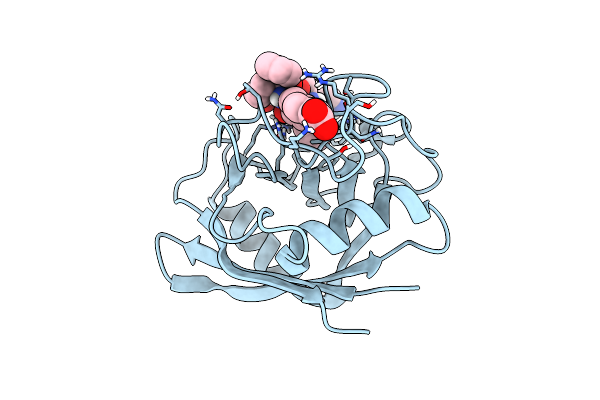
Structure Of Cyclophilin D Peptidyl-Prolyl Isomerase Domain Bound To Macrocyclic Inhibitor B23
Organism: Homo sapiens
Method: X-RAY DIFFRACTION Resolution:1.30 Å Release Date: 2022-08-24 Classification: ISOMERASE Ligands: I5E |
Organism: Homo sapiens
Method: X-RAY DIFFRACTION
Release Date: 2022-08-24
Ligands: I5E
 |
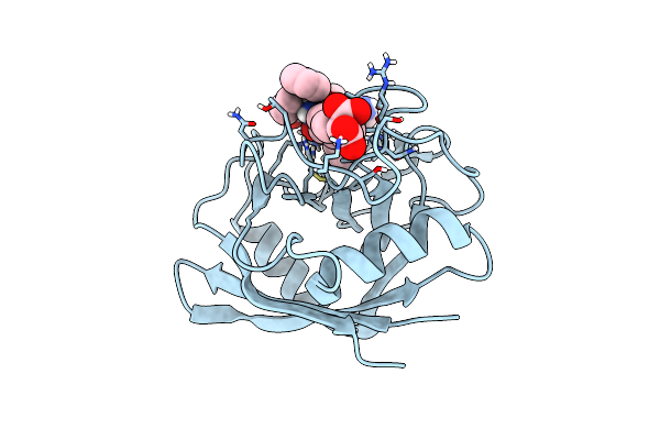
Structure Of Cyclophilin D Peptidyl-Prolyl Isomerase Domain Bound To Macrocyclic Inhibitor B25
Organism: Homo sapiens
Method: X-RAY DIFFRACTION Resolution:1.57 Å Release Date: 2022-08-24 Classification: ISOMERASE Ligands: I4Z |
Organism: Homo sapiens
Method: X-RAY DIFFRACTION
Release Date: 2022-08-24
Ligands: I4Z
 |
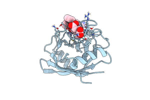
Structure Of Cyclophilin D Peptidyl-Prolyl Isomerase Domain Bound To Macrocyclic Inhibitor B52
Organism: Homo sapiens
Method: X-RAY DIFFRACTION Resolution:1.16 Å Release Date: 2022-08-24 Classification: ISOMERASE Ligands: I4V |
Organism: Homo sapiens
Method: X-RAY DIFFRACTION
Release Date: 2022-08-24
Ligands: I4V
 |
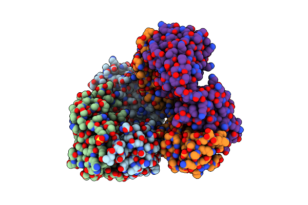
Structure Of Cyclophilin D Peptidyl-Prolyl Isomerase Domain Bound To Macrocyclic Inhibitor B53
Organism: Homo sapiens
Method: X-RAY DIFFRACTION Resolution:1.10 Å Release Date: 2022-08-24 Classification: ISOMERASE/ISOMERASE INHIBITOR Ligands: I4R |
Organism: Homo sapiens
Method: X-RAY DIFFRACTION
Release Date: 2022-08-24
Ligands: I4R
 |
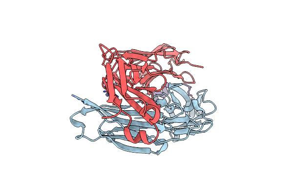
Crystal Structure Of A Fab Fragment Bound To Peptide Ggm
Organism: Oryctolagus cuniculus, Homo sapiens
Method: X-RAY DIFFRACTION Resolution:2.85 Å Release Date: 2021-06-16 Classification: IMMUNE SYSTEM Ligands: SO4 |
Organism: Oryctolagus cuniculus, Homo sapiens
Method: X-RAY DIFFRACTION
Release Date: 2021-06-16
Ligands: SO4
 |
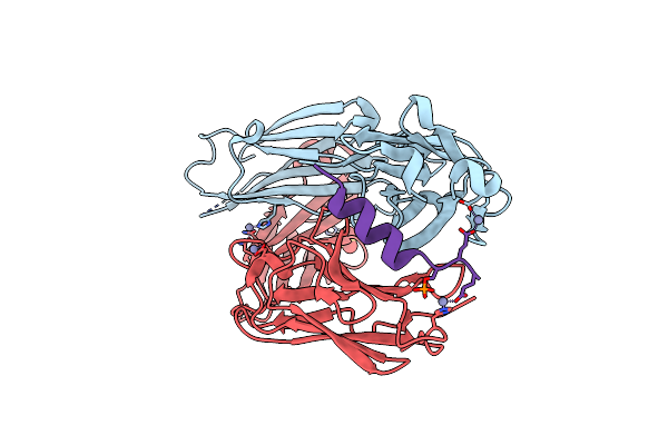
Anti-Pcsk9 Fab 6E2 Bound To The N-Terminal Peptide From Pcsk9, Unmodified
Organism: Mus musculus, Homo sapiens
Method: X-RAY DIFFRACTION Resolution:2.24 Å Release Date: 2019-04-10 Classification: IMMUNE SYSTEM Ligands: ZN, CL |
Organism: Mus musculus, Homo sapiens
Method: X-RAY DIFFRACTION
Release Date: 2019-04-10
Ligands: ZN, CL
 |
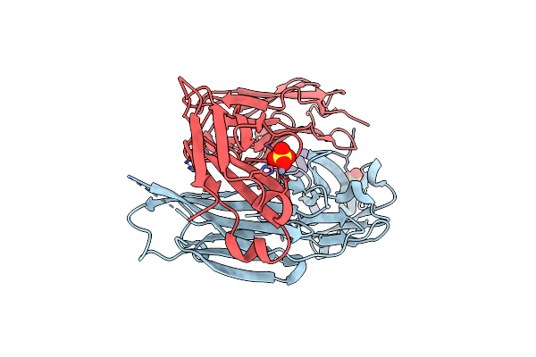
Anti-Pcsk9 Fab 6E2 Bound To The Modified N-Terminal Peptide From Pcsk9
Organism: Mus musculus, Homo sapiens
Method: X-RAY DIFFRACTION Resolution:2.20 Å Release Date: 2019-04-10 Classification: IMMUNE SYSTEM Ligands: ZN, SO4 |
Organism: Mus musculus, Homo sapiens
Method: X-RAY DIFFRACTION
Release Date: 2019-04-10
Ligands: ZN, SO4
 |
Anti-Pcsk9 Fab 6E2 Bound To The N-Terminal Peptide From Pcsk9 (E32K)
Organism: Mus musculus, Homo sapiens
Method: X-RAY DIFFRACTION Resolution:2.10 Å Release Date: 2019-04-10 Classification: IMMUNE SYSTEM Ligands: ZN, CL, SO4 |
Organism: Mus musculus, Homo sapiens
Method: X-RAY DIFFRACTION
Release Date: 2019-04-10
Ligands: ZN, CL, SO4